

Characterization of interlimb interaction via transcutaneous spinal stimulation of cervical and lumbar spinal enlargements
- PMID: 35320019
- PMCID: PMC8993515
- DOI: 10.1152/jn.00456.2021
Characterization of interlimb interaction via transcutaneous spinal stimulation of cervical and lumbar spinal enlargements
Abstract
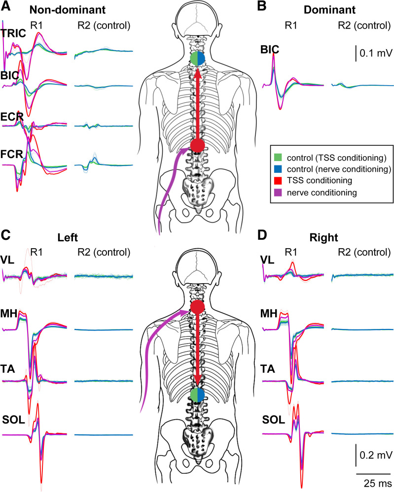
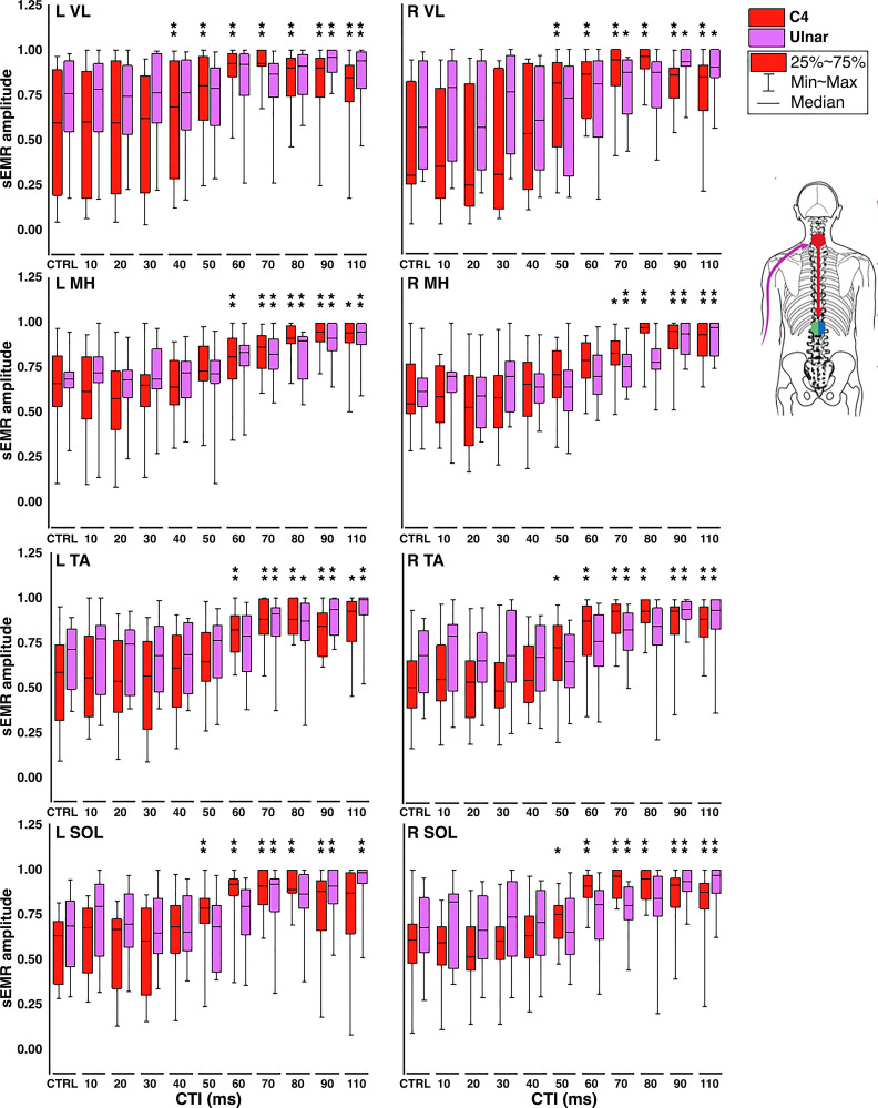
The use of transcutaneous electrical spinal stimulation (TSS) to modulate sensorimotor networks after neurological insult has garnered much attention from both researchers and clinicians in recent years. Although many different stimulation paradigms have been reported, the interlimb effects of these neuromodulation techniques have been little studied. The effects of multisite TSS on interlimb sensorimotor function are of particular interest in the context of neurorehabilitation, as these networks have been shown to be important for functional recovery after neurological insult. The present study utilized a condition-test paradigm to investigate the effects of interenlargement TSS on spinal motor excitability in both cervical and lumbosacral motor pools. Additionally, comparison was made between the conditioning effects of lumbosacral and cervical TSS and peripheral stimulation of the fibular nerve and ulnar nerve, respectively. In 16/16 supine, relaxed participants, facilitation of spinally evoked motor responses (sEMRs) in arm muscles was seen in response to lumbosacral TSS or fibular nerve stimulation, whereas facilitation of sEMRs in leg muscles was seen in response to cervical TSS or ulnar nerve stimulation. The decreased latency between TSS- and peripheral nerve-evoked conditioning implicates interlimb networks in the observed facilitation of motor output. The results demonstrate the ability of multisite TSS to engage interlimb networks, resulting in the bidirectional influence of cervical and lumbosacral motor output. The engagement of interlimb networks via TSS of the cervical and lumbosacral enlargements represents a feasible method for engaging spinal sensorimotor networks in clinical populations with compromised motor function.NEW & NOTEWORTHY Bidirectional interlimb modulation of spinal motor excitability can be evoked by transcutaneous spinal stimulation over the cervical and lumbosacral enlargements. Multisite transcutaneous spinal stimulation engages spinal sensorimotor networks thought to be important in the recovery of function after spinal cord injury.
Keywords: human neurophysiology; interlimb reflexes; propriospinal; spinal cord; transcutaneous spinal stimulation.
Conflict of interest statement
Y.P.G. holds a shareholder interest in NeuroRecovery Technologies and Cosyma. He holds certain inventorship rights on intellectual property licensed by the regents of the University of California to NeuroRecovery Technologies and its subsidiaries. None of the other authors has any conflicts of interest, financial or otherwise, to disclose.
Figures


References
-
- Gill ML, Grahn PJ, Calvert JS, Linde MB, Lavrov IA, Strommen JA, Beck LA, Sayenko DG, Van Straaten MG, Drubach DI, Veith DD, Thoreson AR, Lopez C, Gerasimenko YP, Edgerton VR, Lee KH, Zhao KD. Neuromodulation of lumbosacral spinal networks enables independent stepping after complete paraplegia. Nat Med 24: 1677–1682, 2018. doi: 10.1038/s41591-018-0175-7. - DOI - PubMed
-
- Grahn PJ, Lavrov IA, Sayenko DG, Van Straaten MG, Gill ML, Strommen JA, Calvert JS, Drubach DI, Beck LA, Linde MB, Thoreson AR, Lopez C, Mendez AA, Gad PN, Gerasimenko YP, Edgerton VR, Zhao KD, Lee KH. Enabling task-specific volitional motor functions via spinal cord neuromodulation in a human with paraplegia. Mayo Clin Proc 92: 544–554, 2017. doi: 10.1016/j.mayocp.2017.02.014. - DOI - PubMed
Publication types
MeSH terms
Grants and funding
LinkOut - more resources
Full Text Sources
Medical