. 2022 Dec;11(1):1037-1048.
doi: 10.1080/22221751.2022.2057874.

Peptide microarrays coupled to machine learning reveal individual epitopes from human antibody responses with neutralizing capabilities against SARS-CoV-2

Affiliations

Peptide microarrays coupled to machine learning reveal individual epitopes from human antibody responses with neutralizing capabilities against SARS-CoV-2

Sven-Kevin Hotop et al. Emerg Microbes Infect. 2022 Dec.

Abstract

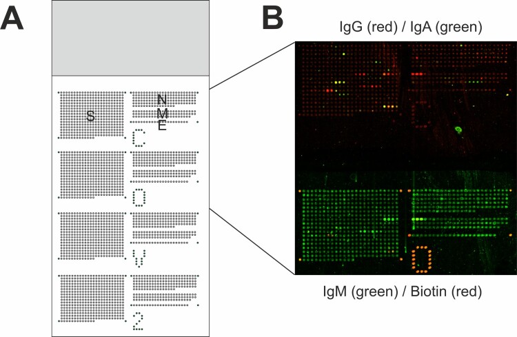
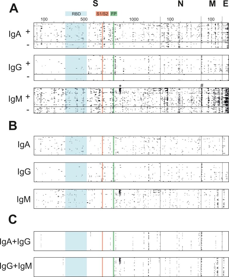
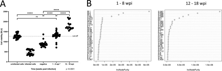
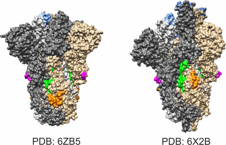
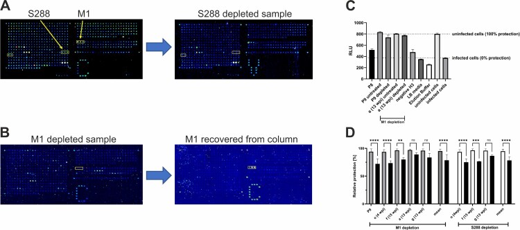
The coronavirus SARS-CoV-2 is the causative agent for the disease COVID-19. To capture the IgA, IgG, and IgM antibody response of patients infected with SARS-CoV-2 at individual epitope resolution, we constructed planar microarrays of 648 overlapping peptides that cover the four major structural proteins S(pike), N(ucleocapsid), M(embrane), and E(nvelope). The arrays were incubated with sera of 67 SARS-CoV-2 positive and 22 negative control samples. Specific responses to SARS-CoV-2 were detectable, and nine peptides were associated with a more severe course of the disease. A random forest model disclosed that antibody binding to 21 peptides, mostly localized in the S protein, was associated with higher neutralization values in cellular anti-SARS-CoV-2 assays. For antibodies addressing the N-terminus of M, or peptides close to the fusion region of S, protective effects were proven by antibody depletion and neutralization assays. The study pinpoints unusual viral binding epitopes that might be suited as vaccine candidates.

Keywords: COVID-19; SARS CoV-2; immunoassays; machine learning; neutralizing antibodies; peptide arrays; serology.


Conflict of interest statement

No potential conflict of interest was reported by the author(s).

Figures

Figure 1.
Figure 1.
Slide layout and exemplary data of fluorescent signals obtained for sample ID f. (A) Each slide holds 2592 peptide sequences and 86 biotin control spots arranged in four sub-grids. Each sub-grid contains 648 15mer peptides with an offset of three amino acids to the next peptide, encompassing the whole sequences of S, N M, and E protein derived from ref. seq. NC_045512. (B) Bound antibodies against SARS-CoV-2 peptide sequences were visualized with isotype-specific anti-human secondary antibodies coupled to different fluorescent dyes or fluorescently labelled streptavidin. In one sub-grid, IgG (red) and IgA (green) antibodies were detected. Antibodies bound to the same peptide sequences produce yellow signals as overlay of red and green signals. In the adjacent sub-grid, IgM (green) and biotin (red) were detected.
Figure 2.
Figure 2.
Distribution of positively reacting peptides across SARS-CoV-2 proteins. (A) Each antibody-bound (positive) peptide spot is given as a black square for each isotype (IgA, IgG, and IgM), with 89 rows for 67 SARS-CoV-2 positive (+; upper rows) and 22 SARS-CoV-2 negative samples (−; lower rows) per isotype, and as many columns, as there are peptides on the chip covering the respective protein. Positive and negative samples are separated by a thick horizontal line. Amino acid numbers are indicated for all four proteins investigated; the receptor-binding domain (RBD; aa 319–514) is given in blue, S1/S2 cleavage site (aa 680–685) in red and fusion peptide (FP; aa 788–806) is given in green; borders between the four proteins are indicated by thin vertical lines. Peptides bound to potentially neutralizing antibodies are marked with * and #. (B) SARS-CoV-2 specific positively reacting peptides were observed in data set. The isotype data sets of all 67 positive samples were depleted from spots that were observed in any negative sample, resulting in SARS-CoV-2 humoral immune answers that were specified in this study. (C) Determination of immunodominant epitopes within the specific data set as depicted in Figure 2(B). Specific peptides are marked if they were detected in at least 10% of the samples in individual sets (IgA, IgG, and IgM).
Figure 3.
Figure 3.
Identification of peptide epitopes contributing to neutralizing capabilities of serum samples. (A) All serum samples (n = 89) were tested for their ability to neutralize SARS-CoV-2 infectivity in Vero E6 cells using an ATP-based viability assay. The mean Relative Light Unit (RLU) level of uninfected cells is visualized as a dotted line, serving as a cut-off for complete protection from viral infection. Positive samples were divided into two groups of 1–8 weeks post-infection (wpi) (n = 51) and 12–18 wpi (n = 16). *No significant differences were observed dividing the group 1–8 wpi into 1–4 wpi and 5–8 wpi (data not shown). ****p < .0001. (B) Detection of peptides contributing to neutralization by a random forest analysis for sera from 1–8 wpi and 12–18 wpi. Peptides are ranked via the mean increase in node purity (IncNodePurity) when splitting on the variable, averaged over all decision trees in the random forest.
Figure 4.
Figure 4.
Mapping of neutralizing antibody binding region on the spike protein. Projection of antibody target site (aa 853–879; green) onto spike trimer (Chain A, B, and C in grey, dark grey, and brown) in closed (PDB: 6ZB5) and open state (6X2B). The binding domain is close to the fusion peptide (aa 788–806; orange). The S1–S2 cleavage site is disordered in crystal structures, but adjacent amino acids were marked in magenta. Amino acids relevant for ACE2 binding are given in blue.
Figure 5.
Figure 5.
Depletion of antibodies against M1 and S288 impairs viral neutralization capacity. (A and B) Control of depletion efficacy by peptide microarrays. (A) Serum f (15 wpi) was depleted from S288 binding antibodies by an S288-functionalized column. Peptide microarrays before (left) and after (right) the depletion step exhibit lower signals in the S288 region. (B) Peptide microarrays after purification of serum a (6 wpi) by an M1-functionalized column (left) and after re-elution from the column (right). A depletion (left) and recovery (right) of M1-binding antibodies is visible. Positions of peptides M1–M3 and S286–S290 on the array are marked by yellow boxes. (C) Neutralization capacity of sera following M1 depletion. Viability of Vero E6 cells is assessed by their ATP content and detected in relative luminescence units (RLU). Uninfected cells were viable, whereas infected cells showed cytopathic effects, leading to lower RLU reads (two right bars). The samples P9 and a (13 wpi) showed a slight reduction of neutralizing capacity compared to the untreated control. All samples were diluted 1:10 and measured four times per experiment. (D) Relative protection as measured in C. As more samples for M1 depletion were available we measured five samples (untreated controls: grey bars vs. depleted samples: black bars) and three of them with known reactivity against S288 were used for depletion (untreated controls: white bars). The obtained readouts showed comparable results ranging from 7.88% to 21.82% with both significant outcomes for mean values (p < .0001).

Similar articles

Cited by

References

    1. Deeks JJ, Dinnes J, Takwoingi Y, et al. . Antibody tests for identification of current and past infection with SARS-CoV-2. Cochrane Database Syst Rev. 2020;6:CD013652. - PMC - PubMed
    1. Kontou PI, Braliou GG, Dimou NL, et al. . Antibody tests in detecting SARS-CoV-2 infection: a meta-analysis. Diagnostics (Basel). 2020;10(5):319. - PMC - PubMed
    1. Hotop SK, Abd El Wahed A, Beutling U.et al. . Serological analysis of Herpes B virus at individual epitope resolution: from two-dimensional peptide arrays to multiplex bead flow assays. Anal Chem. 2019;91(17):11030–11037. - PubMed
    1. Heiss K, Heidepriem J, Fischer N, et al. . Rapid response to pandemic threats: immunogenic epitope detection of pandemic pathogens for diagnostics and vaccine development using peptide microarrays. J Proteome Res. 2020;19(11):4339–4354. - PMC - PubMed
    1. Szymczak LC, Kuo HY, Mrksich M.. Peptide arrays: development and application. Anal Chem. 2018;90(1):266–282. - PMC - PubMed