

Development of a dual antigen lateral flow immunoassay for detecting Yersinia pestis
- PMID: 35320275
- PMCID: PMC8979426
- DOI: 10.1371/journal.pntd.0010287
Development of a dual antigen lateral flow immunoassay for detecting Yersinia pestis
Abstract
Background: Yersinia pestis is the causative agent of plague, a zoonosis associated with small mammals. Plague is a severe disease, especially in the pneumonic and septicemic forms, where fatality rates approach 100% if left untreated. The bacterium is primarily transmitted via flea bite or through direct contact with an infected host. The 2017 plague outbreak in Madagascar resulted in more than 2,400 cases and was highlighted by an increased number of pneumonic infections. Standard diagnostics for plague include laboratory-based assays such as bacterial culture and serology, which are inadequate for administering immediate patient care for pneumonic and septicemic plague.
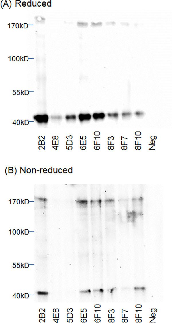
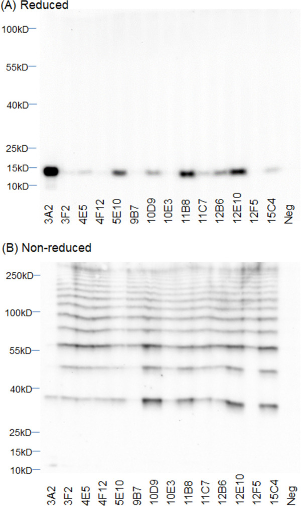
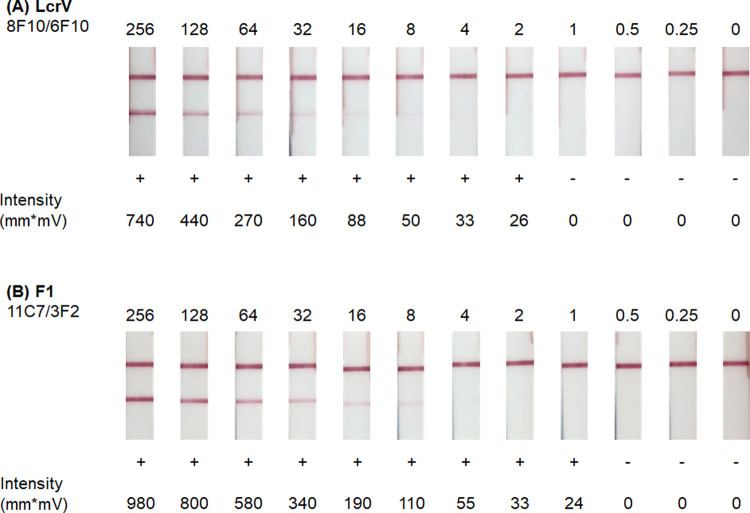
Principal findings: The goal of this study was to develop a sensitive rapid plague prototype that can detect all virulent strains of Y. pestis. Monoclonal antibodies (mAbs) were produced against two Y. pestis antigens, low-calcium response V (LcrV) and capsular fraction-1 (F1), and prototype lateral flow immunoassays (LFI) and enzyme-linked immunosorbent assays (ELISA) were constructed. The LFIs developed for the detection of LcrV and F1 had limits of detection (LOD) of roughly 1-2 ng/mL in surrogate clinical samples (antigens spiked into normal human sera). The optimized antigen-capture ELISAs produced LODs of 74 pg/mL for LcrV and 61 pg/mL for F1 when these antigens were spiked into buffer. A dual antigen LFI prototype comprised of two test lines was evaluated for the detection of both antigens in Y. pestis lysates. The dual format was also evaluated for specificity using a small panel of clinical near-neighbors and other Tier 1 bacterial Select Agents.
Conclusions: LcrV is expressed by all virulent Y. pestis strains, but homologs produced by other Yersinia species can confound assay specificity. F1 is specific to Y. pestis but is not expressed by all virulent strains. Utilizing highly reactive mAbs, a dual-antigen detection (multiplexed) LFI was developed to capitalize on the diagnostic strengths of each target.
Conflict of interest statement
I have read the journal’s policy and the authors of this manuscript have the following competing interests: The goal of this research funded by Naval Research Laboratory contract ND0173-16-C-2003 and the Defense Threat Reduction Agency (DTRA) contract HDTRA1-16-C-0026 was to develop a diagnostic for plague. Currently the project is funded to transition these prototypes into an FDA approved diagnostic for plague through the MCDC contract # MCDC OTA W15QKN-16-9-1002.
Figures
Similar articles
-
Critical Comparison between Large and Mini Vertical Flow Immunoassay Platforms for Yersinia Pestis Detection.Anal Chem. 2021 Jul 13;93(27):9337-9344. doi: 10.1021/acs.analchem.0c05278. Epub 2021 May 14. Anal Chem. 2021. PMID: 33989499
-
Rapid and sensitive detection of Yersinia pestis by lateral-flow assay in simulated clinical samples.BMC Infect Dis. 2018 Aug 14;18(1):402. doi: 10.1186/s12879-018-3315-2. BMC Infect Dis. 2018. PMID: 30107826 Free PMC article.
-
Development of a PCR-lateral flow assay for rapid detection of Yersinia pestis, the causative agent of plague.Acta Trop. 2021 Aug;220:105958. doi: 10.1016/j.actatropica.2021.105958. Epub 2021 May 15. Acta Trop. 2021. PMID: 34004173
-
Plague vaccines and the molecular basis of immunity against Yersinia pestis.Hum Vaccin. 2009 Dec;5(12):817-23. doi: 10.4161/hv.9866. Epub 2009 Dec 1. Hum Vaccin. 2009. PMID: 19786842 Review.
-
[Yersinia pestis and plague - an update].Med Monatsschr Pharm. 2014 Dec;37(12):441-8; quiz 449. Med Monatsschr Pharm. 2014. PMID: 25643450 Review. German.
Cited by
-
Immunogenetics, sylvatic plague and its vectors: insights from the pathogen reservoir Mastomys natalensis in Tanzania.Immunogenetics. 2023 Dec;75(6):517-530. doi: 10.1007/s00251-023-01323-7. Epub 2023 Oct 19. Immunogenetics. 2023. PMID: 37853246 Free PMC article.
-
PepSeq as a highly multiplexed platform for melioidosis antigen discovery and vaccine development.Front Immunol. 2025 Jul 3;16:1605758. doi: 10.3389/fimmu.2025.1605758. eCollection 2025. Front Immunol. 2025. PMID: 40677719 Free PMC article.
-
Functional assays to screen and select monoclonal antibodies that target Yersinia pestis.Hum Vaccin Immunother. 2023 Aug 1;19(2):2216085. doi: 10.1080/21645515.2023.2216085. Epub 2023 Jun 8. Hum Vaccin Immunother. 2023. PMID: 37289480 Free PMC article.
-
Isolation of camel single domain antibodies against Yersinia pestis V270 antigen based on a semi-synthetic single domain antibody library and development of a VHH-based lateral flow assay.Vet Med Sci. 2024 Jul;10(4):e1532. doi: 10.1002/vms3.1532. Vet Med Sci. 2024. PMID: 38952277 Free PMC article.
-
Development of an Antigen Capture Lateral Flow Immunoassay for the Detection of Burkholderia pseudomallei.Diagnostics (Basel). 2024 May 16;14(10):1033. doi: 10.3390/diagnostics14101033. Diagnostics (Basel). 2024. PMID: 38786331 Free PMC article.
References
-
- Benedictow O Jr. The Black Death, 1346–1353: the complete history. Woodbridge, Suffolk, UK; Rochester, N.Y., USA: Boydell Press; 2004. xvi, 433 pages p.
Publication types
MeSH terms
Substances
LinkOut - more resources
Full Text Sources
Medical
