. 2022 Mar 23;17(3):e0265898.
doi: 10.1371/journal.pone.0265898. eCollection 2022.

Observation and motor imagery balance tasks evaluation: An fNIRS feasibility study

Affiliations

Observation and motor imagery balance tasks evaluation: An fNIRS feasibility study

Latifah Almulla et al. PLoS One. .

Abstract

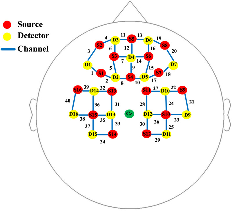
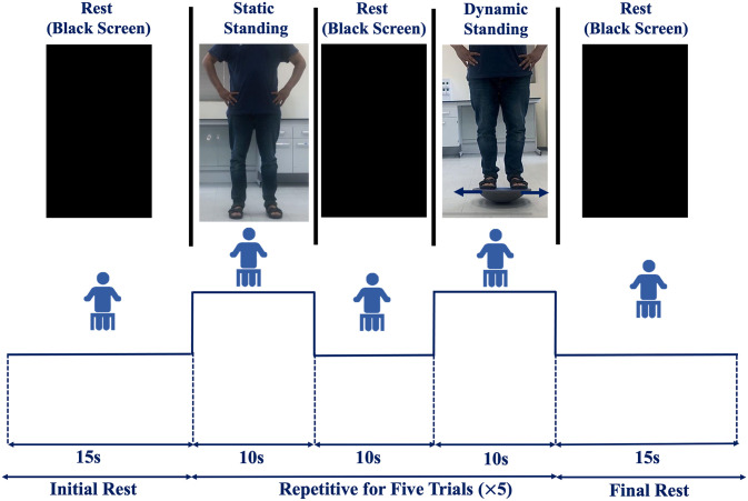
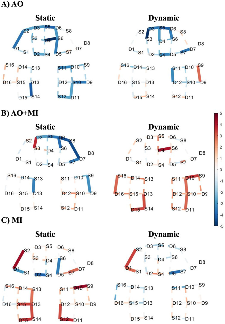
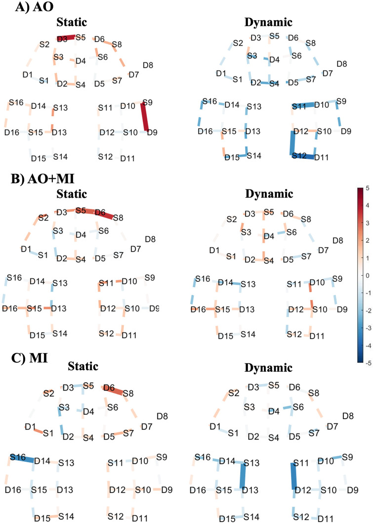
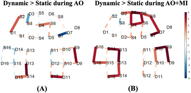
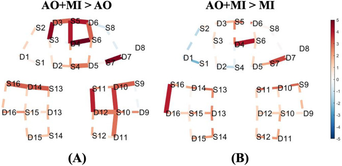
In this study, we aimed at exploring the feasibility of functional near-infrared spectroscopy (fNIRS) for studying the observation and/or motor imagination of various postural tasks. Thirteen healthy adult subjects followed five trials of static and dynamic standing balance tasks, throughout three different experimental setups of action observation (AO), a combination of action observation and motor imagery (AO+MI), and motor imagery (MI). During static and dynamic standing tasks, both the AO+MI and MI experiments revealed that many channels in prefrontal or motor regions are significantly activated while the AO experiment showed almost no significant increase in activations in most of the channels. The contrast between static and dynamic standing tasks showed that with more demanding balance tasks, relative higher activation patterns were observed, particularly during AO and in AO+MI experiments in the frontopolar area. Moreover, the AO+MI experiment revealed a significant difference in premotor and supplementary motor cortices that are related to balance control. Furthermore, it has been observed that the AO+MI experiment induced relatively higher activation patterns in comparison to AO or MI alone. Remarkably, the results of this work match its counterpart from previous functional magnetic resonance imaging studies. Therefore, they may pave the way for using the fNIRS as a diagnostic tool for evaluating the performance of the non-physical balance training during the rehabilitation period of temporally immobilized patients.


Conflict of interest statement

The authors have declared that no competing interests exist.

Figures

Fig 1
Fig 1. Optodes placement with channel numbers configuration placed on prefrontal and motor regions.
Sources are indicated by red, detectors are indicated by yellow, and channels are indicated by blue. Cz point is indicated by green as a reference point.
Fig 2
Fig 2. Experimental paradigm of the three (AO, AO+MI, and MI) experiments.
Subjects watched two videos: static standing balance task (normal standing) and dynamic standing balance task (balancing a mediolateral perturbation), during two experiments: (AO) and while imagining themselves as the person performing the tasks (AO+MI). During the third experiment, participants verbally instructed, through previously recorded voice instructions, to close their eyes and imagine themselves performing static and dynamic balance tasks. Each subject repeated each task five times with a resting period of 10s between the two balance tasks.
Fig 3
Fig 3. T-map of oxy-hemodynamic response (HbO) corresponding to (A) action observation experiment, (B) a combination of motor imagery and action observation experiment, and (C) motor imagery experiment during static standing balance task (left) and dynamic standing balance task (right).
These maps are generated by contrasting each task against the baseline. Significantly activated channels at p < 0.05 (FDR corrected) are indicated by thick and solid lines. The red colour indicates stronger task activity against the baseline. The dashed line shows the channels that were not statistically significant. This map was generated by using NIRS Brain AnalyzIR Toolbox [51]. Fig 2 illustrates the referencing for the brain regions with 10–20 EEG system positions.
Fig 4
Fig 4. T-map of deoxy-hemodynamic response (HbR) corresponding to (A) action observation experiment, (B) a combination of motor imagery and action observation experiment, and (C) motor imagery experiment during static standing balance task (left) and dynamic standing balance task (right).
These maps are generated by contrasting each task against the baseline. Significantly activated channels at p < 0.05 (FDR corrected) are indicated by thick and solid lines. The red colour indicates stronger task activity against the baseline. The dashed line shows the channels that were not statistically significant. This map was generated by using NIRS Brain AnalyzIR Toolbox [51]. Fig 2 illustrates the referencing for the brain regions with 10–20 EEG system positions.
Fig 5
Fig 5. T-map of oxy-hemodynamic response (HbO) corresponding to the contrast between the dynamic standing and static standing balance tasks for (A) action observation experiment, and (B) a combination of motor imagery and action observation experiment.
Significantly activated channels at p < 0.05 (FDR corrected) are indicated by thick, solid lines, red colour indicates stronger activity from the dynamic standing task than the static standing task. The dashed line shows the channels that were not statistically significant. This map was generated by using NIRS Brain AnalyzIR Toolbox [51]. Fig 2 illustrates the referencing for the brain regions with 10–20 EEG system positions.
Fig 6
Fig 6. T-map of oxy-hemodynamic response (HbO) corresponding to the contrast between (A) AO + MI and AO experiments, (B) AO + MI and MI experiments of the dynamic standing task.
Significantly activated channels at p < 0.05 (FDR corrected) are indicated by thick, solid lines, red colour indicates stronger activity from AO + MI experiment than the AO experiment in (A) and MI experiment in (B). The dashed line shows the channels that were not statistically significant. This map was generated by using NIRS Brain AnalyzIR Toolbox [51]. Fig 2 illustrates the referencing for the brain regions with 10–20 EEG system positions.

Similar articles

Cited by

References

    1. Granacher U, Muehlbauer T, Gollhofer A, Kressig RW, Zahner L. An intergenerational approach in the promotion of balance and strength for fall prevention–a mini-review. Gerontology. 2011;57: 304–315. doi: 10.1159/000320250 - DOI - PubMed
    1. Sherrington C, Whitney JC, Lord SR, Herbert RD, Cumming RG, Close JC. Effective exercise for the prevention of falls: a systematic review and meta-analysis. J Am Geriatr Soc. 2008;56: 2234–2243. doi: 10.1111/j.1532-5415.2008.02014.x - DOI - PubMed
    1. Visschedijk J, Achterberg W, Van Balen R, Hertogh C. Fear of falling after hip fracture: a systematic review of measurement instruments, prevalence, interventions, and related factors. J Am Geriatr Soc. 2010;58: 1739–1748. doi: 10.1111/j.1532-5415.2010.03036.x - DOI - PubMed
    1. Gatti R, Sarasso E, Pelachin M, Agosta F, Filippi M, Tettamanti A. Can action observation modulate balance performance in healthy subjects? Arch Physiother. 2019;9: 1. doi: 10.1186/s40945-018-0053-0 - DOI - PMC - PubMed
    1. Mouthon A, Ruffieux J, Wälchli M, Keller M, Taube W. Task-dependent changes of corticospinal excitability during observation and motor imagery of balance tasks. Neuroscience. 2015;303: 535–543. doi: 10.1016/j.neuroscience.2015.07.031 - DOI - PubMed

Publication types