. 2022 May;414(12):3653-3665.
doi: 10.1007/s00216-022-04006-6. Epub 2022 Mar 23.

Changes in the lipid profile of hamster liver after Schistosoma mansoni infection, characterized by mass spectrometry imaging and LC-MS/MS analysis

Affiliations

Changes in the lipid profile of hamster liver after Schistosoma mansoni infection, characterized by mass spectrometry imaging and LC-MS/MS analysis

Katja R Wiedemann et al. Anal Bioanal Chem. 2022 May.

Abstract

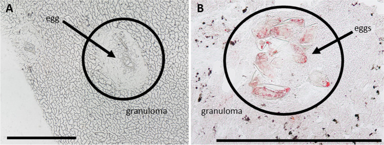
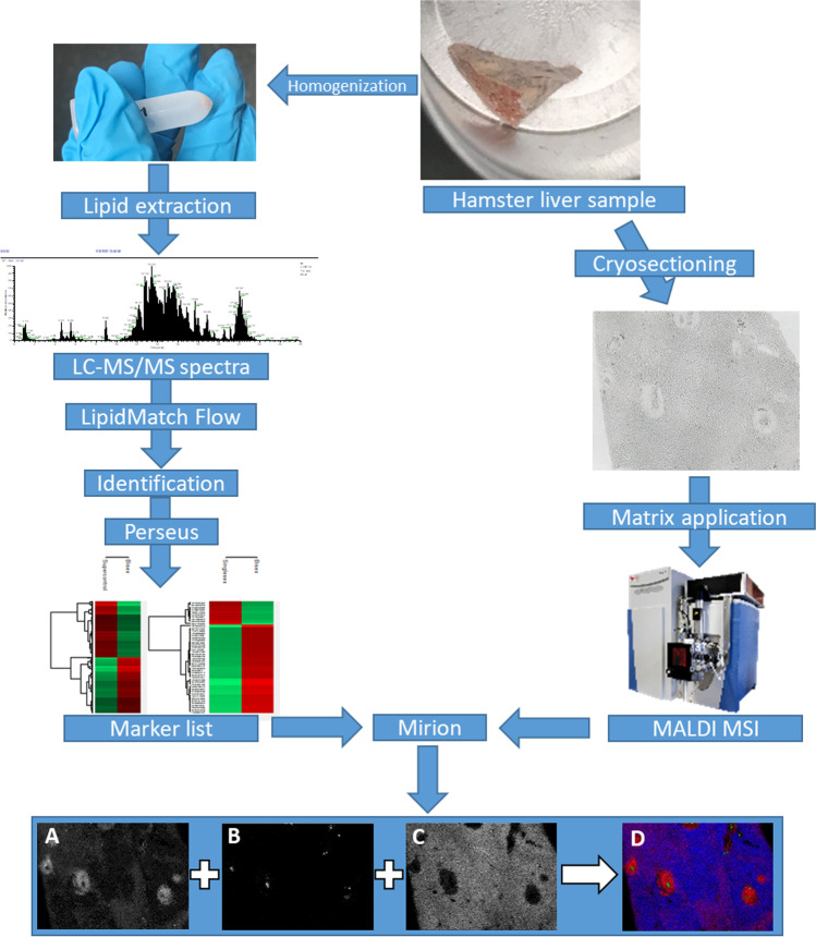
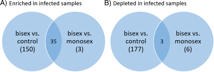
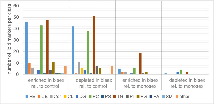
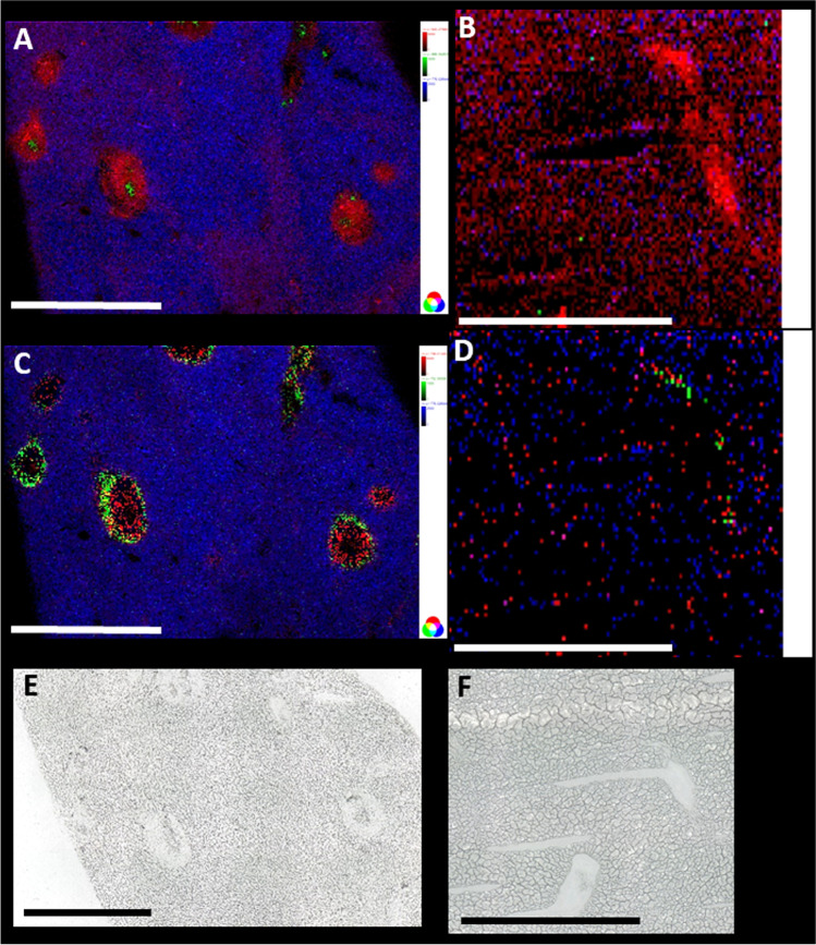
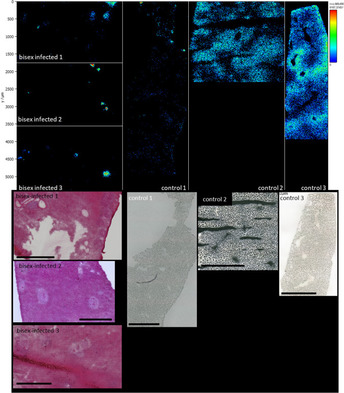
Schistosomiasis, caused by the human parasite Schistosoma mansoni, is one of the WHO-listed neglected tropical diseases (NTDs), and it has severe impact on morbidity and mortality, especially in Africa. Not only the adult worms but also their eggs are responsible for health problems. Up to 50% of the eggs produced by the female worms are not excreted with the feces but are trapped in the host tissue, such as the liver, where they provoke immune responses and a change in the lipid profile. We built up a database with 372 infection markers found in livers of S. mansoni-infected hamsters, using LC-MS/MS for identification, followed by statistical analysis. Most of them belong to the lipid classes of phosphatidylcholines (PCs), phosphatidylethanolamines (PEs), and triglycerides (TGs). We assigned some of these markers to specific anatomical structures by applying high-resolution MALDI MSI to cryosections of hamster liver and generating ion images based on the marker list from the LC-MS/MS experiments. Furthermore, enrichment and depletion of several markers were visualized.

Keywords: AP-SMALDI; Granuloma; Host-parasite interaction; Infection; Mass spectrometry imaging; Parasites; Schistosoma mansoni; Schistosomiasis.


Conflict of interest statement

B.S. and C.G.G. are consultants of TransMIT GmbH, Giessen, Germany. The other authors declare to have no conflicts of interest.

Figures

Fig. 1
Fig. 1
Microscopic images of eggs with granuloma in livers of bisex-infected hamsters. (A) Light microscopic image of unstained tissue. (B) Tissue stained with oil red. Lipid accumulation in granuloma is clearly visible. Scale bars are 500 µm
Fig. 2
Fig. 2
Hamster liver samples were either homogenized or cryosectioned. After homogenization, lipids were extracted, and samples were analyzed using LC–MS/MS. With the help of LipidMatch Flow and Perseus, identified signals were annotated and markers were identified that occurred with statistically determined significance. After cryosectioning, matrix was applied, and samples were measured using MALDI MSI. Using the software Mirion and the marker list from LC–MS/MS experiments, MS images were generated: (A) m/z 500.275684, LPE(20:4), [M − H]; (B) m/z 866.592639, PS(42:4), [M − H]; (C) m/z 776.526946, MMPE(16:0_22:6), [M − H]; (D) RGB overlay of MS images A, B, C
Fig. 3
Fig. 3
Venn diagram showing the numbers of infection markers found. Many more infection markers were found to be enriched when comparing bisex-infected with control samples than with monosex-infected samples (A). The same holds for depleted infection markers (B). The terms “enriched” and “depleted” have to be used carefully since further biological analysis is needed to verify that the eggs actually take up and metabolize the lipids. Here, the terms are meant to describe changes in signal intensities found in LC–MS/MS measurements
Fig. 4
Fig. 4
Enrichment and depletion of lipids of several lipid classes in bisex-infected hamster liver, relative to non-infected controls or monosex-infected hamsters. Significantly more enriched/depleted markers were observed relative to controls than to monosex-infected samples. Markers were found by comparing the groups of bisex-infected samples to monosex-infected or control samples. No individual comparison between the three samples of one sample group was performed. Lipids with a FDR of up to 5% were assumed as infection markers
Fig. 5
Fig. 5
Comparison between liver sections of a bisex-infected hamster (A, C) and a healthy control sample (B, D). For A, m/z 500.275684, LPE(20:4), [M − H]− (red) was found to be a marker for granuloma; m/z 776.526946, MMPE(16:0_22:6), [M − H] (blue) a marker for surrounding tissue; and m/z 866.592639, PS(42:4), [M − H]− (green) a marker for schistosome eggs. For the non-infected sample, B shows the same ions in the same colors, but no characteristic distribution was found. This underlines that statistical markers found by LC–MS/MS also show recognizable and allocatable distributions in the imaging measurements. In image C, m/z 746.511353, plasmenyl-PE(P-16:0/22:6), [M − H]− (red) as a marker for granuloma and m/z 752.555581, plasmanyl-PE(O-18:0/20:4), [M − H]− (green) as a marker for granuloma borders were selected for creating the RGB image. Again, m/z 776.526946 was taken as a marker for non-affected tissue (blue). The same coding as in C was chosen for image D of a non-infected (control) sample, and again, we found no characteristic distribution. Scale bars are 1 mm. Single-ion images are shown in Figure S7 and Figure S8. E and F are the corresponding optical images of the samples
Fig. 6
Fig. 6
Upper part: Distribution comparison of m/z 869.698690 TG(16:0_16:1_18:1) [M + K]+. Liver samples of bisex-infected hamsters are shown on the left side; control samples are grouped on the right side. While the lipid is evenly distributed in the control samples, accumulation in the eggs and the surrounding granuloma area of the bisex sample group is clearly visible. Overall, this triglyceride was found enriched in control samples by LC–MS/MS. Lower part: Corresponding optical images of the samples. Scale bars are 1 mm

Similar articles

Cited by

References

    1. Gryseels B, Polman K, Clerinx J, Kestens L. Human schistosomiasis. Lancet. 2006;368(9541):1106–1118. doi: 10.1016/S0140-6736(06)69440-3. - DOI - PubMed
    1. World Health Organization. Integrating neglected tropical diseases into global health and development: fourth WHO report on neglected tropical diseases. 2017.
    1. World Health Organization. Prevention and control of schistosomiasis and soil-transmitted helminthiasis. 2002. - PubMed
    1. Vale N, Gouveia MJ, Rinaldi G, Brindley PJ, Gartner F, da Costa JMC. Praziquantel for schistosomiasis: single-drug metabolism revisited, mode of action, and resistance. Antimicrob Agents Ch. 2017;61(5):16. doi: 10.1128/AAC.02582-16. - DOI - PMC - PubMed
    1. Caffrey CR. Chemotherapy of schistosomiasis: present and future. Curr Opin Chem Biol. 2007;11(4):433–439. doi: 10.1016/j.cbpa.2007.05.031. - DOI - PubMed

LinkOut - more resources