. 2022 Mar 23;79(4):200.
doi: 10.1007/s00018-022-04241-1.

MDC1 is essential for G2/M transition and spindle assembly in mouse oocytes

Affiliations

MDC1 is essential for G2/M transition and spindle assembly in mouse oocytes

Jiyeon Leem et al. Cell Mol Life Sci. .

Abstract

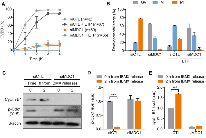
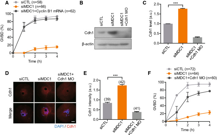
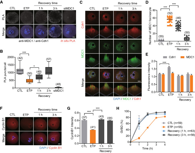
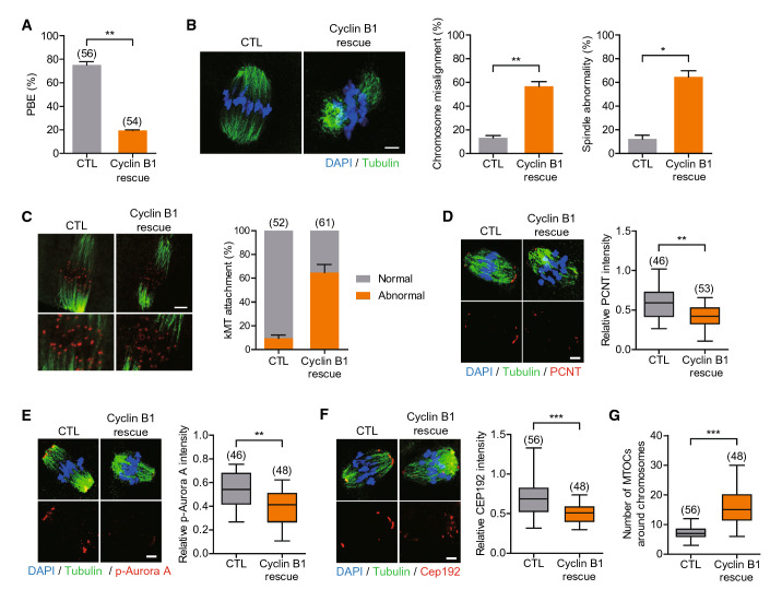
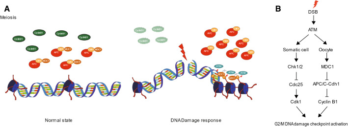
Mammalian oocytes are particularly susceptible to accumulating DNA damage. However, unlike mitotic cells in which DNA damage induces G2 arrest by activating the ATM-Chk1/2-Cdc25 pathway, oocytes readily enter M-phase immediately following DNA damage. This implies a lack of a robust canonical G2/M DNA damage checkpoint in oocytes. Here we show that MDC1 plays a non-canonical role in controlling G2/M transition by regulating APC/C-Cdh1-mediated cyclin B1 degradation in response to DNA damage in mouse oocytes. Depletion of MDC1 impaired M-phase entry by decreasing cyclin B1 levels via the APC/C-Cdh1 pathway. Notably, the APC/C-Cdh1 regulation mediated by MDC1 was achieved by a direct interaction between MDC1 and APC/C-Cdh1. This interaction was transiently disrupted after DNA damage with a concomitant increase in Cdh1 levels, which, in turn, decreased cyclin B1 levels and delayed M-phase entry. Moreover, MDC1 depletion impaired spindle assembly by decreasing the integrity of microtubule organizing centers (MTOCs). Therefore, our results demonstrate that MDC1 is an essential molecule in regulating G2/M transition in response to DNA damage and in regulating spindle assembly in mouse oocytes. These results provide new insights into the regulation of the G2/M DNA damage checkpoint and cell cycle control in oocytes.

Keywords: APC/C-Cdh1; DNA damage; G2/M transition; MDC1; Oocytes; Spindle assembly.


Conflict of interest statement

The authors have no conflict of interests to declare.

Figures

Fig. 1
Fig. 1
MDC1 is required for ATM-mediated DNA damage response in mouse oocytes. A GV oocytes were injected with either MDC1 siRNA (siMDC1) or control siRNA (siCTL) and cultured for 24 h in the presence of IBMX for knockdown. Depletion of MDC1 was confirmed by immunostaining with MDC1 antibodies. Scale bar, 10 μm. B Quantification of MDC1 levels after knockdown. Data are expressed as the mean ± SEM of three independent experiments. CH After treating with etoposide (ETP) or DMSO for 30 min, oocytes were fixed and subjected to TUNEL assay or immunostaining with γ-H2AX and p-ATM antibodies. The fluorescence intensity of TUNEL, γ-H2AX and p-ATM were measured and shown with representative images. Scale bar, 10 μm. Data are expressed as the mean ± SEM of three independent experiments. ***P < 0.0001; ns, not significant
Fig. 2
Fig. 2
MDC1 regulates G2/M transition by stabilizing cyclin B1 in mouse oocytes. A, B Control (siCTL) and siMDC1-injected oocytes were treated with etoposide (ETP). The incidence of GVBD for 4 h and the developmental stage of oocytes after 24-h culture were quantified. C Immunoblotting analysis of cyclin B1 and pY15-Cdk1 at 0 and 2 h after IBMX release in siCTL- or siMDC1-injected oocytes. D, E Levels of pY15-Cdk1 and cyclin B1 were normalized to β-actin, quantified, and expressed in arbitrary unit (a.u.). Data are expressed as the mean ± SEM from three independent experiments. *** P < 0.0001
Fig. 3
Fig. 3
MDC1 regulates APC/C-Cdh1-mediated cyclin B1 degradation during G2/M transition. A The incidence of GVBD was quantified for 4 h after IBMX release in oocytes injected with siCTL, siMDC1 or siMDC1 + cyclin B1 mRNA. BF Oocytes were injected with siCTL, siMDC1 and siMDC1 + Cdh1 MO. After 24-h culture, Cdh1 levels were determined using immunoblotting and immunostaining analyses. B, C Immunoblotting analysis of Cdh1. Levels of Cdh1 were normalized to β-actin, quantified, and expressed in arbitrary unit (a.u.). Data are expressed as the mean ± SEM from three independent experiments. D, E Immunostaining of Cdh1. The fluorescence intensity of Cdh1 was measured and shown with representative images. Scale bar, 10 μm. *** P < 0.0001. F The incidence of GVBD was quantified for 4 h after IBMX release
Fig. 4
Fig. 4
MDC1 interacts with APC/C-Cdh1 in a DNA damage dependent manner. Oocytes were exposed to etoposide (ETP) and cultured in fresh medium for recovery for 1 h and 3 h. Oocytes injected with siMDC1 were used as a negative control. A In situ PLA of oocytes. B The number of PLA puncta per oocyte was quantified. CE Immunostaining analysis of Cdh1 and MDC1. Scale bar, 10 μm. D The number of MDC1 foci was quantified. E The fluorescence intensity of Cdh1 and MDC1 were quantified. F Immunostaining analysis of cyclin B1. Scale bar, 10 μm. G The fluorescence intensity of cyclin B1 was quantified. * P < 0.05; *** P < 0.0001. H The incidence of GVBD was quantified for 4 h after IBMX release
Fig. 5
Fig. 5
MDC1 is essential for MTOC organization during meiotic maturation. Oocytes were injected with cyclin B1 mRNA alone (CTL) or siMDC1 + cycin B1 mRNA (Cyclin B1 rescue). After 24-h culture, oocytes were released from IBMX. A Polar body extrusion (PBE) rate was scored after 16-h culture post-IBMX release. BG After 8 h post-IBMX release, oocytes were fixed and immunostained with α-tubulin, ACA, PCNT, p-Aurora A or CEP192 antibodies. For ACA staining, oocytes were briefly placed on ice before fixation. Scale bar, 10 μm. Chromosome misalignment; spindle abnormality; kMT attachment; and intensity of PCNT, p-Aurora A, and CEP192 were quantified. (G) The number of MTOCs around chromosomes was quantified using CEP192 foci. * P < 0.05; ** P < 0.001; *** P < 0.0001
Fig. 6
Fig. 6
Model of MDC1-mediated regulation of G2/M transition in response to DNA damage. A Working model for the functions of MDC1 during G2/M transition. In the normal state, MDC1 is associated with APC/C-Cdh1 and regulates cyclin B1 to maintain threshold levels. However, in the presence of DNA damage, MDC1 is dissociated from APC/C-Cdh1 and recruited to DNA damage sites. This activity increases APC/C-Cdh1-medidated cyclin B1 degradation. B Comparison of the G2/M DNA damage checkpoint between somatic cells and oocytes

References

    1. Bartek J, Lukas J. DNA damage checkpoints: from initiation to recovery or adaptation. Curr Opin Cell Biol. 2007;19(2):238–245. doi: 10.1016/j.ceb.2007.02.009. - DOI - PubMed
    1. Blackford AN, Jackson SP. ATM, ATR, and DNA-PK: the trinity at the heart of the DNA damage response. Mol Cell. 2017;66(6):801–817. doi: 10.1016/j.molcel.2017.05.015. - DOI - PubMed
    1. Bartek J, Lukas J. Chk1 and Chk2 kinases in checkpoint control and cancer. Cancer Cell. 2003;3(5):421–429. doi: 10.1016/s1535-6108(03)00110-7. - DOI - PubMed
    1. Lou Z, Minter-Dykhouse K, Wu X, Chen J. MDC1 is coupled to activated CHK2 in mammalian DNA damage response pathways. Nature. 2003;421(6926):957–961. doi: 10.1038/nature01447. - DOI - PubMed
    1. Stewart GS, Wang B, Bignell CR, Taylor AM, Elledge SJ. MDC1 is a mediator of the mammalian DNA damage checkpoint. Nature. 2003;421(6926):961–966. doi: 10.1038/nature01446. - DOI - PubMed

Substances