

Diffusion-weighted MRI radiomics of spine bone tumors: feature stability and machine learning-based classification performance
- PMID: 35320464
- PMCID: PMC9098537
- DOI: 10.1007/s11547-022-01468-7
Diffusion-weighted MRI radiomics of spine bone tumors: feature stability and machine learning-based classification performance
Abstract
Purpose: To evaluate stability and machine learning-based classification performance of radiomic features of spine bone tumors using diffusion- and T2-weighted magnetic resonance imaging (MRI).
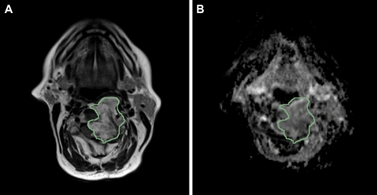
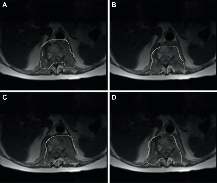
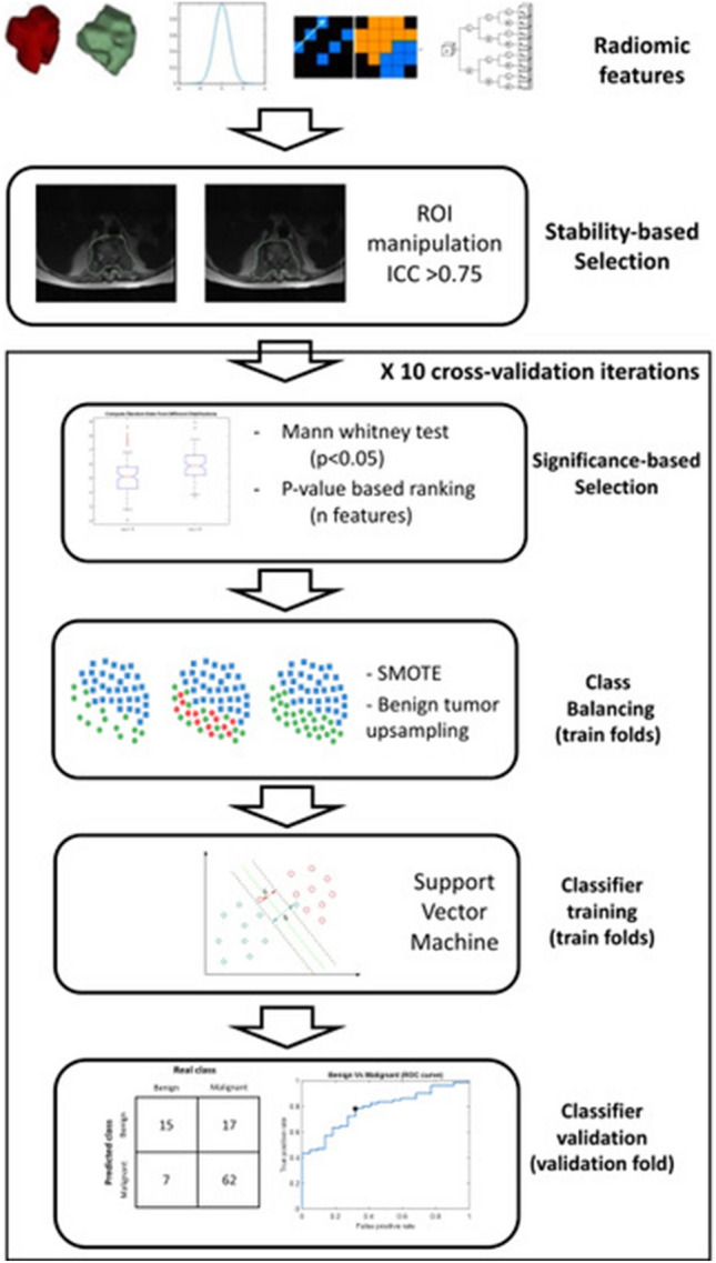
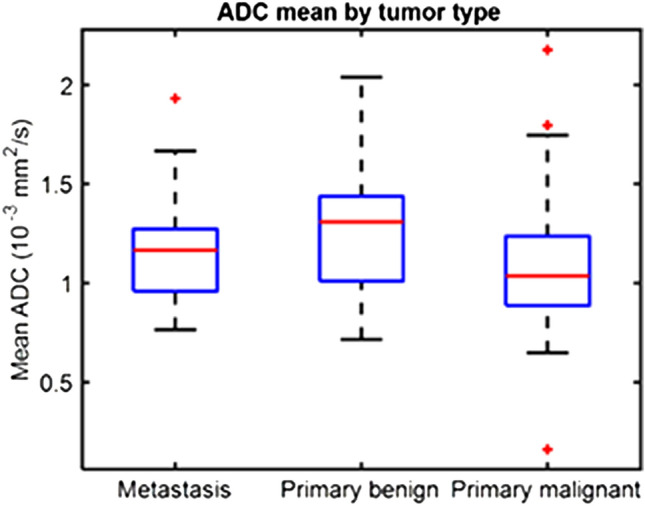
Material and methods: This retrospective study included 101 patients with histology-proven spine bone tumor (22 benign; 38 primary malignant; 41 metastatic). All tumor volumes were manually segmented on morphologic T2-weighted sequences. The same region of interest (ROI) was used to perform radiomic analysis on ADC map. A total of 1702 radiomic features was considered. Feature stability was assessed through small geometrical transformations of the ROIs mimicking multiple manual delineations. Intraclass correlation coefficient (ICC) quantified feature stability. Feature selection consisted of stability-based (ICC > 0.75) and significance-based selections (ranking features by decreasing Mann-Whitney p-value). Class balancing was performed to oversample the minority (i.e., benign) class. Selected features were used to train and test a support vector machine (SVM) to discriminate benign from malignant spine tumors using tenfold cross-validation.
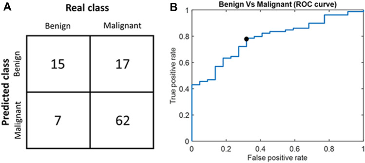
Results: A total of 76.4% radiomic features were stable. The quality metrics for the SVM were evaluated as a function of the number of selected features. The radiomic model with the best performance and the lowest number of features for classifying tumor types included 8 features. The metrics were 78% sensitivity, 68% specificity, 76% accuracy and AUC 0.78.
Conclusion: SVM classifiers based on radiomic features extracted from T2- and diffusion-weighted imaging with ADC map are promising for classification of spine bone tumors. Radiomic features of spine bone tumors show good reproducibility rates.
Keywords: Machine learning; Radiomics; Reproducibility; Spine; Texture analysis; Tumor.
© 2022. The Author(s).
Conflict of interest statement
The authors declare that they have no conflict of interest.
Figures
Similar articles
-
Classification of pulmonary lesion based on multiparametric MRI: utility of radiomics and comparison of machine learning methods.Eur Radiol. 2020 Aug;30(8):4595-4605. doi: 10.1007/s00330-020-06768-y. Epub 2020 Mar 28. Eur Radiol. 2020. PMID: 32222795
-
Magnetic resonance radiomic feature performance in pulmonary nodule classification and impact of segmentation variability on radiomics.Br J Radiol. 2022 Dec 1;95(1140):20220230. doi: 10.1259/bjr.20220230. Epub 2022 Nov 15. Br J Radiol. 2022. PMID: 36367095 Free PMC article.
-
MRI radiomics-based machine-learning classification of bone chondrosarcoma.Eur J Radiol. 2020 Jul;128:109043. doi: 10.1016/j.ejrad.2020.109043. Epub 2020 May 7. Eur J Radiol. 2020. PMID: 32438261
-
Rectal MRI radiomics inter- and intra-reader reliability: should we worry about that?Abdom Radiol (NY). 2022 Jun;47(6):2004-2013. doi: 10.1007/s00261-022-03503-7. Epub 2022 Apr 2. Abdom Radiol (NY). 2022. PMID: 35366088 Free PMC article. Review.
-
A systematic review of radiomics in giant cell tumor of bone (GCTB): the potential of analysis on individual radiomics feature for identifying genuine promising imaging biomarkers.J Orthop Surg Res. 2023 Jun 7;18(1):414. doi: 10.1186/s13018-023-03863-w. J Orthop Surg Res. 2023. PMID: 37287036 Free PMC article.
Cited by
-
The diagnostic value of machine learning for the classification of malignant bone tumor: a systematic evaluation and meta-analysis.Front Oncol. 2023 Sep 7;13:1207175. doi: 10.3389/fonc.2023.1207175. eCollection 2023. Front Oncol. 2023. PMID: 37746301 Free PMC article.
-
Computed tomography radiomics in predicting patient satisfaction after robotic-assisted total knee arthroplasty.Int J Comput Assist Radiol Surg. 2025 Feb;20(2):237-248. doi: 10.1007/s11548-024-03192-1. Epub 2024 Jun 5. Int J Comput Assist Radiol Surg. 2025. PMID: 38836956
-
Advances in imaging modalities for spinal tumors.Neurooncol Adv. 2024 Apr 9;6(Suppl 3):iii13-iii27. doi: 10.1093/noajnl/vdae045. eCollection 2024 Oct. Neurooncol Adv. 2024. PMID: 39430391 Free PMC article. Review.
-
CT and MRI radiomics of bone and soft-tissue sarcomas: an updated systematic review of reproducibility and validation strategies.Insights Imaging. 2024 Feb 27;15(1):54. doi: 10.1186/s13244-024-01614-x. Insights Imaging. 2024. PMID: 38411750 Free PMC article.
-
Radiomics in Spine Surgery.Int J Spine Surg. 2023 Jun;17(S1):S57-S64. doi: 10.14444/8501. Epub 2023 May 16. Int J Spine Surg. 2023. PMID: 37193607 Free PMC article.
