Review
. 2022 Jun 21:91:89-106.
doi: 10.1146/annurev-biochem-032620-105728. Epub 2022 Mar 23.

The Purinosome: A Case Study for a Mammalian Metabolon

Affiliations
Review

The Purinosome: A Case Study for a Mammalian Metabolon

Anthony M Pedley et al. Annu Rev Biochem. .

Abstract

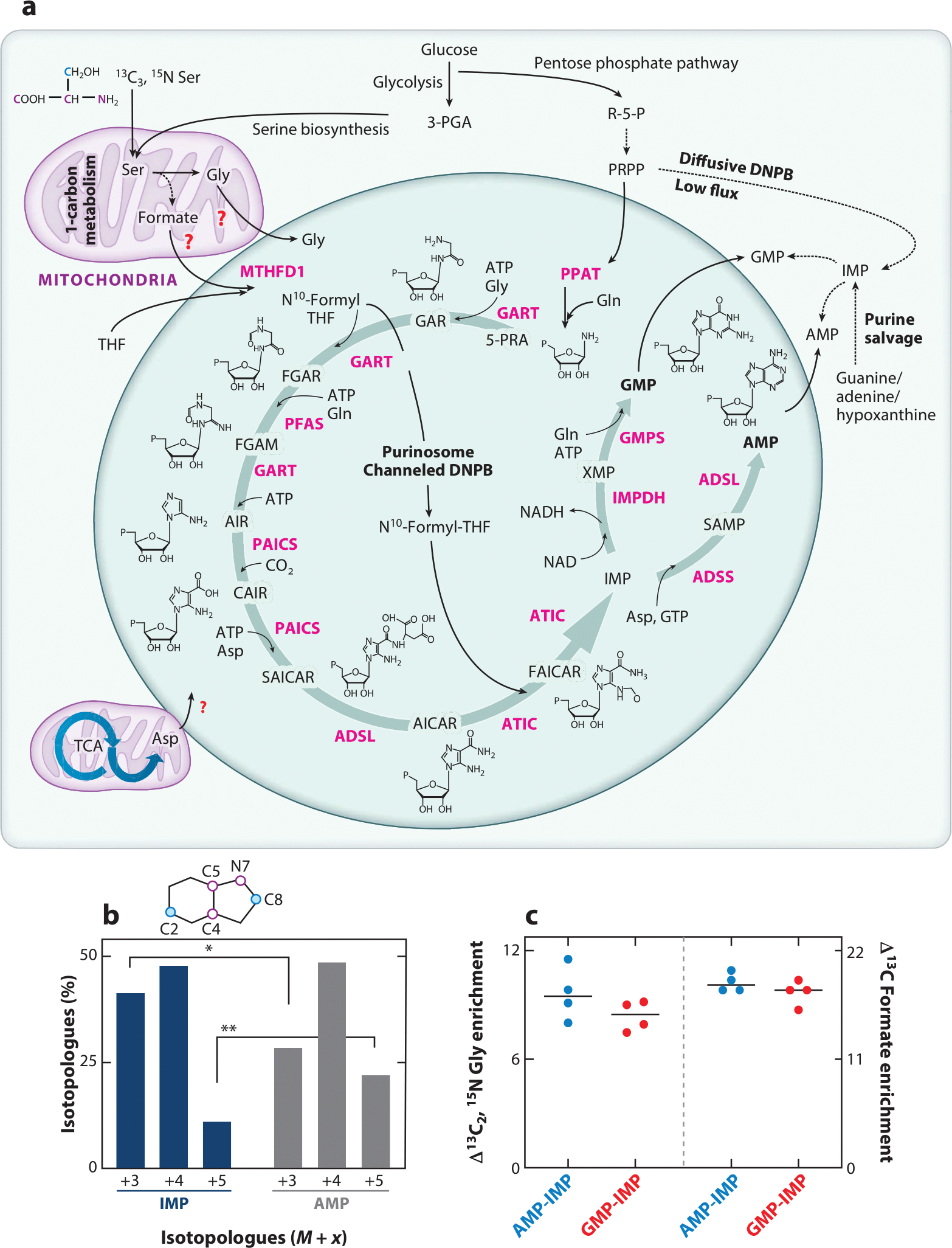
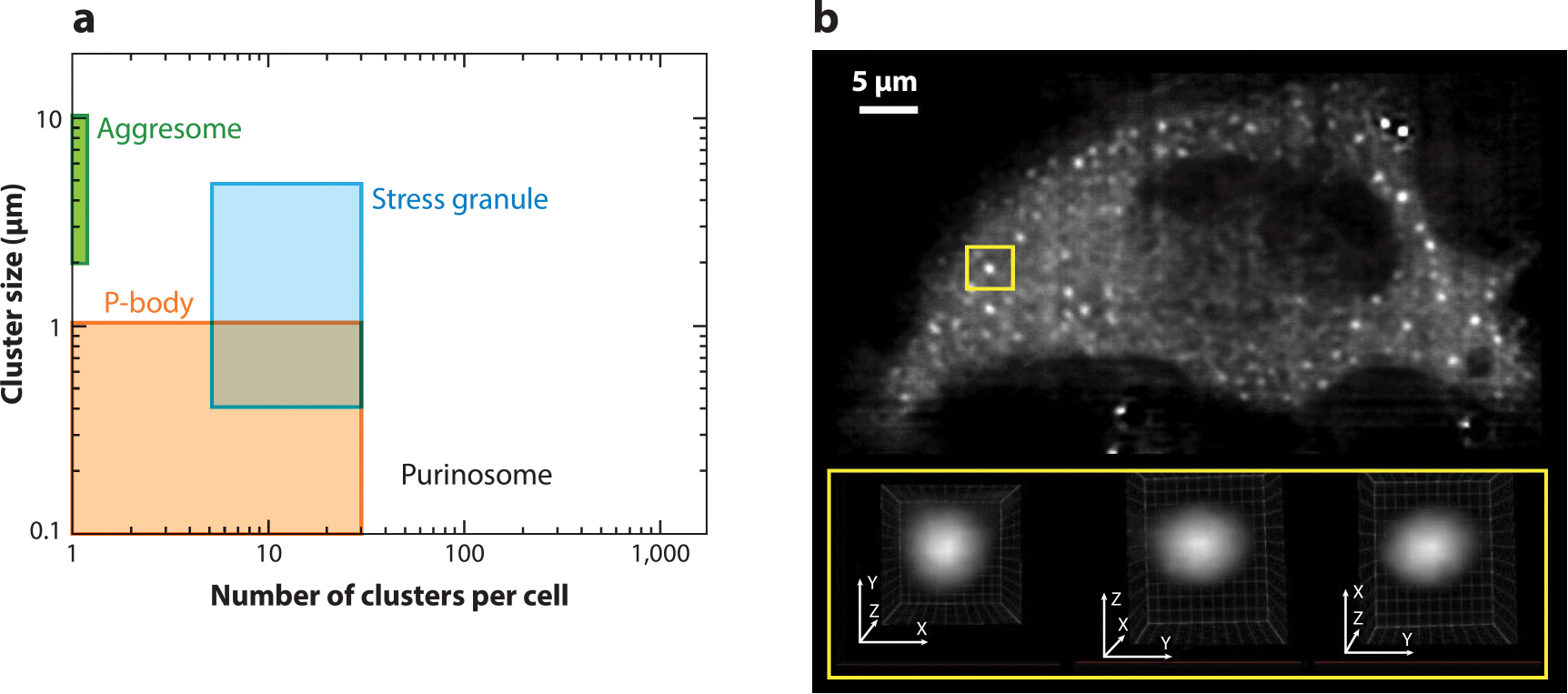
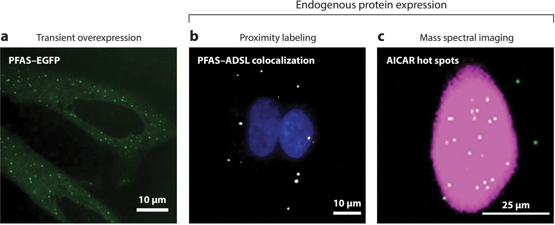
Over the past fifteen years, we have unveiled a new mechanism by which cells achieve greater efficiency in de novo purine biosynthesis. This mechanism relies on the compartmentalization of de novo purine biosynthetic enzymes into a dynamic complex called the purinosome. In this review, we highlight our current understanding of the purinosome with emphasis on its biophysical properties and function and on the cellular mechanisms that regulate its assembly. We propose a model for functional purinosomes in which they consist of at least ten enzymes that localize near mitochondria and carry out de novo purine biosynthesis by metabolic channeling. We conclude by discussing challenges and opportunities associated with studying the purinosome and analogous metabolons.

Keywords: channeling; de novo purine biosynthesis; metabolism; metabolon; protein complex; purinosome.


Figures

Figure 1
Figure 1
Enzymatic composition and function of the purinosome. (a) Channeled DNPB is carried out by the purinosome metabolon, which is composed of at least ten enzymes (pink): PPAT, GART, PFAS, PAICS, ADSL, ATIC, IMPDH, ADSS, GMPS, and MTHFD1. Structures of DNPB intermediates are shown; “P” denotes a phosphate group. Glycolysis, the serine biosynthesis pathway, one-carbon metabolism, the pentose phosphate pathway, and the TCA cycle generate the building block substrates utilized in DNPB. Involvement of mitochondrial Gly, formate, and Asp transporters for the direct uptake of these substrates for DNPB by purinosomes has been proposed though not confirmed yet (red question marks). The stable isotope–labeled positions of Ser are shown for the backbone atoms (pink) and side chain carbon (blue); the positions of these labeled atoms upon incorporation into the purine ring are shown as pink and blue circles in the purine ring diagram (shown in panel b). Alternatively, low-flux DNPB is carried out by the diffusive pool of DNPB enzymes outside purinosomes and by purine salvage enzymes that produce IMP, GMP, and AMP. (b) Upon 13C3, 15N Ser–mediated labeling of de novo synthesized purines, a different isotopologue distribution was discovered in the newly synthesized IMP (blue bars) and AMP (dark gray bars), revealing the two parallel purine-generating pathways (33). The purine ring positions replaced with isotope-labeled atoms are shown as pink and blue circles in the purine ring schematic. The tightly channeled, high-flux DNPB pathway that primarily generates AMP and GMP is carried out by mitochondria-associated purinosomes. Alternatively, the diffusive, low-flux DNPB primarily generates the free IMP pool. (c) The end nucleotides AMP and GMP showed higher incorporation of the mitochondrially derived substrates Gly and formate (33). AMP and GMP showed ~10% higher 13C2, 15N Gly enrichment and ~20% higher 13C formate enrichment compared with diffusively synthesized IMP. The plot shows the difference in isotope incorporation between AMP and IMP (blue circles) and GMP and IMP (red circles). Abbreviations: 3-PGA, 3-phosphoglyceric acid; 5-PRA, 5- phosphoribosylamine; ADSL, adenylosuccinate lyase; ADSS, adenylosuccinate synthetase; AICAR, 5-aminoimidazole-4-carboxamide ribonucleotide; AIR, 5-aminoimidazole ribonucleotide; ATIC, 5-aminoimidazole-4-carboxamide nucleotide formyltransferase/IMP cyclohydroxylase; CAIR, carboxyaminoimidazole ribonucleotide; DNPB, de novo purine biosynthesis; FAICAR, 5-formamidoimidazole-4-carboxamide ribonucleotide; FGAM, formylglycinamidine; FGAR, formylglycinamide ribonucleotide; GAR, glycinamide ribonucleotide; GART, phosphoribosylglycinamide formyltransferase; GMPS, GMP synthetase; IMP, inosine monophosphate; IMPDH, IMP dehydrogenase; MTHFD1, cytosolic methylenetetrahydrofolate dehydrogenase; NAD, nicotinamide adenine dinucleotide oxidized; NADH, nicotinamide adenine dinucleotide reduced; PAICS, phosphoribosyl aminoimidazole succinocarboxamide synthase; PFAS, phosphoribosyl formylglycinimidine transferase; PPAT, PRPP amidotransferase; PRPP, phosphoribosyl pyrophosphate; R-5-P, ribose-5-phosphate; SAICAR, phosphoribosyl aminoimidazole succinocarboxamide; SAMP, succinyladenosine monophosphate; TCA, tricarboxylic acid; THF, tetrahydrofolate; XMP, xanthosine monophosphate.
Figure 2
Figure 2
Biophysical properties of purinosomes. (a) The size and number of PFAS–EGFP clusters in purine-depleted HeLa cells compared to other reported biomolecular condensates. (b) A representative lattice light-sheet fluorescence microscopy image of a purinosome-positive HeLa cell transfected with PFAS–mCherry. Analysis of these clusters showed that they are spherical, as demonstrated by the region of interest (yellow boxes). Panel b modified from the original image published in Pedley et al. (29) with permission from E.L. Kennedy and M. Kyoung. Abbreviations: EGFP, enhanced green fluorescent protein; P-body, processing body; PFAS, phosphoribosyl formylglycinamide synthase.
Figure 3
Figure 3
Imaging methods for purinosome detection. Purinosomes can be visualized by three different imaging methods. (a) Initial studies used the transient overexpression of DNPB enzymes tagged with a fluorescent protein such as PFAS–EGFP in purine-depleted HeLa cells to characterize these enzyme assemblies (green punctate staining) (64). The misidentification of purinosomes due to the possibility of DNPB enzyme self-assembly and aggregation resulted in the development of new methods for detecting the complex using endogenous protein expression. These methods include proximity labeling assays (PLAs) and mass spectral imaging. (b) PLAs detect purinosomes through the colocalization of two DNPB enzymes, such as PFAS and ADSL (white) in purine-depleted HeLa cells (17). Nuclei are shown in blue. (c) A representative GCIB-SIMS image of a HeLa cell showing the total ion current (pink) and the pixels with high AICAR abundance (white) (33). These AICAR hot spots also show high levels of ATP, arising as a result of purinosome activity. Abbreviations: AICAR, 5-aminoimidazole-4-carboxamide ribonucleotide; ADSL, adenylosuccinate lyase; DNPB, de novo purine biosynthesis; EGFP, enhanced green fluorescent protein; GCIB-SIMS, gas cluster ion beam–time of flight–secondary ion mass spectrometry; PFAS, phosphoribosyl formylglycinamide synthase; PLA, proximity labeling assay.

References

    1. Nyhan WL. 2014. Nucleotide synthesis via salvage pathway. In eLS. San Francisco: John Wiley and Sons Ltd.
    1. Yin J, Ren W, Huang X, Deng J, Li T, Yin Y. 2018. Potential mechanisms connecting purine metabolism and cancer therapy. Front. Immunol. 9:1697. - PMC - PubMed
    1. Ashihara H, Ludwig IA, Crozier A. 2020. Salvage pathways of purine nucleotide biosynthesis. In Plant Nucleotide Metabolism—Biosynthesis, Degradation, and Alkaloid Formation, ed. Ashihara H, Ludwig IA, Crozier A, pp. 55–69. Hoboken, NJ: John Wiley & Sons Ltd.
    1. Pareek V, Pedley AM, Benkovic SJ. 2021. Human de novo purine biosynthesis. Crit. Rev. Biochem. Mol. Biol. 56:1–16 - PMC - PubMed
    1. Henderson JF, Khoo KY. 1965. On the mechanism of feedback inhibition of purine biosynthesis de novo in Ehrlich ascites tumor cells in vitro. J. Biol. Chem. 240:3104–9 - PubMed

Publication types

LinkOut - more resources