Review
. 2022 Jun 21:91:705-729.
doi: 10.1146/annurev-biochem-040320-105145. Epub 2022 Mar 23.

Structure and Mechanism of the Lipid Flippase MurJ

Affiliations
Review

Structure and Mechanism of the Lipid Flippase MurJ

Alvin C Y Kuk et al. Annu Rev Biochem. .

Abstract

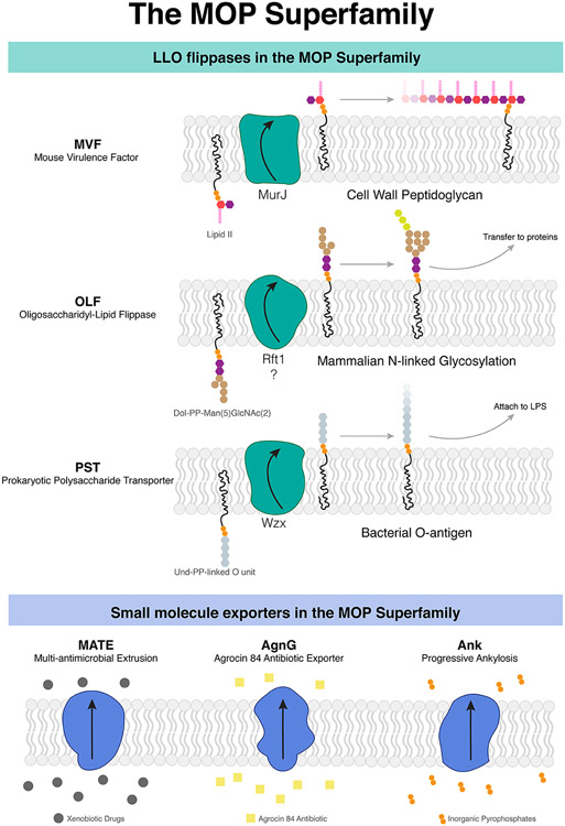
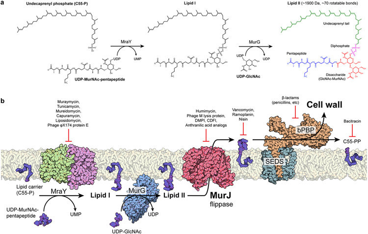
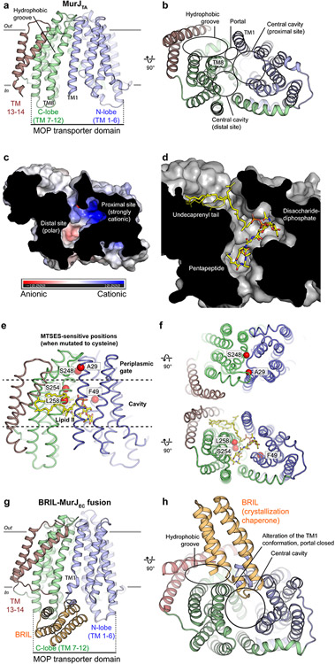
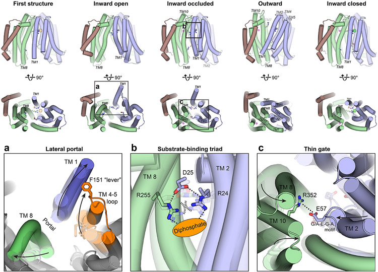
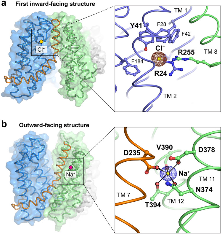
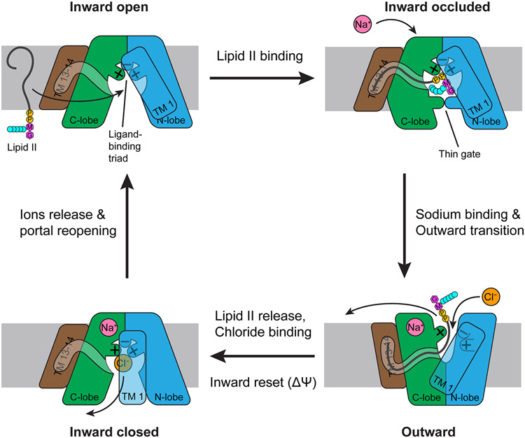
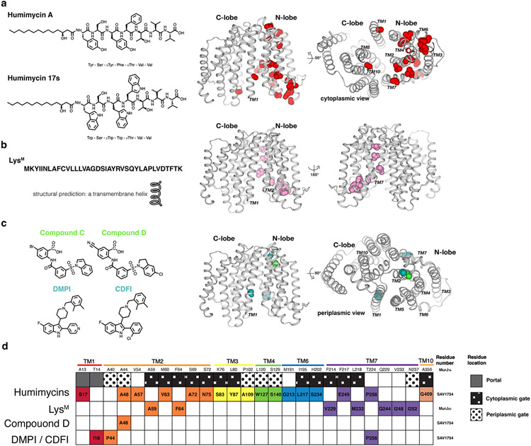
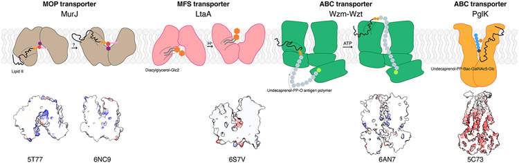
Biosynthesis of many important polysaccharides (including peptidoglycan, lipopolysaccharide, and N-linked glycans) necessitates the transport of lipid-linked oligosaccharides (LLO) across membranes from their cytosolic site of synthesis to their sites of utilization. Much of our current understanding of LLO transport comes from genetic, biochemical, and structural studies of the multidrug/oligosaccharidyl-lipid/polysaccharide (MOP) superfamily protein MurJ, which flips the peptidoglycan precursor lipid II. MurJ plays a pivotal role in bacterial cell wall synthesis and is an emerging antibiotic target. Here, we review the mechanism of LLO flipping by MurJ, including the structural basis for lipid II flipping and ion coupling. We then discuss inhibition of MurJ by antibacterials, including humimycins and the phage M lysis protein, as well as how studies on MurJ could provide insight into other flippases, both within and beyond the MOP superfamily.

Keywords: antibiotic; cell wall; lipid; peptidoglycan; transporter.


Figures

Figure 1.
Figure 1.
MOP superfamily proteins mediate the transport of lipids important for biosynthetic pathways as well as the expulsion of small molecules. (a) Three subfamilies (MVF, OLF, PST) comprise flippases that transport lipid substrates, while another three subfamilies (MATE, AgnG, and Ank) transport small molecules. (b) MurJ is the prototypical MOP flippase belonging to the MVF family. Abbreviations: AgnG, Agrocin 84; Ank, ankylosis; LLO, lipid-linked oligosaccharide; LPS, lipopolysaccharide; MATE, multidrug and toxic compound extrusion; MOP, multidrug/oligosaccharidyl-lipid/polysaccharide; MVF, mouse virulence factor; OLF, oligosaccharidyl-lipid flippase; PST, polysaccharide transporter.
Figure 2.
Figure 2.
Membrane-associated steps of peptidoglycan biosynthesis are hot spots for antibiotic targeting. (a) Soluble cell wall precursors are synthesized in the cytosol and attached to the lipid carrier C55-P by MraY and MurG to form the lipid-linked intermediate lipid II. Lipid II is a large (~1,900 Da), flexible (~70 rotatable bonds), and anionic molecule that requires a dedicated transport protein to flip across the membrane. (b) On the periplasmic side of the cell membrane, a series of glycosyltransferase (SEDS proteins) and transpeptidases (class B PBPs) activities form the cell wall. The flippase MurJ forms the critical link between the cytosolic and periplasmic steps by flipping lipid II from the cytoplasmic side to the periplasmic side. Many of these steps are established targets of known antibiotics. Abbreviations: C55-P, undecaprenyl phosphate; C55-PP, undecaprenyl diphosphate; CDFI, 2-(2-Chlorophenyl)-3- [1-(2,3-dimethylbenzyl)piperidin-4-yl]-5-fluoro-1H-indole; DMPI, 3-{1-[(2,3-Dimethylphenyl)methyl]piperidin-4-yl}-1-methyl-2- pyridin-4-yl-1H-indole; GlcNAc, N-acetylglucosamine; MurNAc, N-acetylmuramic acid; bPBP, class B penicillin-binding protein; SEDS, shape, elongation, division, sporulation; UDP, uridine diphosphate; UMP, uridine monophosphate.
Figure 3.
Figure 3.
Structure of MurJ and putative mode of lipid II binding. (a) MurJTA was captured in an inward-facing conformation not previously seen in other MOP superfamily proteins. MurJ contains the 12-TM MOP transporter domain arranged in two 6-TM bundles (N-lobe and C-lobe) plus two additional C-terminal helices (TMs 13 and 14). (b) TMs 13 and 14 form a hydrophobic groove that enters into the central cavity through a lateral membrane portal between TM1 and TM8. The central cavity can be divided into a proximal site adjacent to the portal and a distal site away from the portal. (c) The proximal site is highly cationic, while the distal site is only polar. (d) Model of lipid II (yellow sticks) docked into the MurJTA structure, placing the undecaprenyl tail in the hydrophobic groove, the disaccharide-diphosphate in the proximal site, and the pentapeptide in the distal site. (e) Previously determined MTSES-sensitive positions in MurJEC that result in cell lysis or cell shape defects are mapped to the MurJTA structure as red spheres. ( f ) Ala29 and Ser248 are located at the periplasmic gate, and adducts likely obstructed the closure of this gate. In contrast, Phe49, Ser254, and Leu258 face toward the central cavity and adducts at these positions could have blocked lipid II binding. (g) The structure of MurJEC was determined by fusion with a crystallization chaperone in the absence of lipid II, whereas lipid II was present in the MurJTA crystallization conditions. The MOP transporter domain of this MurJEC structure is less wide than that of MurJTA. (h) MurJEC also contains a hydrophobic groove and central cavity, but the portal appears to be closed in this structure, possibly due to the BRIL-induced distortion of TM1 and/or the absence of lipid II. Abbreviations: BRIL, thermostabilized cytochrome b562 RIL; MOP, multidrug/oligosaccharidyl-lipid/polysaccharide; MTSES, sodium 2-sulfonatoethyl methanethiosulfonate; MurJEC, Escherichia coli MurJ; MurJTA, Thermosipho africanus MurJ; TM, transmembrane helix.
Figure 4.
Figure 4.
Crystal structures of MurJTA in inward-open, inward-occluded, outward, and inward-closed states elucidate the conformational changes that mediate lipid II flipping. (a) The lateral membrane portal is opened in the inward-open conformation by the bending of TM1 out into the membrane and concomitant rearrangements in TM8 and the TM4–5 loop (orange), specifically those involving the Phe151 lever, allowing entry of the large lipid II headgroup into the cavity. Translucent outlines denote the other inward-facing conformations. (b) In the inward-occluded structure, Arg24 and Arg255 are brought into unusual proximity at the apex of the cavity, their electrostatic repulsion possibly stabilized by Asp25 and the anionic diphosphate moiety of lipid II (orange oval). TM1 is hidden in this panel for clarity. Translucent sticks denote these residues in the other inward-facing conformations. Arg255 is disengaged from this Arg24-Asp25-Arg255 triad in the outward-facing conformation. (c) Bending of TM2 and closing of the C-lobe forms a thin gate between Glu57 of the N-lobe and Arg352 of the C-lobe, possibly to occlude the lipid II headgroup from the cytosol. Translucent sticks denote these residues in the other inward-facing conformations. Abbreviation: MurJTA, Thermosipho africanus MurJ; TM, transmembrane helix.
Figure 5.
Figure 5.
Ions captured in the MurJTA structure that might be involved in lipid II transport. (a) A chloride ion was observed in the first inward-facing (2.0-Å resolution, shown) and the inward-closed structures. Chloride was coordinated by Arg24, Tyr41, and Arg255, with the last being the only residue from the C-lobe and the rest from the N-lobe. Phe28, Phe42, and Phe184 are also in orientations suitable to form anion-quadrupole interactions with chloride. Anomalous difference electron density from a bromide soak is shown in brown mesh contoured to 4.5 σ. (b) A sodium ion was observed in the outward-facing (1.8-Å resolution, shown), inward-occluded, and inward-closed structures. Sodium was coordinated in trigonal bipyramidal geometry by Asn374, Asp378, Val390 (backbone carbonyl), Thr394, and Asp235, with the last being the only residue from the hinge helix TM7 (orange) and the rest from TM11/12. Omit electron density for sodium is shown in purple mesh contoured to 4.5 σ. Abbreviations: MurJTA, Thermosipho africanus MurJ; TM, transmembrane helix.
Figure 6.
Figure 6.
Mechanism of lipid II flipping by MurJ. Lipid II enters the central cavity through a lateral portal. The Arg24/Asp25/Arg255 triad in the cavity binds the diphosphate moiety of lipid II, while the undecaprenyl tail fits into the hydrophobic groove formed by TMs 13 and 14. Lipid II is occluded from the cytosol by the Glu57-Arg352 thin gate, poised for outward transition. Upon sodium binding to the C-lobe, MurJ transits to the outward-facing state, disengaging Arg255 from Arg24 and Asp25. Because the outward-facing cleft is too narrow to accommodate the lipid II headgroup, lipid II is released. Chloride binding reassociates the Arg24/Asp25/Arg255 triad, resetting MurJ to an inward-facing apo state where the portal is closed. Membrane potential might also facilitate this inward reset. Release of sodium and chloride ions into the cytosol mediates reopening of the portal, completing the transport cycle. The net reaction is the export of lipid II, uptake of one sodium ion, and uptake of one chloride ion. Abbreviation: TM, transmembrane helix.
Figure 7.
Figure 7.
Inhibitors of MurJ. (a) Chemical structures of humimycins. The locations of resistance mutations mapped to the MurJTA structure are indicated as spheres. (b) Peptide sequence of LysM and resistance mutations to LysM mapped to the MurJTA structure. (c) Chemical structures of the putative small-molecule inhibitors DMPI, CDFI, Compound C, and Compound D. Resistance mutations to each were mapped to the MurJTA structure. (d) A barcode tabular summary of resistance mutations for each inhibitor and their locations on MurJ. Humimycins appeared to target the portal and cytoplasmic gate of MurJ, while LysM putatively targets the TM2–TM7 region of MurJ facing outside, toward the membrane. The mechanism of small-molecule inhibitors is less clear due to the small number of resistant mutants that have been isolated. Abbreviations: CDFI, 2-(2-Chlorophenyl)-3-[1-(2,3-dimethylbenzyl)piperidin-4-yl]-5-fluoro-1H-indole; DMPI, 3-{1-[(2,3-Dimethylphenyl)methyl]piperidin-4-yl}-1-methyl-2-pyridin-4-yl-1H-indole; LysM, phage M lysis protein; MurJTA, Thermosipho africanus MurJ; TM, transmembrane helix.
Figure 8.
Figure 8.
Diversity of LLO flipping mechanisms beyond the MOP superfamily. Cartoon representations of LLO flippases MurJ, LtaA, Wzm-Wzt and PglK with their flipping mechanisms. Dashed lines indicate a hydrophobic groove or opening. MurJ and LtaA utilize alternating access. Wzm-Wzt forms a continuous channel inside the transmembrane region to transport substrate. PglK was proposed to use an unconventional mechanism in which only the outer-facing conformation is involved in flipping. The lipid tail does not enter into the protein but interacts only with the helix on the periplasmic side while the headgroup enters the positive charged cavity. Shown below are electrostatic surface representations highlighting the cavity where substrates bind, with cationic surfaces in blue and anionic surfaces in red. Abbreviations: ABC, ATP-binding cassette; LLO, lipid-linked oligosaccharide; MFS, major facilitator superfamily; MOP, multidrug/oligosaccharidyl-lipid/polysaccharide; PDB ID, Protein Data Bank identifier.

References

    1. Rush JS. 2015. Role of Flippases in Protein Glycosylation in the Endoplasmic Reticulum. Lipid Insights 8: 45–53 - PMC - PubMed
    1. Ruiz N. 2015. Lipid Flippases for Bacterial Peptidoglycan Biosynthesis. Lipid Insights 8: 21–31 - PMC - PubMed
    1. Segawa K, Nagata S. 2015. An Apoptotic 'Eat Me' Signal: Phosphatidylserine Exposure. Trends Cell Biol 25: 639–50 - PubMed
    1. Henderson JC, Zimmerman SM, Crofts AA, Boll JM, Kuhns LG, et al. 2016. The Power of Asymmetry: Architecture and Assembly of the Gram-Negative Outer Membrane Lipid Bilayer. Annu Rev Microbiol 70: 255–78 - PubMed
    1. Pomorski T, Menon AK. 2006. Lipid flippases and their biological functions. Cell Mol Life Sci 63: 2908–21 - PMC - PubMed

Publication types

MeSH terms

LinkOut - more resources