Review
. 2022 Mar 7:9:839391.
doi: 10.3389/fcvm.2022.839391. eCollection 2022.

Non-cardiac Manifestations in Adult Patients With Mucopolysaccharidosis

Affiliations
Review

Non-cardiac Manifestations in Adult Patients With Mucopolysaccharidosis

Karolina M Stepien et al. Front Cardiovasc Med. .

Abstract

Mucopolysaccharidoses (MPS) are a heterogeneous group of disorders that results in the absence or deficiency of lysosomal enzymes, leading to an inappropriate storage of glycosaminoglycans (GAGs) in various tissues of the body such as bones, cartilage, heart valves, arteries, upper airways, cornea, teeth, liver and nervous system. Clinical manifestations can become progressively exacerbated with age and affect their quality of life. Developments in advanced supportive treatment options such as enzyme replacement therapy (ERT), hematopoietic stem cell transplantation (HSCT) may have improved patients' life span. Adult MPS patients require specialist clinical surveillance long-term. In many cases, in addition to the MPS-related health problems, they may develop age-related complications. Considering the complexity of their clinical manifestations and lack of guidelines on the management of adult MPS disorders, multispecialty and multidisciplinary teams' care is essential to diagnose and treat health problems that are likely to be encountered. This review presents non-cardiac clinical manifestations, their pathophysiology, management and long-term outcomes in adult MPS patients.

Keywords: adult mucopolysaccharidosis; life span; long-term complications; long-term outcomes; mortality.


Conflict of interest statement

The authors declare that the research was conducted in the absence of any commercial or financial relationships that could be construed as a potential conflict of interest.

Figures

Figure 1
Figure 1
Oral cavity in MPSII; shows bulky tongue, small dysplastic teeth, mallampati grade IV, lack of curvature of the hard palate.
Figure 2
Figure 2
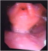
Nasendoscopy in MPS 2; bulky supraglottis and glottis with narrow glottic inlet.
Figure 3
Figure 3
Nasendoscopy in MPS IV; large epiglottis and bulky anterior glottis.
Figure 4
Figure 4
Three-dimensional reconstruction of airway in MPS VI, showing tortuous trachea.
Figure 5
Figure 5
Mechanisms of respiratory involvement in adult MPS patients.
Figure 6
Figure 6
Progression of Sleep Disordered Breathing related hypoventilation in adult MPS patients.
Figure 7
Figure 7
Cross sectional thoracic CT scan shows bronchomalacia in adult MPS IV.
Figure 8
Figure 8
Cross sectional thoracic CT scan shows bronchial dilation and early bronchiectasis in adult MPS type IV.
Figure 9
Figure 9
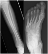
AP radiographs of the tibia and fibula and an AP view of the foot in a 22- year-old male with MPS I. The tibia shows epiphyseal dysplasia on the lateral side of the ankle and fibula hypoplasia causing valgus wedging with a decreased LDTA, tipping the hindfoot into valgus. The foot shows dysplastic and shortened metatarsals with widened proximal metaphyses.
Figure 10
Figure 10
X-rays of both feet and ankles of a 20-year-old MPS IV patient with distal tibial epiphyseal dysplasia and valgus wedging of the tibial articular surface. Avascular necrosis, collapse and fragmentation of the talus and navicular can be seen bilaterally. Collapse of the 1st metatarsal head and arthritis of the 1st metatarso-phalangeal joint was also seen on the CT.
Figure 11
Figure 11
Clinical photograph of a 26- year- old male with MPS VI in fixed equinus contracture, caused by gastro-soleus spasticity secondary to spinal cord compression at the level of the conus medullaris. Radiographs of the left foot and ankle reveals distal tibial epiphyseal dysplasia with valgus wedging, decreased LDTA, syndesmotic widening with lateral talar shift and increased medial clear space suggestive of ligamentous deficiency, shortened “bullet” metatarsals and an hypoplastic intermediate cuneiform.
Figure 12
Figure 12
demonstrating pathophysiology of spinal compression in MPS (190).
Figure 13
Figure 13
Atlanto-axial instability in adult MPS VI adult MPS VI patient demonstrating atlanto-axial instability with hypoplasia of the dens (yellow arrow), reduction in spinal canal dimensions between posterior body of C2 and posterior arch of C (red arrow), exacerbated by GAG accumulation in the anterior spinal space (red asterix).
Figure 14
Figure 14
Cervical cord stenosis. Sagittal MRI of adult with MPS I demonstrating cervical cord stenosis at C2–C4 and hypertrophied ligament, with effacement of anterior and posterior CSF spaces (red arrows). Also note the abnormal vertebrae at C7 and T1 with degenerative disc and hypertrophied ligament (yellow arrow) causing cervical-thoracic kyphosis and effacement of anterior CSF spaces.
Figure 15
Figure 15
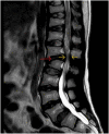
Thoraco-lumbar kyphosis. T2-weighted sagittal MRI of the adult spine with MPS VI, demonstrating abnormal vertebra body shape at L2, resulting in kyphosis at L1-L2 (red arrow) and early spinal canal compromise (yellow arrows).

References

    1. Wraith JE. The mucopolysaccharidoses: a clinical review and guide to management. Arch Dis Child. (1995) 72:263–7. 10.1136/adc.72.3.263 - DOI - PMC - PubMed
    1. Braunlin EA, Harmatz PR, Scarpa M, Furlanetto B, Kampmann C, Loehr JP, et al. . Cardiac disease in patients with mucopolysaccharidosis: presentation, diagnosis and management. J Inherited Metabolic Dis. (2011) 34:1183–97. 10.1007/s10545-011-9359-8 - DOI - PMC - PubMed
    1. Neufeld E, Muenzer J. The mucopolysaccharidoses. In: Scriver CR, Beaudet AL, Sly WS, Valle D, Childs R, Kinzler KW. editors. The Metabolic and Molecular Bases of Inherited Diseases. 8 ed. New York, NY: McGraw-Hill; (2001). p. 3421–52.
    1. Valayannopoulos V, Nicely H, Harmatz P, Turbeville S. Mucopolysaccharidosis vi. Orphanet J Rare Dis. (2010) 5:5. 10.1186/1750-1172-5-5 - DOI - PMC - PubMed
    1. Giugliani R. The Mucopolysaccharidoses. In: Mehta A, Winchester B. editor, Lysosomal Storage Disorders: A Practical Guide. London, UK: John Wiley & Sons, Ltd; (2012) p.94–100.

LinkOut - more resources