. 2022 Feb;89(1):73-100.
doi: 10.1177/00243639211059245. Epub 2021 Dec 6.

Fetal Pain in the First Trimester

Affiliations

Fetal Pain in the First Trimester

Bridget Thill. Linacre Q. 2022 Feb.

Abstract

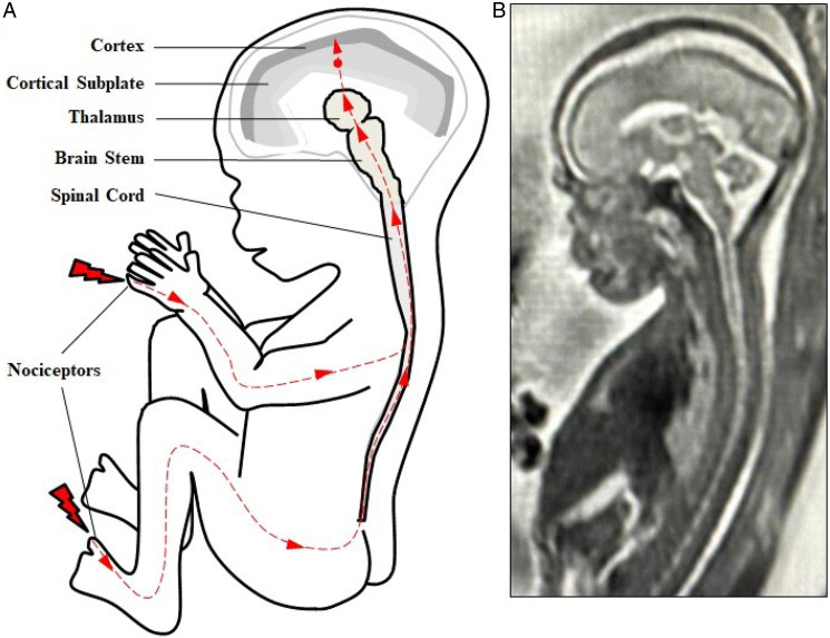
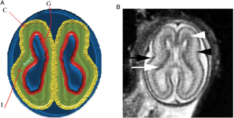
Fetal pain perception has important implications for fetal surgery, as well as for abortion. Current neuroscientific evidence indicates the possibility of fetal pain perception during the first trimester (<14 weeks gestation). Evidence for this conclusion is based on the following findings: (1) the neural pathways for pain perception via the cortical subplate are present as early as 12 weeks gestation, and via the thalamus as early as 7-8 weeks gestation; (2) the cortex is not necessary for pain to be experienced; (3) consciousness is mediated by subcortical structures, such as the thalamus and brainstem, which begin to develop during the first trimester; (4) the neurochemicals in utero do not cause fetal unconsciousness; and (5) the use of fetal analgesia suppresses the hormonal, physiologic, and behavioral responses to pain, avoiding the potential for both short- and long-term sequelae. As the medical evidence has shifted in acknowledging fetal pain perception prior to viability, there has been a gradual change in the fetal pain debate, from disputing the existence of fetal pain to debating the significance of fetal pain. The presence of fetal pain creates tension in the practice of medicine with respect to beneficence and nonmaleficence.

Keywords: fetal analgesia; fetal anesthesia; fetal awareness; fetal nociception; fetal pain.


Conflict of interest statement

Declaration of Conflicting Interest: The author(s) declared that there is no potential conflict of interest with respect to the research, authorship, and/or publication of this article.

Figures

Figure 1.
Figure 1.
(Fetal nociceptive pathways. (A) Nociception involves neural pathways extending from peripheral nociceptors to the spinal cord to the brainsteam and thalamus then to cortical structures. Projections from the thalamus (thalamocortical fibers) first invade the cortical subplate beginning at 12 weeks gestation (red dot). Thalamocortical projections to the cortex begin at approximately 24 weeks gestation. (B) T2-weighted magnetic resonance imaging (MRI) of a fetus at 21 weeks gestation.
Figure 2.
Figure 2.
Normal multi-layered magnetic resonance imaging (MRI) appearance of fetal brain early in gestation. (A) A diagram representing the fetal brain at 19 weeks of gestation shows surface and multi-layered appearance of the parenchyma with an inner germinal matrix (G), intermediate layer (l), and a developing cortex. (C). The small arrows point to the direction of the migrating neurons from germinal matrix to the developing cortex. (B) Axial balanced fast field echo MR image of a normal brain at 19 weeks of gestation shows a smooth surface and multi-layered parenchyma with an inner hypointense germinal matrix (white arrow), an intermediate layer, and an outer hypointense developing cortex (black arrow). Two additional sublayers can be identified: subventricular zone (white arrowhead) and subplate (black arrowhead). Subventricular zone is thick in the frontal region and shows slightly hypointense signal as it contains germinal matrix with increased cell pro-duction. The subplate zone appears slightly hyperintense as it has high water content, because of extracellular matrix. Reproduced by permission from SAGE Publications (Saleem 2013).
Figure 3.
Figure 3.
(A) Initial items from neonatal facial coding system and 2 supplementary items. 1. Brow lowering. 2. Eyes squeezed shut. 3. Deepening of the nasolabial furrow. 4. Open lips. 5. Horizontal mouth stretch. 6. Vertical mouth stretch. 7. Lip purse. 8. Taut tongue. 9. Tongue protrusion. 10. Chin quiver. 11. Neck deflection. 12. Yawning. (B) Final items from the Fetal-5 Scale. 1. Brow lowering. 2. Eyes squeezed shut. 3. Deepening of the nasolabial furrow. 4. Open lips. 5. Horizontal mouth stretch. 6. Vertical mouth stretch. 7. Neck deflection. Reproduced by permission from IASP (Bernardes et al. 2021b).

Similar articles

Cited by

References

    1. AboEllail M. A. M., Hata T. 2017. “Fetal face as important indicator of fetal brain function.” Journal of Perinatal Medicine 45 (6): 729‐736. doi: 10.1515/jpm-2016-0377. - DOI - PubMed
    1. Adzick N. S. 2020. “The IFMSS Tower Lecture: Fetal Surgery Past, Fetal Surgery Present, and Fetal Surgery Yet to Come!.” Fetal Diagnosis and Therapy 47 (12): 859‐864. doi: 10.1159/000507349. - DOI - PubMed
    1. Ahmad K. A., Frey C. S., Fierro M. A., Kenton A. B., Placencia F. X. 2017. “Two-Year Neurodevelopmental Outcome of an Infant Born at 21 Weeks’ 4 Days’ Gestation.” Pediatrics 140 (6): e20170103. doi: 10.1542/peds.2017-0103. - DOI - PMC - PubMed
    1. Aleman B., Merker B. 2014. “Consciousness Without Cortex: A Hydranencephaly Family Survey.” Acta Paediatrica 103 (10): 1057‐1065. doi: 10.1111/apa.12718. - DOI - PubMed
    1. American Academy of Pediatrics Task Force on Circumcision . 2012. “Circumcision Policy Statement.” Pediatrics 130 (3): 585‐586. 10.1542/peds.2012-1989. - DOI - PubMed