

Effects of Electroacupuncture on the Gut Microbiome in Cisplatin-Induced Premature Ovarian Failure Mice
- PMID: 35321505
- PMCID: PMC8938064
- DOI: 10.1155/2022/9352833
Effects of Electroacupuncture on the Gut Microbiome in Cisplatin-Induced Premature Ovarian Failure Mice
Abstract
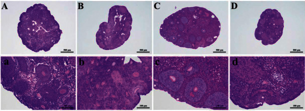
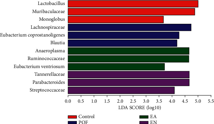
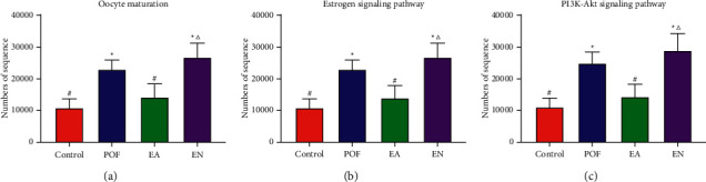
Growing evidence showed that the gut microbiota was associated with premature ovarian failure (POF). Many clinical types of research had shown that electroacupuncture was effective in the treatment of POF. However, there was little research on regulating the gut microbiome of POF mice by electroacupuncture. Therefore, this study attempted to verify whether electroacupuncture could regulate the gut microbiome in POF mice. POF mice were established by being injected intraperitoneally with cisplatin (2 mg/kg) for 2 weeks. Guanyuan (CV4) and Sanyinjiao (SP6) were selected in the electroacupuncture-at-the-acupoints group (EA group). Nonacupoints around CV4 and SP6 were selected in the electroacupuncture-at-the-nonacupoints group (EN group). The EA group and EN group were treated for 3 weeks. The ovarian function was evaluated by histopathological and molecular assays. Meanwhile, the gut microbiome of all mice was detected by 16S rDNA sequencing. The results showed that EA could restore the estrous cycle and reduce the number of atresia follicles in POF mice. The levels of serum follicle-stimulating hormone and luteinizing hormone were decreased by EA. As well, the levels of serum estradiol, anti-Mullerian hormone, and β-glucuronidase were increased by EA. The relative expressions of PI3K, AKT, and mTOR were increased to promote the proliferation of ovarian cells in the EA group. According to the results of 16S rDNA sequencing, the abundance and diversity of the gut microbiome could be regulated by EA. The relative abundance of beneficial bacteria was increased by EA. The KEGG pathway analysis showed that the gut microbiome associated with the estrogen signaling pathway, oocyte maturation, and PI3K-AKT signaling pathway was regulated by EA.
Copyright © 2022 Qi-da He et al.
Conflict of interest statement
The authors declare that there are no conflicts of interest regarding the publication of this paper.
Figures


Similar articles
-
[Effect of electroacupuncture at different acupoints on follicle development and related factors in serum and ovary tissues of PCOS rats].Zhen Ci Yan Jiu. 2019 Oct 25;44(10):740-6. doi: 10.13702/j.1000-0607.190041. Zhen Ci Yan Jiu. 2019. PMID: 31657164 Chinese.
-
Electroacupuncture May Inhibit Oxidative Stress of Premature Ovarian Failure Mice by Regulating Intestinal Microbiota.Oxid Med Cell Longev. 2022 Aug 30;2022:4362317. doi: 10.1155/2022/4362317. eCollection 2022. Oxid Med Cell Longev. 2022. PMID: 36082082 Free PMC article.
-
Electro-acupuncture attenuates the mice premature ovarian failure via mediating PI3K/AKT/mTOR pathway.Life Sci. 2019 Jan 15;217:169-175. doi: 10.1016/j.lfs.2018.11.059. Epub 2018 Dec 3. Life Sci. 2019. PMID: 30521869
-
Electroacupuncture attenuates ac4C modification of P16 mRNA in the ovarian granulosa cells of a mouse model premature ovarian failure.Acupunct Med. 2023 Feb;41(1):27-37. doi: 10.1177/09645284221085284. Epub 2022 Apr 27. Acupunct Med. 2023. PMID: 35475376
-
[Clinical research and the effect mechanism on premature ovarian failure treated with acupuncture in recent 20 years].Zhongguo Zhen Jiu. 2018 May 12;38(5):5653-70. doi: 10.13703/j.0255-2930.2018.05.031. Zhongguo Zhen Jiu. 2018. PMID: 29797923 Review. Chinese.
Cited by
-
Acupuncture influences multiple diseases by regulating gut microbiota.Front Cell Infect Microbiol. 2024 Jul 8;14:1371543. doi: 10.3389/fcimb.2024.1371543. eCollection 2024. Front Cell Infect Microbiol. 2024. PMID: 39040602 Free PMC article. Review.
-
Electro-Acupuncture Regulates Metabolic Disorders of the Liver and Kidney in Premature Ovarian Failure Mice.Front Endocrinol (Lausanne). 2022 Jul 25;13:882214. doi: 10.3389/fendo.2022.882214. eCollection 2022. Front Endocrinol (Lausanne). 2022. PMID: 35957829 Free PMC article.
-
Isoacids supplementation improves growth performance and feed fiber digestibility associated with ruminal bacterial community in yaks.Front Microbiol. 2023 Jun 15;14:1175880. doi: 10.3389/fmicb.2023.1175880. eCollection 2023. Front Microbiol. 2023. PMID: 37396385 Free PMC article.
-
Notch2 improves granulosa cell functions in premature ovarian failure by activating the Wnt2/β-catenin pathway.J Ovarian Res. 2025 Jul 30;18(1):169. doi: 10.1186/s13048-025-01745-9. J Ovarian Res. 2025. PMID: 40739650 Free PMC article.
-
Signaling pathway intervention in premature ovarian failure.Front Med (Lausanne). 2022 Nov 25;9:999440. doi: 10.3389/fmed.2022.999440. eCollection 2022. Front Med (Lausanne). 2022. PMID: 36507521 Free PMC article. Review.
References
LinkOut - more resources
Full Text Sources
Research Materials
Miscellaneous
