Review
. 2022 Mar 23;22(1):112.
doi: 10.1186/s12883-022-02645-x.

Primary intra- and extradural extramedullary mesenchymal chondrosarcoma with isolated punctate calcification: case report and literature review

Affiliations
Review

Primary intra- and extradural extramedullary mesenchymal chondrosarcoma with isolated punctate calcification: case report and literature review

Meigui Chen et al. BMC Neurol. .

Abstract

Background: Mesenchymal chondrosarcoma (MCS) is an ultra-rare, high-grade subtype of chondrosarcoma affecting both bone and soft tissues. Extra-skeletal MCS rarely occurs in intra- and extradural regions.

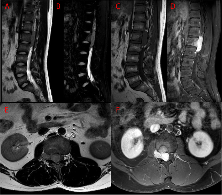
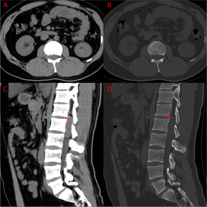
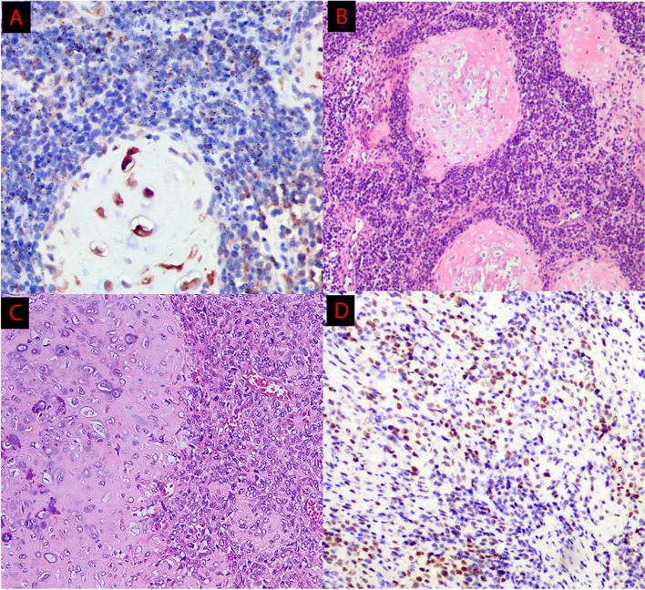
Case presentation: We presented a case of intraspinal dumbbell-shaped MCS at the T12-L2 level with isolated punctate calcification in a 19-year-old male complaining of progressive lower back pain. Surgical treatment for removal of the tumor was performed. The postoperative pathological result confirmed MCS. The patient showed symptomatic improvement and follow-up MRI showed no evidence of recurrence or metastasis for nearly 1 year after surgery.

Conclusions: CT and MRI play an important role in differential diagnosis for intraspinal MCS. MCS should be added to the differential diagnosis of intraspinal dumbbell-shaped tumors, especially when radiological examinations reveal punctate calcification in a homogeneous enhanced tumor without dural tail sign. However, the final diagnosis depends on histopathological results. Despite the good prognosis of intraspinal MCS, close follow-up after operation is still necessary.

Keywords: Calcification; Case report, Mesenchymal chondrosarcoma, intra- and extradural; Dumbbell-shape.


Conflict of interest statement

The authors declare that there is no conflict of interests.

Figures

Fig. 1
Fig. 1
Preoperative MR images. A Sagittal T2WI, B Sagittal T2WI with fat suppression, C Sagittal T1WI, D Sagittal T1WI with gadolinium enhancement, E Axial T1WI, F Axial T1WI with gadolinium enhancement. Dumbbell tumor could be visualized at the L1 level
Fig. 2
Fig. 2
Preoperative CT images. A, B Axial images. C, D Sagittal images. CT images revealed isolated punctate calcification in the mass (arrows)
Fig. 3
Fig. 3
Postoperative histopathological findings. A The chondroid component stained positive for S-100 protein. B Round or spindle-shaped undifferentiated cells with island-like regions of cartilage C A biphasic pattern. D spindle-shaped undifferentiated cells stained positive for myogenin

References

    1. Chan H, Turner-Gomes S, Chuang S, Fitz C, Daneman A, Martin D, et al. A rare cause of spinal cord compression in childhood from intraspinal mesenchymal chondrosarcoma. A report of two cases and review of the literature. Neuroradiology. 1984;26:323–327. doi: 10.1007/BF00339779. - DOI - PubMed
    1. Reif J, Graf N. Intraspinal mesenchymal chondrosarcoma in a three-year-old boy. Neurosurg Rev. 1987;10:311–314. doi: 10.1007/BF01781958. - DOI - PubMed
    1. Di Lorenzo N, Palatinsky E, Artico M, Palma L. Dural mesenchymal chondrosarcoma of the lumbar spine. Case report. Surg Neurol. 1989;31:470–472. doi: 10.1016/0090-3019(89)90095-5. - DOI - PubMed
    1. Rushing E, Armonda R, Ansari Q, Mena H. Mesenchymal chondrosarcoma: a clinicopathologic and flow cytometric study of 13 cases presenting in the central nervous system. Cancer. 1996;77:1884–1891. doi: 10.1002/(SICI)1097-0142(19960501)77:9<1884::AID-CNCR19>3.0.CO;2-W. - DOI - PubMed
    1. Bae G, Choi S, Youm J, Kim S. Primary spinal dumbbell-shaped mesenchymal chondrosarcoma located intradurally and extradurally. J Korean Neurosurg Soc. 2011;50:468–471. doi: 10.3340/jkns.2011.50.5.468. - DOI - PMC - PubMed

LinkOut - more resources