

Patients with isolated posterior cruciate ligament rupture had a higher posterior intercondylar eminence
- PMID: 35321666
- PMCID: PMC8943983
- DOI: 10.1186/s12891-022-05189-w
Patients with isolated posterior cruciate ligament rupture had a higher posterior intercondylar eminence
Abstract
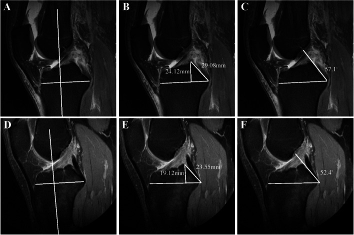
Background: To evaluate the anatomic geometry of the posterior intercondylar eminence and its association with PCL injury risk.
Methods: Patients who underwent primary PCL reconstruction from 2015 to 2018 were retrospectively analyzed. The control group included inpatients diagnosed with ACL rupture because of a sports-related accident during the same period, matched by age, gender, height, weight, and side of injury. Measurements of the height of the apex of the posterior intercondylar eminence (HPIE), the slope length (SLPIE) and the slope angle (SAPIE) of the posterior intercondylar eminence were performed using conventional MRI scans assessed by 2 blinded, independent raters. Intraclass correlation coefficients (ICCs) was used to evaluate the consistency of measurement results. Independent sample t tests, Chi-square tests, and logistic analyses were used to compare the two group, with P < 0.05 considered statistically significant.
Results: Fifty-five patients with PCL rupture met the inclusion criteria and 55 PCL-intact matched controls were included. There were no significant differences between the groups in gender (P = 1.000), limb side (P = 0.848), age (P = 0.291), BMI (P = 0.444) or height (P = 0.290). Inter-observer reproducibility was excellent agreement in HPIE, SLPIE and SAPIE of case and control groups (ICC: HPIE = 0.81, SLPIE = 0.77, SAPIE = 0.85). Patients with PCL rupture had significantly greater HPIE, SAPIE (both P < 0.001), and SLPIE (P < 0.05) than PCL-intact patients. The multivariable analysis showed that HPIE (OR, 1.62 [95% CI, 1.24-2.11], P < 0.001) and SAPIE (OR, 1.17 [95% CI, 1.05-1.31], P < 0.001) were independent factors associated with PCL rupture.
Conclusion: Through this retrospective observational study, we found that patients with PCL rupture may have a higher posterior intercondylar eminence compared to PCL-intact patients.
Level of evidence: III.
Keywords: MRI; PCL risk factors; Posterior cruciate ligament; Posterior intercondylar eminence.
© 2022. The Author(s).
Conflict of interest statement
The authors declare that they have no competing interests.
Figures

References
-
- DePhillipo NN, Cinque ME, Godin JA, Moatshe G, Chahla J, LaPrade RF. Posterior Tibial Translation Measurements on Magnetic Resonance Imaging Improve Diagnostic Sensitivity for Chronic Posterior Cruciate Ligament Injuries and Graft Tears. Am J Sports Med. 2018;46(2):341–347. doi: 10.1177/0363546517734201. - DOI - PubMed
-
- Logan CA, Beaulieu-Jones BR, Sanchez G, Chahla J, Kennedy NI, Cinque ME, LaPrade RF, Whalen JM, Vopat BG, Price MD, et al. Posterior Cruciate Ligament Injuries of the Knee at the National Football League Combine: An Imaging and Epidemiology Study. Arthroscopy. 2018;34(3):681–686. doi: 10.1016/j.arthro.2017.08.304. - DOI - PubMed
Publication types
MeSH terms
Grants and funding
LinkOut - more resources
Full Text Sources
Medical
Research Materials
Miscellaneous
