. 2022 Mar 23;22(1):312.
doi: 10.1186/s12885-022-09376-9.

PARP inhibitors chemopotentiate and synergize with cisplatin to inhibit bladder cancer cell survival and tumor growth

Affiliations

PARP inhibitors chemopotentiate and synergize with cisplatin to inhibit bladder cancer cell survival and tumor growth

Sayani Bhattacharjee et al. BMC Cancer. .

Abstract

Background: Management of bladder cancer (BLCA) has not changed significantly in the past few decades, with platinum agent chemotherapy being used in most cases. Chemotherapy reduces tumor recurrence after resection, but debilitating toxicities render a large percentage of patients ineligible. Recently approved immunotherapy can improve outcomes in only a third of metastatic BLCA patients. Therefore, more options for therapy are needed. In this study, we explored the efficacy of PARP inhibitors (PARPi) as single agents or as combinations with platinum therapy.

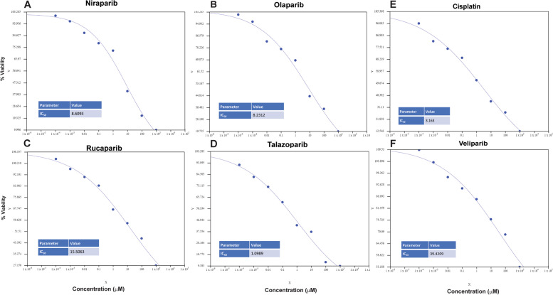
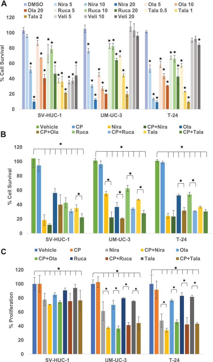
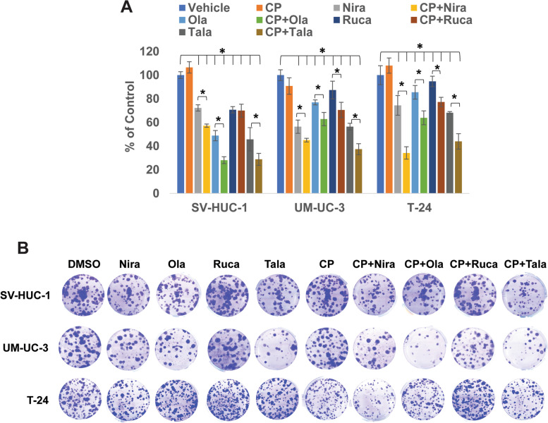
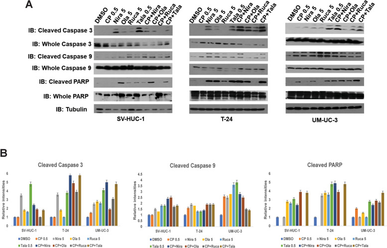
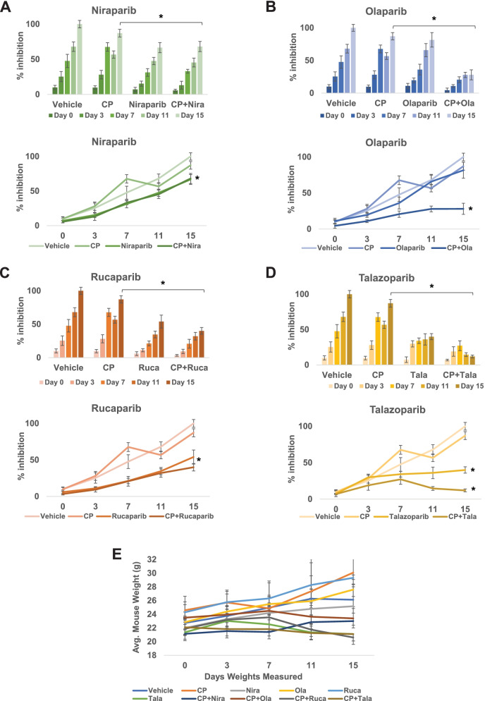
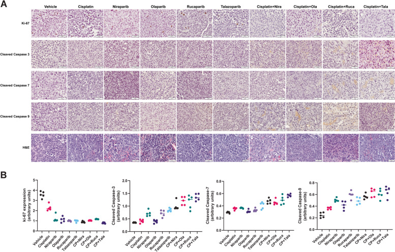
Methods: We treated BLCA cells with PARPi (olaparib, niraparib, rucaparib, veliparib, or talazoparib) alone or as the combination of cisplatin with PARPi. We then measured their survival, proliferation, apoptosis, as well as their ability to form colonies. BLCA xenografts in male SCID mice were treated similarly, followed by the assessment of their growth, proliferation, and apoptosis.

Results: PARPi niraparib and talazoparib were effective in reducing BLCA cell survival as single agents. Combinations of Cisplatin with talazoparib and niraparib effectively reduced the survival of BLCA cells, while veliparib was not effective even at high concentrations. In vivo, the combinations of cisplatin with niraparib, rucaparib, or talazoparib reduced BLCA xenograft growth significantly.

Conclusions: We provide evidence that PARPi can be effective against BLCA as single agents or as combinatorial therapy with cisplatin.

Keywords: Bladder cancer; Cisplatin; Combination therapy; DNA damage repair; PARP inhibition; Urothelial carcinoma.


Conflict of interest statement

The authors declare that they have no relevant competing interests.

Figures

Fig. 1
Fig. 1
UM-UC-3 cells were treated with 0, 0.0001, 0.001, 0.01, 0.1, 1, 10, 100, or 1000 μM concentrations of niraparib, olaparib, rucaparib, talazoparib, veliparib, or cisplatin for 72 h. Cell survival was reported as % cells surviving compared with vehicle-treated control. IC50s were calculated using the AAT Bioquest Online IC50 calculator
Fig. 2
Fig. 2
PARPi suppress the cell survival and proliferation of BLCA cells in vitro. A The BLCA cell lines UM-UC-3 and T-24 and the normal urothelial cells SV-HUC-1 were treated with varying concentrations of PARPi (niraparib, olaparib, rucaparib, talazoparib, or veliparib) for 72 h to determine the effective concentrations to be used in subsequent assays. Cell survival was measured as the percentage of cells surviving in comparison with the DMSO vehicle control in each cell line. B The BLCA cell lines UM-UC-3 and T-24 and the normal urothelial cells SV-HUC-1 were treated with sub-IC50 concentrations of niraparib, olaparib, rucaparib (5 μM each), talazoparib (0.5 μM) either singly or in combination with sub-IC50 concentration of cisplatin (0.5 μM) for 72 h. Cell survival was measured as the percentage of cells surviving in comparison with DMSO vehicle control in each cell line. C The BLCA cell lines UM-UC-3 and T-24 and the normal urothelial cells SV-HUC-1 were treated with sub-IC50 concentrations of niraparib, olaparib, rucaparib (5 μM each), talazoparib (0.5 μM) either singly or in combination with sub-IC50 concentration of cisplatin (0.5 μM) for 72 h. Cell proliferation was measured as the percentage of cells proliferating in comparison with DMSO vehicle control in each cell line. All results are presented as means±SD of 3 independent experiments with triplicates. P ≤ 0.05 was considered significant (*)
Fig. 3
Fig. 3
PARPi suppress the clonogenic ability of BLCA cells in vitro. A The BLCA cell lines UM-UC-3 and T-24 and the normal urothelial cells SV-HUC-1 were treated with sub-IC50 concentrations of niraparib, olaparib, rucaparib (5 μM each), talazoparib (0.5 μM) either singly or in combination with sub-IC50 concentration of cisplatin (0.5 μM) for 72 h. Cells were plated at low densities (400 cells/well) in 6-well plates and incubated at 37oC in a 5% CO2-incubator for 10-14 days. At the end of the experiment, colonies were stained with 0.5% crystal violet in buffered formalin and counted using the Colony Counter plug-in of ImageJ. Results are presented as means±SD of 3 independent experiments with triplicates. P ≤ 0.05 was considered significant (*). B Representative images of colonies formed by SV-HUC-1, UM-UC-3, and T-24 cells after treatment
Fig. 4
Fig. 4
PARPi induce apoptosis in BLCA cells in vitro. A The BLCA cell lines UM-UC-3 and T-24 and the normal urothelial cells SV-HUC-1 were treated with sub-IC50 concentrations of niraparib, olaparib, rucaparib (5 μM each), talazoparib (0.5 μM) either singly or in combination with sub-IC50 concentration of cisplatin (0.5 μM) for 72 h. The resulting cell lysates were subjected to Western blotting with antibodies against the apoptotic markers cleaved/whole caspase 3, cleaved/whole caspase 9, or cleaved/whole PARP. Representative images of 3 independent experiments with duplicates are presented. B Relative band intensities (compared to whole caspase/PARP and Tubulin) were measured using ImageJ and are presented for Cleaved caspases 3 and 9 and cleaved PARP
Fig. 5
Fig. 5
PARPi suppress the growth of BLCA xenografts in vivo. Xenografts of UM-UC-3 cells in SCID mice were generated by injecting 2x10⁶ cells into both flanks of SCID mice (n = 5/group). When the tumor volumes reached ~100 mm3, mice were treated with the PARPi niraparib (A), olaparib (B), rucaparib (C), talazoparib (D) or a combination of PARPi with cisplatin as detailed in the Methods section. Tumor growth was monitored using digital calipers. Toxicity was assessed by weighing the mice twice a week. At the end of the experiment, tumor tissues were harvested, and tumor inhibition was calculated as percentage tumor growth inhibition compared with vehicle control. E Average body weights±SD of all mice in each group are shown
Fig. 6
Fig. 6
PARPi induce apoptosis and suppress proliferation in BLCA xenografts in vivo. Xenografts of UM-UC-3 cells in SCID mice were generated by injecting 2x10⁶ cells into both flanks of SCID mice. When the tumor volumes reached ~100 mm3, mice were treated with the PARPi niraparib, olaparib, rucaparib, talazoparib, or a combination of PARPi with cisplatin as detailed in the Methods section. At the end of the experiment, tumor tissues were harvested, and sections prepared for immunohistochemistry with antibodies against the proliferation marker ki-67, and the apoptotic markers cleaved caspases 3, 7, or 9. A Representative images from 10 tumors in each group are presented. The scale bar at the lower right corner in each panel represents 50 μm. B Staining intensities were calculated as detailed in Methods using the ImageJ Fiji software. Dot plots showing staining intensities in arbitrary units are presented

References

    1. Pinto IG. Systemic therapy in bladder cancer. Indian J Urol. 2017;33(2):118–126. doi: 10.4103/iju.IJU_294_16. - DOI - PMC - PubMed
    1. Antoni S, Ferlay J, Soerjomataram I, Znaor A, Jemal A, Bray F. Bladder Cancer Incidence and Mortality: A Global Overview and Recent Trends. Eur Urol. 2017;71(1):96–108. doi: 10.1016/j.eururo.2016.06.010. - DOI - PubMed
    1. Ploeg M, Aben KKH, Kiemeney LA. The present and future burden of urinary bladder cancer in the world. World J Urol. 2009;27(3):289–293. doi: 10.1007/s00345-009-0383-3. - DOI - PMC - PubMed
    1. Kaufman DS, Shipley WU, Feldman AS. Bladder cancer. Lancet. 2009;374(9685):239–249. doi: 10.1016/S0140-6736(09)60491-8. - DOI - PubMed
    1. Witjes JA, Compérat E, Cowan NC, De Santis M, Gakis G, Lebret T, et al. EAU Guidelines on Muscle-invasive and Metastatic Bladder Cancer: Summary of the 2013 Guidelines. Eur Urol. 2014;65(4):778–792. doi: 10.1016/j.eururo.2013.11.046. - DOI - PubMed

Substances