. 2022 Mar 23;19(1):16.
doi: 10.1186/s12979-022-00273-0.

Telomerase deficiency reflects age-associated changes in CD4+ T cells

Affiliations

Telomerase deficiency reflects age-associated changes in CD4+ T cells

Diana M Matthe et al. Immun Ageing. .

Abstract

Background: Amongst other systemic changes, aging leads to an immune dysfunction. On the molecular level, a hallmark of aging is telomere shortening. The functional relevance of telomerase, an enzyme capable of elongating telomeres in T cells upon antigen stimulation, is not fully understood. Studying the impact of telomere shortening on CD4+ T cells and especially Th1 effector function can provide a better understanding on immune dysfunctions in elderly.

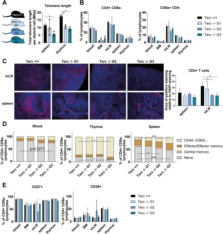
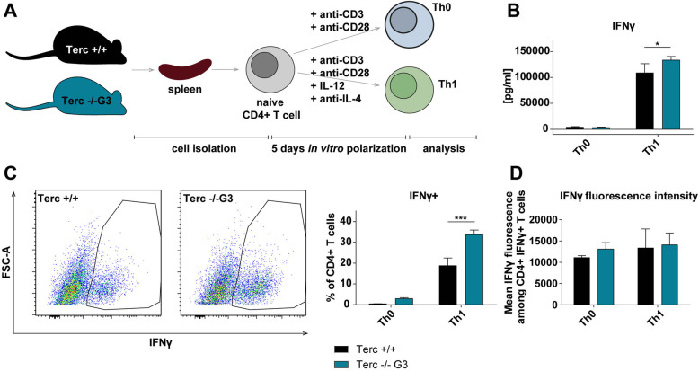
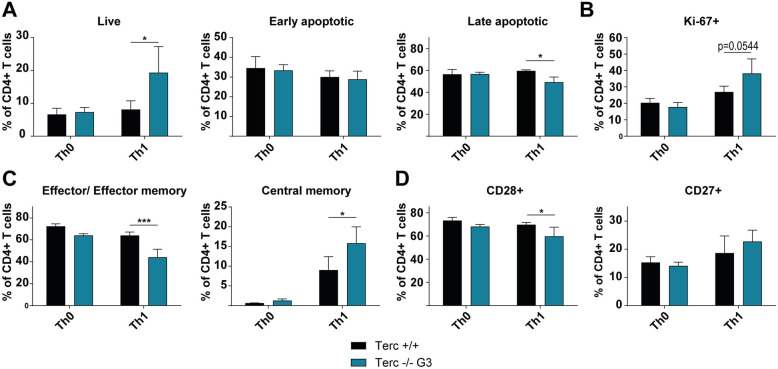
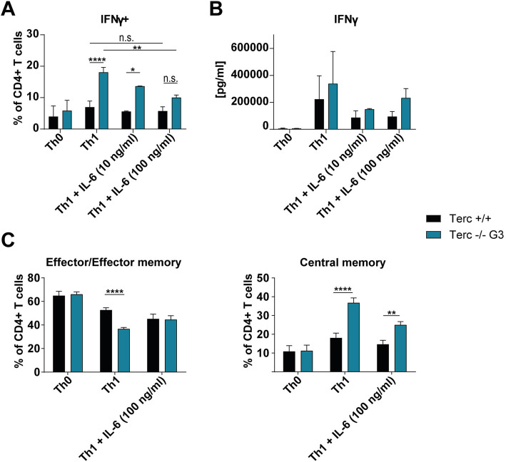
Results: We investigated T cell numbers and differentiation in telomerase-deficient (mTerc-/-) mice under steady-state conditions and the functional role of telomerase in CD4+ T cells using in vitro stimulation and Th1 polarization protocols by comparing T cells from mTerc-/- and control mice. We report reduced relative CD4+ T cell numbers in blood and secondary lymphoid organs and a relative decline in the naïve T cell population in thymus, blood and spleen of mTerc-/- mice compared to control mice. Importantly, after in vitro polarization, mTerc-/- G3 CD4+ T cells showed higher numbers of IFNγ-producing cells and reduced expression of CD28. Notably, telomerase-deficient T cells were more susceptible to inhibition of Th1 polarization by IL-6 in vitro. These results demonstrate that telomerase deficiency recapitulates several changes of CD4+ T cells seen in aged humans regarding the naïve T cell population, expression of CD28 and cytokine production.

Conclusion: Our data suggest that telomere shortening could play a key role in the aging of T cell immunity, with clinical implications for immune diseases and tumor development and that mTerc-/- mice are a suitable model to study aging-related defects of adaptive immunity.

Keywords: Aging; CD4-positive T-lymphocytes; Telomerase; Telomere shortening; Th1 cells.


Conflict of interest statement

The authors declare that they have no competing interests.

Figures

Fig. 1
Fig. 1
Characterization of CD4+ T cells from telomerase-deficient mice under steady state conditions. A Schematic representation of the different generations of mTerc−/− mice and validation of the telomere loss. Generation 2 (G2) animals were obtained by crossing mTerc−/− G1 animals, G3 animals are offspring of mTerc−/− G2 mice. Telomere length was quantified by a qPCR-based method in Terc+/+, Terc−/− G1 and Terc−/− G3 mice (n = 3 per group, except thymus of Terc−/− G3: n = 2). B Flow cytometric analysis of CD4+ and CD8a + cells from blood and lymphoid organs of mTerc−/− mice (n = 3) under steady-state conditions. C Immunohistochemical stainings of CD4+ T cells (red) in mesenteric lymph node (mLNs, n = 3 except Terc −/− G2: n = 2) and spleen (n = 3) of mTerc−/− mice. Nuclei were counterstained with DAPI (blue). Quantification was done with ImageJ software by calculation of the area of positive CD4 staining relative to the area of DAPI staining from non-overlapping pictures (≥ 5 pictures per mLN and ≥ 7 pictures per spleen). D Composition of the CD4+ T cell pool with regards to memory populations. Cells were defined as naïve (CD44- CD62L+), central memory (CD44+ CD62L+) and effector/effector memory (CD44+ CD62L-). E Relative numbers of CD4+ T cells expressing costimulatory molecules CD28 and CD27 in lymphoid organs from telomerase-deficient mice. If not otherwise indicated, the experiment was performed with n = 3 mice from each generation. Graphs show the mean ± SD, * adjusted p ≤ 0.05, ** adjusted p ≤ 0.01, *** adjusted p ≤ 0.001
Fig. 2
Fig. 2
mTerc−/− CD4+ T cells show functional differences after in vitro polarization. A Schematic representation of the experimental setup of in vitro polarization. CD4+ T cells were isolated and stimulated in vitro for five days with anti-CD3 and anti-CD28 only (Th0) or with anti-CD3, anti-CD28, IL-12 and anti-IL-4 (Th1). On the day of analysis, the cells were treated with eBioscience™ Cell Stimulation Cocktail (plus protein transport inhibitors) (500X) (Invitrogen) for 3.5 h before analysis. B Quantification of IFNγ in the medium by ELISA. C and D Flow cytometric analysis of IFNγ-producing CD4+ T cells. Graphs show the mean ± SD, * adjusted p ≤ 0.05, ** adjusted p ≤ 0.01, *** adjusted p ≤ 0.001
Fig. 3
Fig. 3
mTerc−/− CD4+ T cells show functional differences after in vitro polarization. A and (B Flow cytometric quantification of proliferating (Ki-67+), live (Annexin V- PI-), early apoptotic (Annexin V+ PI-) and late apoptotic (Annexin V+ PI+) cells. C Functional analysis of memory populations (central memory: CD44+ CD62L+; effector/ effector memory: CD44+ CD62L-) after in vitro polarization measured by flow cytometry. D Expression of costimulatory molecules CD28 and CD27 among in vitro polarized CD4+ T cells as determined by flow cytometry. All graphs show the mean ± SD, * adjusted p ≤ 0.05, ** adjusted p ≤ 0.01, *** adjusted p ≤ 0.001
Fig. 4
Fig. 4
mTerc−/− CD4+ T cells are more susceptible to IL-6-mediated inhibition of Th1 differentiation. CD4+ T cells were isolated from mTerc−/− mice as before and stimulated in vitro for five days with anti-CD3 and anti-CD28 only (Th0) or with anti-CD3, anti-CD28, IL-12 and anti-IL-4 (Th1). In addition, IL-6 was added to the Th1-inducing mixture in the indicated concentrations. A Flow cytometric analysis of IFNγ-producing cells after in vitro polarization under IL-6 treatment. The figure depicts pooled data from two independent experiments. B Quantification of IFNγ in the medium after in vitro polarization of mTerc−/− CD4+ T cells. The figure shows pooled data from two independent experiments. C Memory populations after in vitro polarization as determined by flow cytometry (effector/ effector memory: CD44+ CD62L-; central memory: CD44+ CD62L+). Data from one measurement is shown. All graphs show the mean ± SD, * adjusted p ≤ 0.05, ** adjusted p ≤ 0.01, *** adjusted p ≤ 0.001, **** adjusted p ≤ 0.0001

References

    1. Kubben N, Misteli T. Shared molecular and cellular mechanisms of premature ageing and ageing-associated diseases. Nat Rev Mol Cell Biol. 2017;18(10):595–609. doi: 10.1038/nrm.2017.68. - DOI - PMC - PubMed
    1. López-Otín C, Blasco MA, Partridge L, Serrano M, Kroemer G. The hallmarks of aging. Cell. 2013;153(6):1194–1217. doi: 10.1016/j.cell.2013.05.039. - DOI - PMC - PubMed
    1. Goronzy JJ, Weyand CM. Understanding immunosenescence to improve responses to vaccines. Nat Immunol. 2013;14(5):428–436. doi: 10.1038/ni.2588. - DOI - PMC - PubMed
    1. Nikolich-Žugich J. The twilight of immunity: emerging concepts in aging of the immune system. Nat Immunol. 2018;19(1):10–19. doi: 10.1038/s41590-017-0006-x. - DOI - PubMed
    1. Montecino-Rodriguez E, Berent-Maoz B, Dorshkind K. Causes, consequences, and reversal of immune system aging. J Clin Investig. 2013;123(3):958–965. doi: 10.1172/JCI64096. - DOI - PMC - PubMed