. 2022 Mar 24;18(1):37.
doi: 10.1186/s13007-022-00860-8.

Simple and efficient isolation of plant genomic DNA using magnetic ionic liquids

Affiliations

Simple and efficient isolation of plant genomic DNA using magnetic ionic liquids

Miranda N Emaus et al. Plant Methods. .

Abstract

Background: Plant DNA isolation and purification is a time-consuming and laborious process relative to epithelial and viral DNA sample preparation due to the cell wall. The lysis of plant cells to free intracellular DNA normally requires high temperatures, chemical surfactants, and mechanical separation of plant tissue prior to a DNA purification step. Traditional DNA purification methods also do not aid themselves towards fieldwork due to the numerous chemical and bulky equipment requirements.

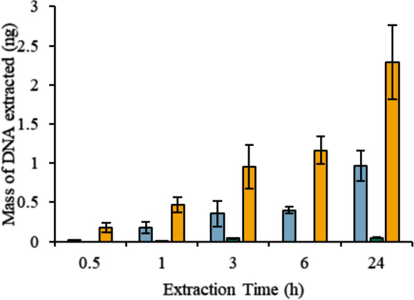
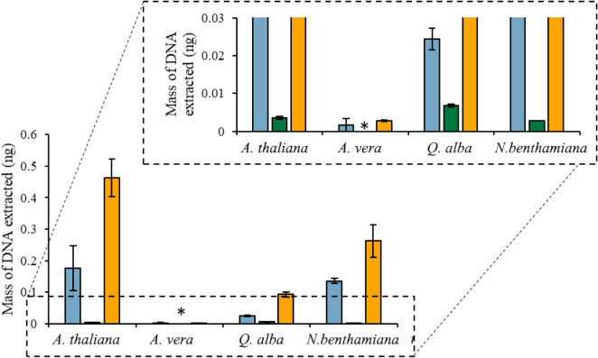
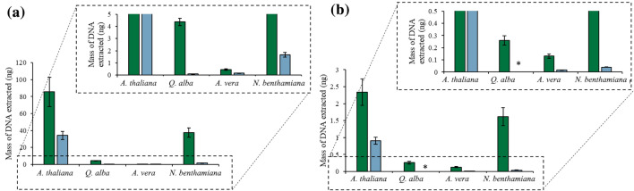
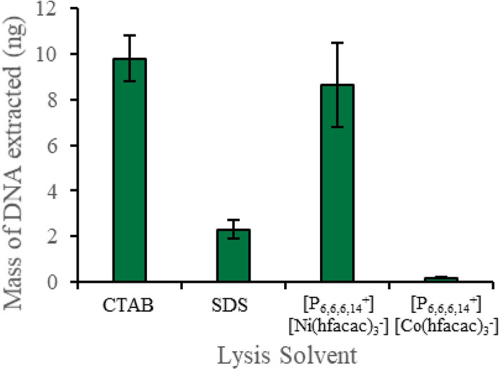
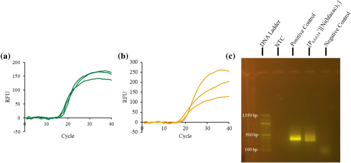
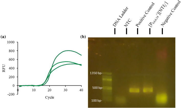
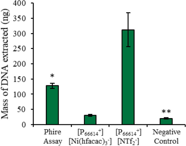
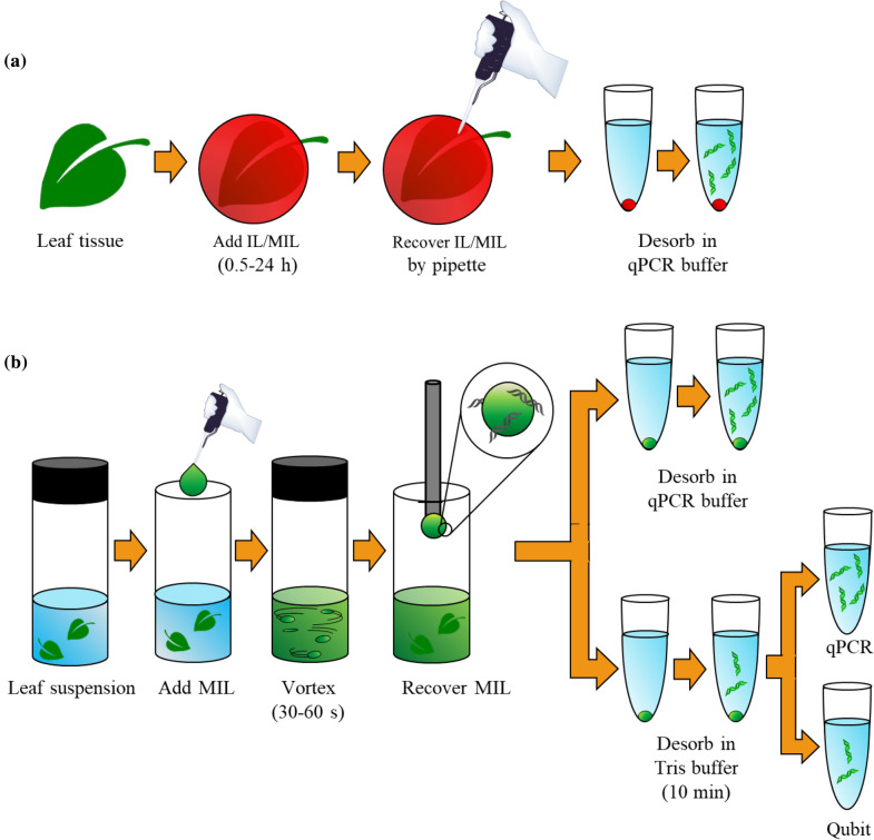
Results: In this study, intact plant tissue was coated by hydrophobic magnetic ionic liquids (MILs) and ionic liquids (ILs) and allowed to incubate under static conditions or dispersed in a suspension buffer to facilitate cell disruption and DNA extraction. The DNA-enriched MIL or IL was successfully integrated into the qPCR buffer without inhibiting the reaction. The two aforementioned advantages of ILs and MILs allow plant DNA sample preparation to occur in one minute or less without the aid of elevated temperatures or chemical surfactants that typically inhibit enzymatic amplification methods. MIL or IL-coated plant tissue could be successfully integrated into a qPCR assay without the need for custom enzymes or manual DNA isolation/purification steps that are required for conventional methods.

Conclusions: The limited amount of equipment, chemicals, and time required to disrupt plant cells while simultaneously extracting DNA using MILs makes the described procedure ideal for fieldwork and lab work in low resource environments.

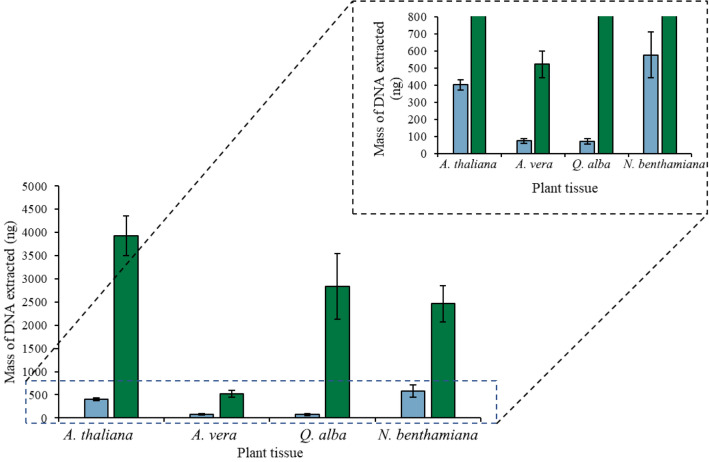
Keywords: Aloe vera L.; Arabidopsis thaliana (L.) Heynh.; Ionic liquids; Nicotiana benthaminana Domin; One-pot qPCR; One-step cell lysis; Plant DNA isolation; Quercus alba L..


Conflict of interest statement

The authors declare that they have no competing interests.

Figures

Fig. 1
Fig. 1
Mass of DNA recovered overtime after placing 6 µL of (blue) [P6,6,6,14+][Ni(hfacac)3] MIL, (green) [P6,6,6,14+][Co(hfacac)3] MIL, and (orange) [P6,6,6,14+][NTf2] IL on 40 mg of A. thaliana tissue
Fig. 2
Fig. 2
DNA extracted by placing 6 µL of (blue) [P6,6,6,14+][Ni(hfacac)3], (green) [P6,6,6,14+][Co(hfacac)3], and (orange) [P6,6,6,14+][NTf2] on 40 mg of plant tissue for 1 h. *DNA recovered could not be quantified
Fig. 3
Fig. 3
Amount of DNA recovered from the (green) [P6,6,6,14+][Ni(hfacac)3] and (blue) [P6,6,6,14+][Co(hfacac)3] MIL using the optimized dispersive lysis and DNA extraction method. Buffer volume: 0.5 mL; buffer composition with the [P6,6,6,14+][Ni(hfacac)3] MIL: 2 mM Tris, 50 mM EDTA; buffer composition with the [P6,6,6,14+][Co(hfacac)3] MIL: 2 mM Tris; mass of plant tissue: 40 mg; volume of MIL dispersed: 6 µL; dispersion time for the [P6,6,6,14+][Ni(hfacac)3] MIL: 30 s; dispersion time for the [P6,6,6,14+][Co(hfacac)3] MIL: 60 s. *DNA recovered could not be quantified
Fig. 4
Fig. 4
The amount of DNA recovered from a plant cell lysate generated using CTAB, SDS, [P6,6,6,14+][Ni(hfacac)3] MIL, and [P6,6,6,14+][Co(hfacac)3] MIL. Buffer volume: 0.5 mL; buffer composition with the [P6,6,6,14+][Ni(hfacac)3] MIL: 2 mM Tris, 50 mM EDTA; buffer composition with the [P6,6,6,14+][Co(hfacac)3] MIL: 2 mM Tris; mass of plant tissue: 40 mg; volume of MIL dispersed: 6 µL; dispersion time for the [P6,6,6,14+][Ni(hfacac)3] MIL: 30 s; dispersion time for the [P6,6,6,14+][Co(hfacac)3] MIL: 60 s
Fig. 5
Fig. 5
Amplification of 1 mg A. thaliana tissue and 6 µL [P6,6,6,14+][Ni(hfacac)3] within the qPCR buffer a with 20 ng of ITS amplicon added to the assay and without the ITS spike with b qPCR and c gel agarose detection
Fig. 6
Fig. 6
Fluorescence detection via a qPCR and b agarose gel electrophoresis with 1 mg of A. thaliana tissue and 6 µL of [P6,6,6,14+][NTf2] IL integrated into the custom-qPCR assay
Fig. 7
Fig. 7
Integration of 0.5 mg of one of four plant tissues into a qPCR assay with 6 µL of (green) [P6,6,6,14+][Ni(hfacac)3] MIL and (orange) [P6,6,6,14+][NTf2] IL to facilitate cell lysis and prevent reaction inhibition
Fig. 8
Fig. 8
Comparison of the amount of DNA lysed during the different one-pot PCR assays. *Assay is not qPCR compatible. **Initial qPCR assay was unsuccessful
Fig. 9
Fig. 9
Standard curves generated with 6 µL of a [P6,6,6,14+][Ni(hfacac)3] MIL and b [P6,6,6,14+][NTf2] IL and 1 mg of A. thaliana plant tissue in the qPCR assay with a non-target DNA sequence spiked into the assay
Fig. 10
Fig. 10
Schematic illustrating the a static and b dispersive one-step lysis and DNA extraction method using MILs

Similar articles

Cited by

References

    1. Li X, Yang Y, Henry RJ, Rossetto M, Wang Y, Chen S. Plant DNA barcoding: from gene to genome. Biol Rev Camb Philos Soc. 2015;90:157–166. doi: 10.1111/brv.12104. - DOI - PubMed
    1. Nezhad AS. Future of portable devices for plant pathogen diagnosis. Lab Chip. 2014;14:2887–2904. doi: 10.1039/c4lc00487f. - DOI - PubMed
    1. Fraiture M-A, Herman P, Taverniers I, De Loose M, Dieter D, Roosens NH. Current and new approaches in GMO Detectin: challenges and solutons. Biomed Res. Int. 2015 doi: 10.1155/2016/9160375. - DOI - PMC - PubMed
    1. Meyer R. Development and application of DNA analytical methods for the detection of GMOs in food. Food Control. 1999;10:391–399. doi: 10.1016/S0956-7135(99)00081-X. - DOI
    1. Emaus MN, Varona M, Eitzmann DR, Hsieh SA, Zeger VR, Anderson JL. Nucleic acid extraction: fundamentals of sample preparation methodologies, current advancements, and future endeavors. TrAC - Trends Anal Chem. 2020;130:115985. doi: 10.1016/j.trac.2020.115985. - DOI