

Mandible shape variation and feeding biomechanics in minks
- PMID: 35322070
- PMCID: PMC8943020
- DOI: 10.1038/s41598-022-08754-4
Mandible shape variation and feeding biomechanics in minks
Abstract
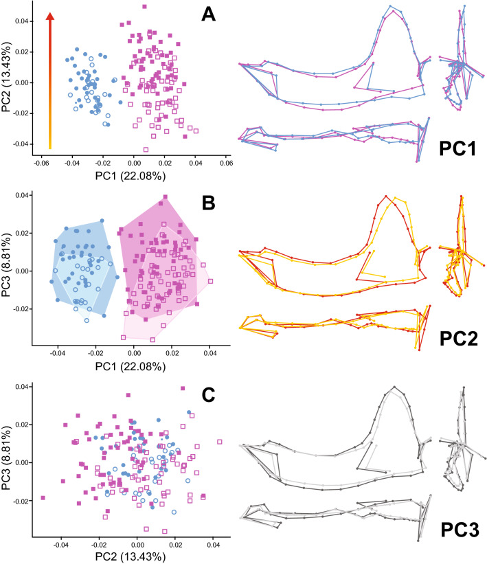
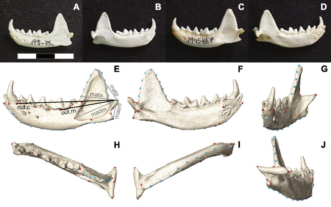
European and American minks are very similar in ecology, behavior and morphology. Both species hunt terrestrial vertebrates and aquatic prey, but the American mink is a more generalist predator which, among other factors, allows it to outcompete the European mink in areas where it has been introduced. We used 3D geometric morphometrics and estimates of muscle mechanical advantage to assess the degree of variation in mandibular morphology, and to determine whether such variation reflects dietary differences between the two species. The three main axes of variation represented interspecific differences, a common allometric trajectory between species and sexes, and the interspecific effect of sexual size dimorphism, with males having overall stronger bites than females. Differences in mandible shape and biomechanical parameters suggest that American minks are better equipped for preying on terrestrial vertebrates, while the features seen in European mink could be related to tougher prey, fish capture, or both. Additionally, within each species, the larger specimens of each sex present indicators of a higher percentage of terrestrial prey in their diet. These results indicate a low potential dietary overlap between both species, suggesting that factors other than prey competition may have a role in the decline of the European mink.
© 2022. The Author(s).
Conflict of interest statement
The authors declare no competing interests.
Figures

References
-
- Jordan MJR. Dietary analysis for mammals and birds: a review of field techniques and animal-management applications. Int. Zoo Yb. 2005;39:108–116.
-
- Mbizah MM, Marino J, Groom RJ. Diet of four sympatric carnivores in Savé Valley Conservancy, Zimbabwe: implications for conservation of the African wild dog (Lycaon pictus) S. Afr. J. Wildl. Res. 2012;42:94–103.
-
- Mills MGL. A comparison of methods used to study food habits of large African carnivores. In: McCullough DR, Barrett RH, editors. Wildlife 2001: Populations. Berlin: Springer; 1992. pp. 1112–1124.
-
- Klare U, Kamler JF, Macdonald DW. A comparison and critique of different scat-analysis methods for determining carnivore diet. Mammal Rev. 2011;41:294–312.
-
- Shehzad W, et al. Carnivore diet analysis based on next-generation sequencing: application to the leopard cat (Prionailurus bengalensis) in Pakistan. Mol. Ecol. 2012;21:1951–1965. - PubMed
Publication types
MeSH terms
LinkOut - more resources
Full Text Sources
