. 2022 Mar 23;12(1):5031.
doi: 10.1038/s41598-022-09002-5.

DNA methylation in Friedreich ataxia silences expression of frataxin isoform E

Affiliations

DNA methylation in Friedreich ataxia silences expression of frataxin isoform E

Layne N Rodden et al. Sci Rep. .

Abstract

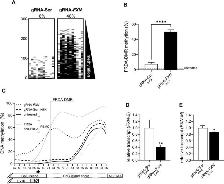
Epigenetic silencing in Friedreich ataxia (FRDA), induced by an expanded GAA triplet-repeat in intron 1 of the FXN gene, results in deficiency of the mitochondrial protein, frataxin. A lesser known extramitochondrial isoform of frataxin detected in erythrocytes, frataxin-E, is encoded via an alternate transcript (FXN-E) originating in intron 1 that lacks a mitochondrial targeting sequence. We show that FXN-E is deficient in FRDA, including in patient-derived cell lines, iPS-derived proprioceptive neurons, and tissues from a humanized mouse model. In a series of FRDA patients, deficiency of frataxin-E protein correlated with the length of the expanded GAA triplet-repeat, and with repeat-induced DNA hypermethylation that occurs in close proximity to the intronic origin of FXN-E. CRISPR-induced epimodification to mimic DNA hypermethylation seen in FRDA reproduced FXN-E transcriptional deficiency. Deficiency of frataxin E is a consequence of FRDA-specific epigenetic silencing, and therapeutic strategies may need to address this deficiency.


Conflict of interest statement

The authors declare no competing interests.

Figures

Figure 1
Figure 1
Frataxin-E is encoded by the FXN-E transcript that originates in intron 1 of the FXN gene, and its deficiency is related to the expanded GAA triplet-repeat in FRDA. (A) Schematic of the proximal portion of the FXN gene, showing the expanded GAA triplet-repeat (triangle), and the FRDA-DMR in intron 1 (dashed box; vertical lines represent the 11 CpGs, numbered 72–82). Basic gene set from GENCODE shows an alternate first exon in intron 1 of the FXN gene (Ex1b; located close to the FRDA-DMR), coincident with a DNase hypersensitive site (HS) and a curated promoter, per EPDnew (FXN_2), which is distinct from the canonical FXN promoter upstream of Exon 1 (FXN_1). (B) RT-PCR with a forward primer in Ex1b (F2) and reverse primer in Ex2 (R2) showing three spliceforms (IIa, IIb, and IIc). RT-PCR with a forward primer in Ex1 (F1) and reverse primer in Ex2 (R1) showing the canonical FXN transcript formed by splicing Exon 1 and 2. Note: Uncropped gel images are included in Fig. S14. (C) FXN-E is formed via splicing of Ex1b to Ex2 via three alternate splice donor sites to form three spliceforms (IIa, IIb, and IIc), as shown. Isoforms IIb and IIc contain an extra 18 (gray) or 30 (hatched) nucleotides of intron 1 sequence, respectively, at the 3′ end of Ex1b. Isoforms IIb and IIc use the uncommon “GC” splice donor. Isoforms IIa, IIb, and IIc are together referred to as FXN-E. FXN-M, which encodes the mitochondrial form of frataxin, is shown. (D) All isoforms of FXN-E are predicted to encode the extra-mitochondrial isoform, frataxin-E. Sequence of Ex1b (lowercase) is shown spliced to exon 2 (uppercase). The solid and dashed boxes mark the + 18 and + 30 nucleotides in isoforms IIb and IIc. The ATG initiation codon, M76 for frataxin-M and M1 for frataxin-E (underlined) is shown. The cleavage site in the second step of processing via mitochondrial processing peptidase (MPP #2) is marked by an arrow, hence frataxin-E lacks most of the mitochondrial targeting sequence.
Figure 2
Figure 2
Frataxin-E is deficient in FRDA. Western blot analysis utilizing (A) a commercial anti-frataxin monoclonal antibody and (B) a specific anti-frataxin-E monoclonal antibody. Blood samples from n = 5 non-FRDA controls (C1 to C5; 0.3 mL) and n = 5 FRDA patients (F1 to F5; 0.5 mL) were immunopurified with anti-frataxin antibody and analyzed via western blot. Lanes “E” and “M” show results for histidine-tagged/purified frataxin-E and frataxin-M protein, respectively. Arrows and labels (right-hand side) show location of proteins of interest. Although there is a small difference in size (Δ 685 Da) between endogenous frataxin-E (MW = 14,953 Da) and frataxin-M (14,268 Da), these proteins were resolved on PAGE using MES and SDS as the running buffer. Frataxin-E and frataxin M together with their His-tagged standards were detected by the anti-frataxin antibody; whereas, only frataxin-E and His-frataxin E were detected by the anti-frataxin-E mAb. Protein plus protein dual color standards were run on each gel and visualized in black and white by the ImageQuant LAS 4000 camera. Western blots of the frataxin proteins were visualized separately by the camera using the SuperSignal West femto luminol enhancement reagent. The two separate images were then combined.
Figure 3
Figure 3
Deficiency of frataxin-E correlates with the length of the expanded GAA triplet repeat. (A) Frataxin-E protein in erythrocytes is deficient in FRDA (FRDA [n = 32] vs non-FRDA [n = 11]; mean ± SD; two-tailed unpaired t test, p < 0.0001, t = 14.86, df = 41). (B) Frataxin E protein is inversely correlated with the shorter of the two expanded GAA alleles (GAA1) in FRDA. (C) Deficiency of FXN-E transcript in FRDA lymphoblastoid cells (FRDA [n = 4] vs non-FRDA [n = 2]; mean ± SD; two-tailed, unpaired t test, p = 0.0001, t = 10.98, df = 5).
Figure 4
Figure 4
Deficiency of frataxin-E correlates with FXN DNA hypermethylation in FRDA. (A) DNA methylation is plotted as a percentage of 1000 sequencing reads, at each of 39 CpG dinucleotides (numbered 57–95) that map between the 3′ end of the CpG island and the Alu repeat element that contains the expanded GAA repeat. The data are displayed as LOWESS trendlines for FRDA (n = 32; black lines) and non-FRDA controls (n = 14; purple lines). The FRDA-DMR is outlined with a gray box, and the relative locations of the CpG island, CpG island shore, Alu/GAA, and Ex1b are indicated. (B) FRDA-DMR hypermethylation among the FRDA patients in our cohort (n = 32); the solid and dotted lines within the violin plot indicate median and 25th/75th percentiles, respectively. (C) Frataxin-E protein is inversely correlated with FRDA-DMR methylation. (D) Bar graph showing R2 values (Pearson) for correlation of frataxin-E levels in the 32 patients with methylation at each of the 39 CpG sites. Note the clustering of CpG sites showing significant correlation (blue bars; with Benjamini and Hochberg correction for multiple comparisons) in the FRDA-DMR (gray box). Gray bars indicate correlations that are not significant (n.s.). The relative locations of the CpG island, CpG island shore, Alu/GAA, and Ex1b are displayed below the graph.
Figure 5
Figure 5
FRDA-DMR hypermethylation and deficiency of FXN-E transcript in iPS-derived proprioceptive neurons in FRDA. (A) Proprioceptive neurons derived from non-FRDA control iPS lines express FXN-M (isoform I) and FXN-E (isoforms IIa, IIb, and IIc). Note: Uncropped gel images are included in Fig. S14. (B) DNA methylation is plotted as a percentage of 1000 sequencing reads, at each of 39 CpG dinucleotides (numbered 57–95) that map between the 3′ end of the CpG island and the Alu repeat element that contains the expanded GAA repeat. Data are displayed as LOWESS trendlines for FRDA (neurons [n = 3]; neuronal progenitors [n = 3]; PBMCs [n = 32]) and non FRDA controls (neurons [n = 1]; neuronal progenitors [n = 1]; PBMCs [n = 14]). The FRDA-DMR is outlined with a gray box, and the relative locations of the CpG island, CpG island shore, Alu/GAA, and Ex1b are indicated. (C) FRDA-DMR methylation assayed as a single amplicon to test each of the n = 11 CpG dinucleotides in cis displayed as n = 300 sequencing reads stacked vertically for both neuronal progenitors and proprioceptive neurons from FRDA and non-FRDA controls (black dash = methylated CpG; reads are sorted with highest methylation at the bottom). (D) FXN-E and (E) FXN-M transcript levels in non-FRDA neuronal progenitors (n = 1) and proprioceptive neurons (n = 1) are shown as dotted horizontal lines. Transcript levels in FRDA neuronal progenitors (n = 3) and proprioceptive neurons (n = 3) are shown as black bars (mean ± SD); for FXN-E: p = 0.7953, t = 0.2773, df = 4, and for FXN-M: p = 0.0107, t = 4.511, df = 4.
Figure 6
Figure 6
FRDA-DMR hypermethylation and deficiency of FXN-E transcript in tissues from the YG8sR humanized mouse model of FRDA. (A) Cerebellum (CBL), heart, and skeletal muscle (SkM) from the non-FRDA control mouse (Y47R; with 9 GAA triplets) express FXN-M (isoform I) and FXN-E isoforms (IIa, IIb, and IIc). Note: Uncropped gel images are included in Fig. S14. (B) Relative FXN-E transcript levels in CBL, heart, and SkM in Y47R mice (n = 3) and in the FRDA mouse model (YG8sR; with 480 GAA repeats; n = 3). (C) Relative FXN-M transcript levels in CBL, heart, and SkM in Y47R (n = 3) and YG8sR (n = 3) mice. (D) FRDA-DMR methylation levels in CBL, heart, and SkM from the Y47R (GAA-9; n = 3) and YG8sR (GAA-480; n = 3) mice. All graphs show mean ± SD; 2way ANOVA with Tukey’s multiple comparisons; *p < 0.05, **p < 0.01, ***p < 0.001, ****p < 0.0001, n.s. = not significant.
Figure 7
Figure 7
DNA hypermethylation of the FRDA-DMR silences FXN-E. (A) FRDA-DMR methylation assayed as a single amplicon to test each of the n = 11 CpG dinucleotides in cis displayed as n = 300 sequencing reads stacked vertically for HEK293T cells transfected with dCas9-DNMT3A targeted to intron 1 of FXN via a guide RNA (gRNA-FXN) or with a scramble guide used as a negative control (gRNA-Scr). Black dash = methylated CpG; reads are sorted with highest methylation at the bottom and percent methylation in the FRDA-DMR is displayed above the respective panels. (B) FRDA-DMR methylation (mean ± SD) for n = 3 experiments (****p < 0.0001, t = 21.78, df = 4). FRDA-DMR methylation for untreated HEK293T cells is shown as a dashed line for comparison. (C) DNA methylation is plotted as a percentage of 1000 sequencing reads, at each of 39 CpG dinucleotides (numbered 57–95) that map between the 3′ end of the CpG island and the Alu repeat element that contains the expanded GAA repeat. Data are displayed as LOWESS trendlines for HEK293T cells either untreated or treated with dCas9-DNMT3A+ gRNA-FXN (to target the FRDA-DMR) or dCas9- DNMT3A+ gRNA-Scr (negative/scramble control). Non-FRDA and FRDA PBMCs are shown for comparison. The FRDA-DMR is outlined with a gray box, and the relative locations of the CpG island, CpG island shore, Alu/GAA, and Ex1b are indicated. The location of the gRNA is indicated by the rightward pointing arrow alongside the X-axis. (D) Relative FXN-E transcript levels and (E) Relative FXN-M transcript levels for n = 4 transfections with either dCas9-DNMT3A + gRNA-FXN (to target the FRDA-DMR; black bars) or dCas9-DNMT3A+ gRNA-Scr (negative/scramble control; white bars). Transcript levels are shown as mean ± SD; Two-tailed, unpaired t test; for FXN-E: p = 0.0038, t = 4.579, df = 6, and for FXN-M: p = 0.0106, t = 3.656, df = 6. Note: despite the statistical significance of FXN-M, the magnitude of change is not considered biologically significant, and FXN-M was not significantly altered in another independent experiment (see Fig. S13).

Similar articles

Cited by

References

    1. Bidichandani, S. I. & Delatycki, M. B. Friedreich ataxia. In GeneReviews((R)) (editors Adam, M. P., et al.) (Seattle, WA, 1993).
    1. Tsou AY, Paulsen EK, Lagedrost SJ, et al. Mortality in Friedreich ataxia. J. Neurol. Sci. 2011;307(1–2):46–49. - PubMed
    1. Durr A, Cossee M, Agid Y, et al. Clinical and genetic abnormalities in patients with Friedreich's ataxia. N. Engl. J. Med. 1996;335(16):1169–1175. - PubMed
    1. Regner SR, Wilcox NS, Friedman LS, et al. Friedreich ataxia clinical outcome measures: Natural history evaluation in 410 participants. J. Child. Neurol. 2012;27(9):1152–1158. - PMC - PubMed
    1. Metz G, Coppard N, Cooper JM, et al. Rating disease progression of Friedreich's ataxia by the International Cooperative Ataxia Rating Scale: Analysis of a 603-patient database. Brain. 2013;136(Pt 1):259–268. - PMC - PubMed

Publication types