

Saffron extract and crocin exert anti-inflammatory and anti-oxidative effects in a repetitive mild traumatic brain injury mouse model
- PMID: 35322143
- PMCID: PMC8943204
- DOI: 10.1038/s41598-022-09109-9
Saffron extract and crocin exert anti-inflammatory and anti-oxidative effects in a repetitive mild traumatic brain injury mouse model
Abstract
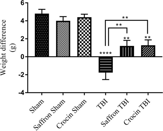
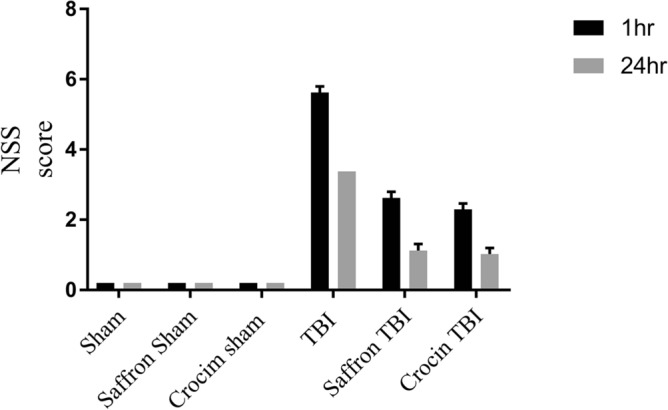
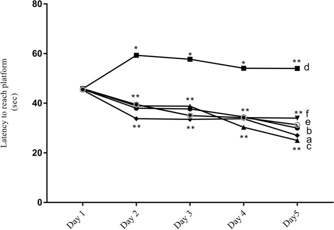
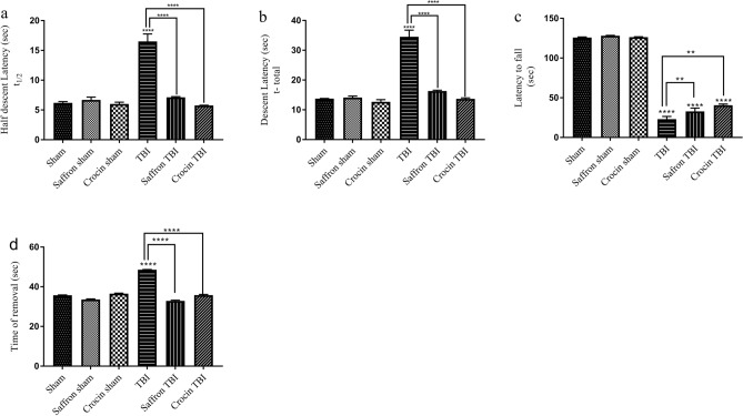
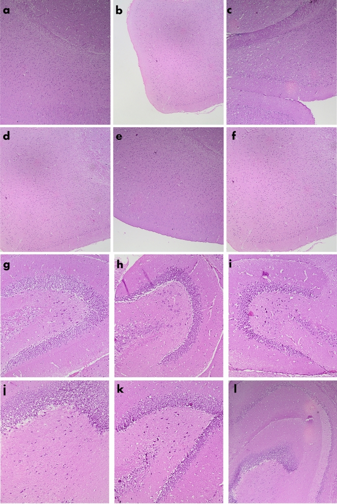
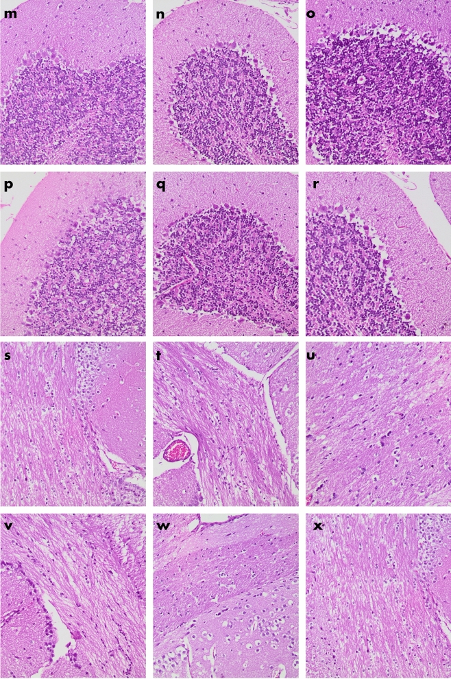
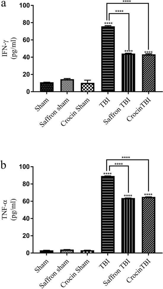
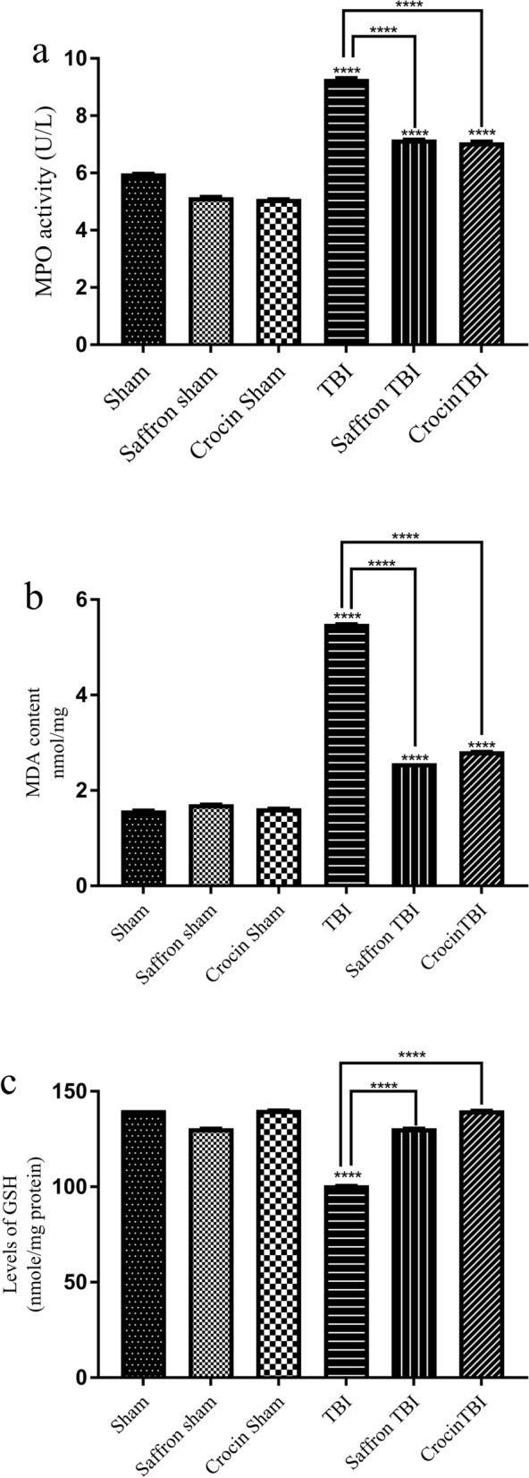
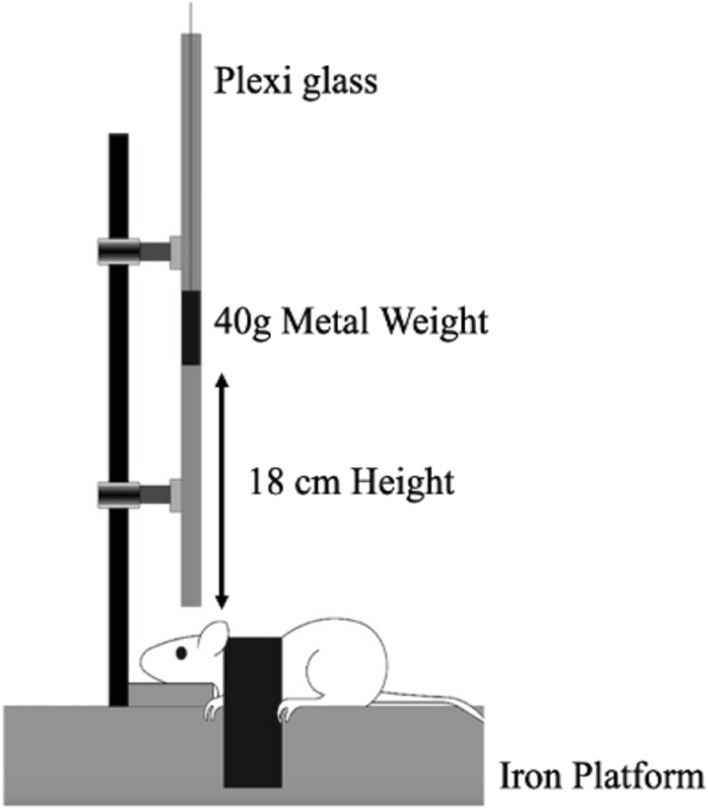
Saffron Crocus sativus L. (C. sativus) is a flower from the iridaceous family. Crocin, saffron's major constituent, and saffron have anti-oxidative and anti-inflammatory activities. In this work, the neuroprotective effects of saffron and crocin are being investigated in a repetitive mild traumatic brain injury (rmTBI) mouse model. A weight drop model setup was employed to induce mild brain injury in male albino BABL/c mice weighing 30-40 g. Saffron (50 mg/kg) and crocin (30 mg/kg) were administrated intraperitoneally 30 min before mTBI induction. Behavioral tests were conducted to assess behavioral deficits including the modified neurological severity score (NSS), Morris water maze (MWM), pole climb test, rotarod test, and adhesive test. The levels of TNF alpha (TNF-α), interferon-gamma (IFN-γ), myeloperoxidase activity (MPO), malonaldehyde (MDA), and reduced glutathione (GSH) were measured. Histological analysis of different brain parts was performed. Both saffron and crocin demonstrated marked improved neurological, cognitive, motor, and sensorimotor functions. Besides, both compounds significantly reduced the oxidative stress and inflammatory processes. No abnormal histological features were observed in any of the injured groups. Saffron extract and crocin provide a neuroprotective effect in a mouse model of rmTBI by decreasing oxidative stress, inflammatory responses, and behavioral deficits.
© 2022. The Author(s).
Conflict of interest statement
The authors declare no competing interests.
Figures
Similar articles
-
Evidence of neuroprotective effects of saffron and crocin in a Drosophila model of parkinsonism.Neurotoxicology. 2016 Jan;52:230-42. doi: 10.1016/j.neuro.2015.12.010. Epub 2015 Dec 17. Neurotoxicology. 2016. PMID: 26705857
-
Saffron and crocin ameliorate prenatal valproic acid-induced autistic-like behaviors and brain oxidative stress in the male offspring rats.Metab Brain Dis. 2023 Oct;38(7):2231-2241. doi: 10.1007/s11011-023-01275-7. Epub 2023 Aug 11. Metab Brain Dis. 2023. PMID: 37566156
-
Protective effects of saffron extract and its active constituent crocin against oxidative stress and spatial learning and memory deficits induced by chronic stress in rats.Eur J Pharmacol. 2011 Sep 30;667(1-3):222-9. doi: 10.1016/j.ejphar.2011.05.012. Epub 2011 May 18. Eur J Pharmacol. 2011. PMID: 21616066
-
Saffron (Crocus sativus L.) in Ocular Diseases: A Narrative Review of the Existing Evidence from Clinical Studies.Nutrients. 2019 Mar 18;11(3):649. doi: 10.3390/nu11030649. Nutrients. 2019. PMID: 30889784 Free PMC article. Review.
-
Therapeutic potential of saffron in brain disorders: From bench to bedside.Phytother Res. 2024 May;38(5):2482-2495. doi: 10.1002/ptr.8169. Epub 2024 Mar 6. Phytother Res. 2024. PMID: 38446350 Review.
Cited by
-
Gut Microbiota as a Modifier of Huntington's Disease Pathogenesis.J Huntingtons Dis. 2024;13(2):133-147. doi: 10.3233/JHD-240012. J Huntingtons Dis. 2024. PMID: 38728199 Free PMC article. Review.
-
NAMPT‑NAD+ is involved in the senescence‑delaying effects of saffron in aging mice.Exp Ther Med. 2024 Feb 1;27(3):123. doi: 10.3892/etm.2024.12411. eCollection 2024 Mar. Exp Ther Med. 2024. PMID: 38410190 Free PMC article.
-
Therapy of traumatic brain injury by modern agents and traditional Chinese medicine.Chin Med. 2023 Mar 11;18(1):25. doi: 10.1186/s13020-023-00731-x. Chin Med. 2023. PMID: 36906602 Free PMC article. Review.
-
Therapeutic effects of saffron and its components on neurodegenerative diseases.Heliyon. 2024 Jan 10;10(2):e24334. doi: 10.1016/j.heliyon.2024.e24334. eCollection 2024 Jan 30. Heliyon. 2024. PMID: 38298664 Free PMC article. Review.
-
Crocin Improves Cognitive Impairment in LPS-treated Rats through Anti-Apoptotic, Anti-Inflammatory, and Antioxidant Activities.Mol Neurobiol. 2025 May;62(5):5804-5815. doi: 10.1007/s12035-024-04638-y. Epub 2024 Dec 4. Mol Neurobiol. 2025. PMID: 39630406
References
-
- Aubry M, et al. Summary and agreement statement of the first International Conference on Concussion in Sport, Vienna 2001. Phys. Sportsmed. 2002;36:6–10. - PubMed
-
- American Congress of Rehabilitation Medicine Definition of mild traumatic brain injury. J. Head Trauma Rehabil. 1993;8:86–87.
MeSH terms
Substances
LinkOut - more resources
Full Text Sources
Medical
Research Materials
Miscellaneous
