. 2022 Mar 23;12(1):5034.
doi: 10.1038/s41598-022-09060-9.

A toolkit for recombinant production of seven human EGF family growth factors in active conformation

Affiliations

A toolkit for recombinant production of seven human EGF family growth factors in active conformation

Arthur Schveitzer Ferreira et al. Sci Rep. .

Abstract

Epidermal growth factors (EGF) play a wide range of roles in embryogenesis, skin development, immune response homeostasis. They are involved in several pathologies as well, including several cancer types, psoriasis, chronic pain and chronic kidney disease. All members share the structural EGF domain, which is responsible for receptor interaction, thereby initiating transduction of signals. EGF growth factors have intense use in fundamental research and high potential for biotechnological applications. However, due to their structural organization with three disulfide bonds, recombinant production of these factors in prokaryotic systems is not straightforward. A significant fraction usually forms inclusion bodies. For the fraction remaining soluble, misfolding and incomplete disulfide bond formation may affect the amount of active factor in solution, which can compromise experimental conclusions and biotechnological applications. In this work, we describe a reliable procedure to produce seven human growth factors of the EGF family in Escherichia coli. Biophysical and stability analyses using limited proteolysis, light scattering, circular dichroism and nanoDSF show that the recombinant factors present folded and stable conformation. Cell proliferation and scratch healing assays confirmed that the recombinant factors are highly active at concentrations as low as 5 ng/ml.


Conflict of interest statement

The authors declare no competing interests.

Figures
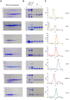
Figure 1
Figure 1
Purification of the EGF growth factors. (A) Images of Coomassie stained SDS–polyacrylamide gels showing the peak fractions of the initial affinity chromatography (1 to 10). (B) Images of Coomassie stained SDS–polyacrylamide gels showing the result of thrombin protease digestion (lane 1), elution fraction from the purification of Trx-his6 after thrombin cleavage (lane 2) and peak fractions after the size exclusion chromatography (SEC) (lanes 3–6). Sizes (kDa) of the molecular mass markers (M) are indicated on the left. Only the relevant parts of the gels are shown. Original uncropped images of the gels are presented in the Supplementary Information file, Figure S1. (C) Elution profiles of the size exclusion chromatography analyses on a Superdex 75 10/300 GL column. Brackets indicate the region of the peak analyzed by SDS-PAGE in (B). Each row represents the purification of the EGF growth factor indicated on the right.
Figure 2
Figure 2
Stability analysis by limited proteolysis using trypsin. The proteolytic assays were performed with samples both untreated and treated with the reducing agent DTT and incubated for 30 and 60 min. Control non-reduced and reduced samples without trypsin treatment were incubated under the same conditions. (A) The panels show Coomassie stained SDS–polyacrylamide gels used for the analysis of the proteolysis products and control reactions of the EGF growth factors as identified on the right side of each panel. M, molecular mass markers. -, non-reducing condition. R, reducing condition. Only the relevant parts of the gels are shown. Original uncropped images of the gels are presented in the Supplementary Information file, Figure S2. (B) Sequences of the EGF domains with the basic residues target of trypsin marked in red and the cysteine residues marked in green. Green lines indicate the disulfide bonds. The sequence identified by (linker) represents the part of the linker encoded by the vector which remains attached to the growth factors after thrombin digestion. The underlined EPR region indicates the segment of the hEPR propeptide required for stable expression of this EGF growth factor.
Figure 3
Figure 3
Radius size and aggregation analysis by DLS. Profiles of radius size distribution of the purified EGF growth factors under reducing and non-reducing conditions as determined by dynamic light scattering analyses. The dashed vertical lines represent the center of the cumulative radius of each sample. All assays were performed at 20 °C. Times and treatment are indicated in the figure. The graphics show the intensity distribution (%) as a function of particle radius (nm).
Figure 4
Figure 4
Particle size distributions (nm) for the seven growth factors as a relation of temperature increase from 20 to 95 °C as determined by DLS using non-reduced proteins.
Figure 5
Figure 5
Circular dichroism spectra of the seven EGF growth factors. The blue lines correspond to the spectra acquired at 20 °C, the red lines to the spectra acquired after heating to the indicated temperatures and the green lines correspond to the spectra acquired after the temperature was reduced to 20 °C. The far-UV circular dichroism spectra (195–260 nm) were recorded using a J-815 spectropolarimeter with five accumulations and using a 1 mm path length cell.
Figure 6
Figure 6
Activity of the seven EGF growth factors on cell proliferation. (A) Induction of HeLa cell proliferation by the seven EGF growth factors. Triplicates of HeLa cell cultures were treated with 50 ng/mL of each growth factor and the fold change of the proliferation rate relative to untreated cells (control) was determined after 48 h. The cells were quantified by flow cytometry and normalized using the CountBright™ Absolute Counting Beads method. *p < 0.05. (B) HeLa cell proliferation induced by the seven EGF growth factors as determined by the MTT assay. Triplicate cultures of HeLa cells were treated with the indicated amounts of the respective EGF growth factors for 48 h and quantified using the MTT assay. *p < 0.05. (C) Cell cultures absorbance as a function of the growth factor concentration. The mean values were derived from the assay described in (B) and curves were fitted with nonlinear regression.
Figure 7
Figure 7
Scratch wound closure induced by the seven EGF growth factors. (A) Confluent cultures of NDFH cells were scratched with a sterile pipette tip and treated with 50 ng/mL of each EGF growth factor for 48 h. For quantification of the gap closure rates, the area of the gap at 48 h was compared to the area of the initial gap. Gap areas were determined with the ImageJ software using microscopic images as described in the Methods section. *p < 0.05. (B) Representative microscopy images at 100 X magnification acquired at time 0 (unstained living cells after the scratch) and after 48 h of fixed cells stained with violet crystal. The bar size is 100 µm.
Figure 8
Figure 8
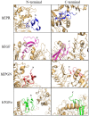
Analysis of the position of the N- and C-terminal ends of EGF growth factors bound to EGF receptors according to the crystal structures of the complexes available at PDB. The EGF receptors are shown in light orange. EGFR and EPR (blue) from PDB: 5WB7. EGFR and EGF (pink) from PDB: 1NQL.EPGN (red) from PDB: 5WB8. EGFR and TGFα (green) from PDB: 1MOX. The N- and C-terminal of the ligands are indicated. The images shown that the N- and C-terminals of the ligands are exposed to the solvent and not involved in the interaction with the receptor.

Similar articles

Cited by

References

    1. Yang CC, Chang KW. Eicosanoids and HB-EGF/EGFR in cancer. Cancer Metastasis Rev. 2018;37:385–395. - PubMed
    1. Zhao J, et al. Betacellulin enhances ovarian cancer cell migration by up-regulating Connexin43 via MEK-ERK signaling. Cell. Signal. 2020;65:109439. - PubMed
    1. Carvalho S, et al. An antibody to amphiregulin, an abundant growth factor in patients’ fluids, inhibits ovarian tumors. Oncogene. 2016;35:438–447. - PubMed
    1. Verma V, et al. The dichotomous role of epiregulin in pain. Pain. 2020;161:1052–1064. - PMC - PubMed
    1. Liu Y. Epidermal growth factor on the healing of human subacute tympanic membrane perforation. Am. J. Otolaryngol. 2020;41:102399. - PubMed

MeSH terms

Substances