. 2022 May;19(5):634-643.
doi: 10.1038/s41423-022-00849-2. Epub 2022 Mar 23.

Histone methyltransferase Nsd2 ensures maternal-fetal immune tolerance by promoting regulatory T-cell recruitment

Affiliations

Histone methyltransferase Nsd2 ensures maternal-fetal immune tolerance by promoting regulatory T-cell recruitment

Le Zhang et al. Cell Mol Immunol. 2022 May.

Abstract

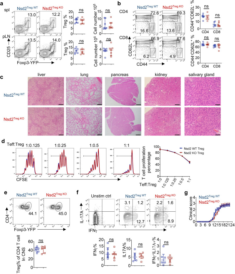
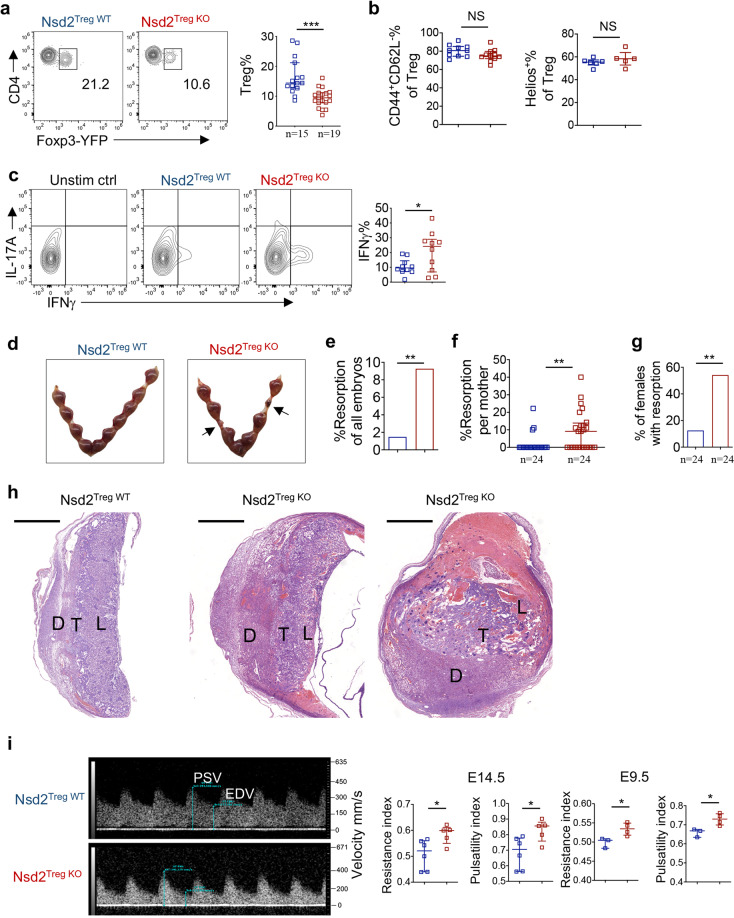
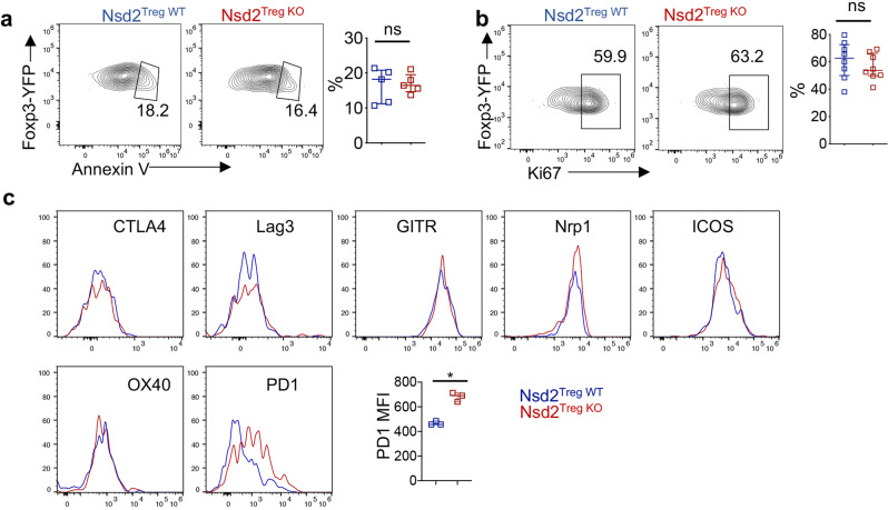
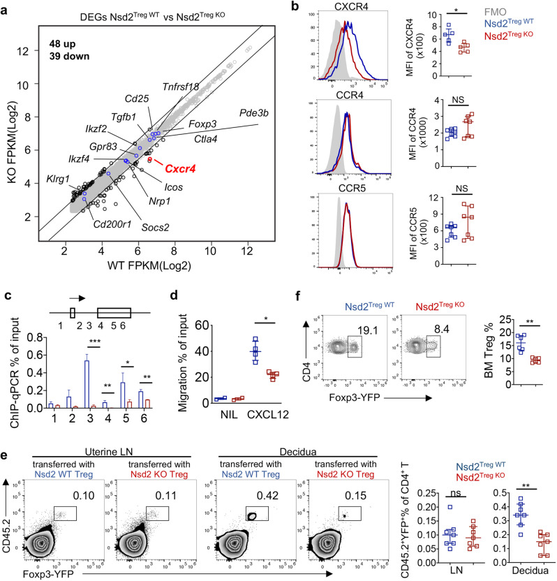
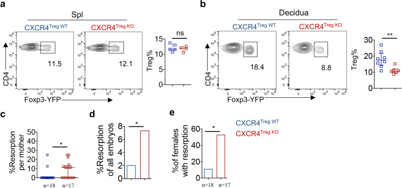
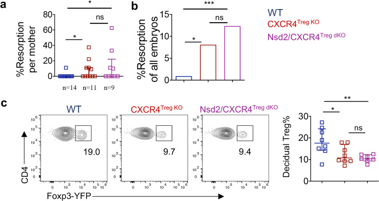
Regulatory T cells (Tregs) are fundamentally important for maintaining systemic immune homeostasis and are also required for immune tolerance at the maternal-fetal interface during pregnancy. Recent studies have suggested that epigenetic regulation is critically involved in Treg development and function. However, the role of H3K36me has not yet been investigated. Here, we found that the H3K36me2 methyltransferase Nsd2 was highly expressed in Tregs. Although loss of Nsd2 did not impair systemic Treg development or function, the level of Tregs at the maternal-fetal interface was significantly decreased in pregnant Nsd2 conditional knockout mice. Consequently, maternal-fetal immune tolerance was disrupted in the absence of Nsd2 in Tregs, and the pregnant mice showed severe fetal loss. Mechanistically, Nsd2 was found to upregulate CXCR4 expression via H3K36me2 modification to promote Treg cell recruitment into the decidua and suppress the anti-fetal immune response. Overall, our data identified Nsd2 as a critical epigenetic regulator of Treg recruitment for maternal-fetal tolerance.

Keywords: Cell migration; Immune tolerance; Regulatory T cell.


Conflict of interest statement

The authors declare no competing interests.

Figures

Fig. 1
Fig. 1
Nsd2 is dispensable for Treg development and function. a Flow cytometric analysis of the Treg (CD4+CD25+YFP+) percentage and number in the spleen (Spl) and peripheral lymph nodes (pLNs, pooled from inguinal and axillary LNs) of 8-week-old Nsd2Treg WT and Nsd2Treg KO mice. n = 7. b Flow cytometric analysis of the frequencies of naïve (CD44loCD62Lhi) and memory-like (CD44hiCD62Llo) CD4+ and CD8+ T cells in total splenocytes from 8-week-old Nsd2Treg WT and Nsd2Treg KO mice. n = 7. c Representative images of hematoxylin and eosin staining of the indicated tissues from Nsd2Treg WT and Nsd2Treg KO mice at the age of 4 months. Scale bars, 200 µm. d In vitro Treg suppression assay measuring the percentage of divided CD4+ T cells (Teff) by CFSE dilution after coculture for 3 days with the indicated ratio of Teff/Treg cells. Data are representative of two experiments. e Flow cytometric analysis of YFP+ Tregs among CD4+ cells infiltrating the CNS of Nsd2Treg WT and Nsd2Treg KO mice. n = 6. f Flow cytometric analysis of IFNγ+, IL-17A+, and IFNγ+IL-17A+ cells among CD4+ cells infiltrating the CNS of Nsd2Treg WT and Nsd2Treg KO mice. n = 6. g The clinical severity of EAE in Nsd2Treg WT and Nsd2Treg KO mice was monitored for 22 days after immunization with the MOG peptide. n = 12. ns not significant
Fig. 2
Fig. 2
Nsd2 deficiency results in reduced Tregs at the maternal–fetal interface and in fetal resorption. a Representative flow cytometric analysis of YFP+ Treg cells in the decidua of Nsd2Treg WT and Nsd2Treg KO mice previously mated with male BALB/c mice on Days E13.5–E14.5. b Percentage of CD44+CD62Llow and Helios+ Tregs determined by flow cytometry in the decidua of Nsd2Treg WT and Nsd2Treg KO mice previously mated with male BALB/c mice on Days E13.5–E14.5. c Flow cytometric analysis of IFNγ and IL-17A expression in decidual CD4+ T cells. n = 10. d Representative image of resorption of allogeneic embryos in the uterus of Nsd2Treg WT and Nsd2Treg KO mice on Day E14.5. Arrows indicate resorptions. e Percentage of resorbed embryos in all Nsd2Treg WT and Nsd2Treg KO pregnancies resulting from matings with male BALB/c mice. Two-sided Fisher’s exact test was used to assess the significance. f Percentage of resorption observed in individual mothers with the indicated genotype. g Incidence of pregnancies with at least one resorption. Two-sided Fisher’s exact test was used to assess the significance. h Histopathological evaluation of placentas from female Nsd2Treg WT (left) and Nsd2Treg KO (right, two examples) mice mated with male BALB/c mice; low-magnification survey of representative H&E-stained sections of placenta. D decidua, T trophoblast, L labyrinths. Scale bars, 1000 mm. i Representative Doppler image of the uterine artery (left, E14.5), which was used to determine the uterine artery resistance and pulsatility indices (right) of Nsd2Treg WT and Nsd2Treg KO female mice on Days E14.5 and E9.5. *P ≤ 0.05; **P ≤ 0.01; ***P ≤ 0.001
Fig. 3
Fig. 3
Nsd2 deficiency did not affect the apoptosis or proliferation of decidual Tregs. a Cell-surface staining for Annexin V on decidual Tregs isolated from Nsd2Treg WT and Nsd2Treg KO mice. n = 5. b Flow cytometric analysis of Ki67 expression in decidual Tregs from Nsd2Treg WT and Nsd2Treg KO mice. n = 8. c Histogram overlay of CTLA-4, LAG-3, GITR, Nrp1, ICOS, OX40 and PD-1 expression and plots of the PD-1 (n = 3) mean fluorescence intensity (MFI) of decidual Treg cells from Nsd2Treg WT and Nsd2Treg KO mice. ***P ≤ 0.001; ns not significant
Fig. 4
Fig. 4
Nsd2 promotes Treg recruitment into the decidua by regulating CXCR4 expression. a RNA-seq analysis of CD25+ YFP+ Treg cells from Nsd2Treg WT and Nsd2Treg KO mice. The examples of functionally relevant Treg genes without significant differential expression are labeled with blue dots. b Flow cytometric analysis of CXCR4, CCR4, and CCR5 expression in splenic Tregs. n = 5–7. c ChIP-qPCR for H3K36me2 modifications on the CXCR4 locus. Primers specific for the six indicated regions were used for the analysis. d Transwell migration assay performed with Tregs from Nsd2Treg WT and Nsd2Treg KO mice of the indicated genotype in response to medium alone (nil) and 1 µg/ml SDF, shown as the percentage of input cells. e A total of 2 × 106 CD25+YFP+ Tregs from Nsd2Treg WT or Nsd2Treg KO mice were adoptively transferred into pregnant CD45.1+ BoyJ mice on Day E12.5. The cell percentages of transferred Tregs in the uterine-draining lymph nodes and decidua were examined by flow cytometry 48 h after injection. n = 7. f Flow cytometric analysis of the Treg (CD4+YFP+) percentage in the bone marrow (BM) of 8-week-old Nsd2Treg WT and Nsd2Treg KO mice. n = 7. *P ≤ 0.05; **P ≤ 0.01; ***P ≤ 0.001; ns not significant
Fig. 5
Fig. 5
Treg-specific CXCR4 conditional KO mice displayed decidual Treg reduction and fetal abortion phenotypes similar to those of Nsd2Treg KO mice. a Flow cytometric analysis of the Treg (CD4+YFP+) percentage and number in the spleen (Spl) of 8-week-old CXCR4Treg WT (n = 5) and CXCR4Treg KO mice (n = 4). b Representative flow cytometric analysis of YFP+ Treg cells in the decidua of CXCR4Treg WT and CXCR4Treg KO mice previously mated with BALB/c males on Days E13.5–E14.5. n = 6–8. c Percentage of resorption observed in individual mothers with the indicated genotype. d Percentage of resorbed embryos in all CXCR4Treg WT and CXCR4Treg KO pregnancies resulting from mating with male BALB/c mice. Two-sided Fisher’s exact test was used to assess the significance. e Incidence of pregnancies with at least one resorption. Two-sided Fisher’s exact test was used to assess the significance. *P ≤ 0.05; **P ≤ 0.01; ns not significant
Fig. 6
Fig. 6
Nsd2 promotes Treg migration into the decidua primarily by regulating CXCR4 expression. a Percentage of resorption observed in individual WT, CXCR4Treg KO, and Nsd2/CXCR4Treg dKO pregnancies resulting from mating with male BALB/c mice. b Percentage of resorbed embryos in all WT, CXCR4Treg KO, and Nsd2/CXCR4Treg dKO pregnancies resulting from mating with male BALB/c mice. Two-sided Fisher’s exact test was used to assess the significance. c Representative flow cytometric analysis of YFP+ Tregs in the decidua and analysis of the decidua of female WT, CXCR4Treg KO and Nsd2/CXCR4Treg dKO mice mated with male BALB/c mice and analyzed on Days E13.5–E14.5. n = 8. *P ≤ 0.05; **P ≤ 0.01; ***P ≤ 0.001; ns not significant

Similar articles

Cited by

References

    1. Josefowicz SZ, Lu L-F, Rudensky AY. Regulatory T cells: mechanisms of differentiation and function. Annu Rev Immunol. 2012;30:531–64. doi: 10.1146/annurev.immunol.25.022106.141623. - DOI - PMC - PubMed
    1. Sakaguchi S, Mikami N, Wing JB, Tanaka A, Ichiyama K, Ohkura N. Regulatory T cells and human disease. Annu Rev Immunol. 2020;38:541–66. doi: 10.1146/annurev-immunol-042718-041717. - DOI - PubMed
    1. Savage PA, Klawon DEJ, Miller CH. Regulatory T cell development. Annu Rev Immunol. 2020;38:421–53. doi: 10.1146/annurev-immunol-100219-020937. - DOI - PubMed
    1. Feuerer M, Hill JA, Mathis D, Benoist C. Foxp3+ regulatory T cells: differentiation, specification, subphenotypes. Nat Immunol. 2009;10:689–95. doi: 10.1038/ni.1760. - DOI - PubMed
    1. DuPage M, Chopra G, Quiros J, Rosenthal WL, Morar MM, Holohan D, et al. The chromatin-modifying enzyme Ezh2 is critical for the maintenance of regulatory T cell identity after activation. Immunity. 2015;42:227–38. doi: 10.1016/j.immuni.2015.01.007. - DOI - PMC - PubMed

Publication types

Substances