

Cryo-EM structure of transmembrane AAA+ protease FtsH in the ADP state
- PMID: 35322207
- PMCID: PMC8943139
- DOI: 10.1038/s42003-022-03213-2
Cryo-EM structure of transmembrane AAA+ protease FtsH in the ADP state
Abstract
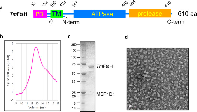
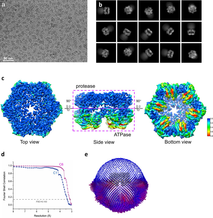
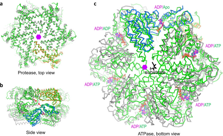
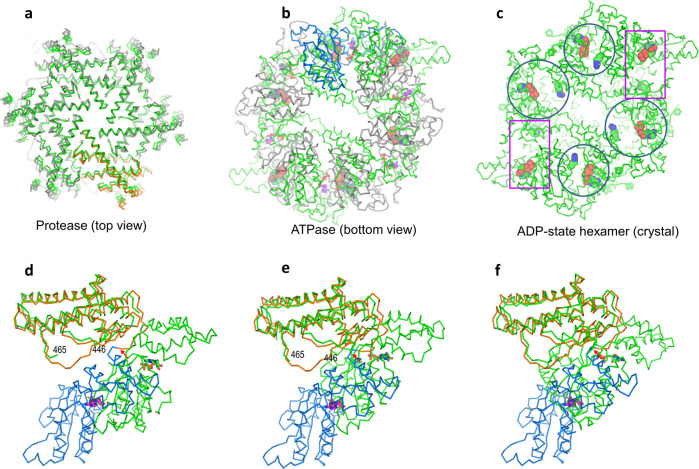
AAA+ proteases regulate numerous physiological and cellular processes through tightly regulated proteolytic cleavage of protein substrates driven by ATP hydrolysis. FtsH is the only known family of membrane-anchored AAA+ proteases essential for membrane protein quality control. Although a spiral staircase rotation mechanism for substrate translocation across the FtsH pore has been proposed, the detailed conformational changes among various states have not been clear due to absence of FtsH structures in these states. We report here the cryo-EM structure for Thermotoga maritima FtsH (TmFtsH) in a fully ADP-bound symmetric state. Comparisons of the ADP-state structure with its apo-state and a substrate-engaged yeast YME1 structure show conformational changes in the ATPase domains, rather than the protease domains. A reconstruction of the full-length TmFtsH provides structural insights for the dynamic transmembrane and the periplasmic domains. Our structural analyses expand the understanding of conformational switches between different nucleotide states in ATP hydrolysis by FtsH.
© 2022. The Author(s).
Conflict of interest statement
The authors declare no competing interests.
Figures


Similar articles
-
ATPase and Protease Domain Movements in the Bacterial AAA+ Protease FtsH Are Driven by Thermal Fluctuations.J Mol Biol. 2018 Oct 26;430(22):4592-4602. doi: 10.1016/j.jmb.2018.07.023. Epub 2018 Jul 22. J Mol Biol. 2018. PMID: 30044948
-
The crystal structure of apo-FtsH reveals domain movements necessary for substrate unfolding and translocation.Proc Natl Acad Sci U S A. 2009 Dec 22;106(51):21579-84. doi: 10.1073/pnas.0910708106. Epub 2009 Dec 2. Proc Natl Acad Sci U S A. 2009. PMID: 19955424 Free PMC article.
-
Cryo-EM structure of the entire FtsH-HflKC AAA protease complex.Cell Rep. 2022 May 31;39(9):110890. doi: 10.1016/j.celrep.2022.110890. Cell Rep. 2022. PMID: 35649372
-
AAA+ ATPases in Protein Degradation: Structures, Functions and Mechanisms.Biomolecules. 2020 Apr 18;10(4):629. doi: 10.3390/biom10040629. Biomolecules. 2020. PMID: 32325699 Free PMC article. Review.
-
Stairway to translocation: AAA+ motor structures reveal the mechanisms of ATP-dependent substrate translocation.Protein Sci. 2020 Feb;29(2):407-419. doi: 10.1002/pro.3743. Epub 2019 Oct 17. Protein Sci. 2020. PMID: 31599052 Free PMC article. Review.
Cited by
-
Maintaining the Integral Membrane Proteome: Revisiting the Functional Repertoire of Integral Membrane Proteases.Chembiochem. 2025 May 5;26(9):e202500048. doi: 10.1002/cbic.202500048. Epub 2025 Mar 18. Chembiochem. 2025. PMID: 40056010 Free PMC article. Review.
-
Transforming an ATP-dependent enzyme into a dissipative, self-assembling system.Nat Chem Biol. 2025 Jun;21(6):883-893. doi: 10.1038/s41589-024-01811-1. Epub 2025 Jan 13. Nat Chem Biol. 2025. PMID: 39806067
-
FtsH degrades dihydrofolate reductase by recognizing a partially folded species.Protein Sci. 2022 Sep;31(9):e4410. doi: 10.1002/pro.4410. Protein Sci. 2022. PMID: 36630366 Free PMC article.
-
Diverse nature of ClpX degradation motifs in Streptococcus mutans.Microbiol Spectr. 2024 Jan 11;12(1):e0345723. doi: 10.1128/spectrum.03457-23. Epub 2023 Dec 5. Microbiol Spectr. 2024. PMID: 38051052 Free PMC article.
-
Deciphering the mechanism and function of Hsp100 unfoldases from protein structure.Biochem Soc Trans. 2022 Dec 16;50(6):1725-1736. doi: 10.1042/BST20220590. Biochem Soc Trans. 2022. PMID: 36454589 Free PMC article.
References
-
- Sauer RT, Baker TA. AAA+ proteases: ATP-fueled machines of protein destruction. Annu. Rev. Biochem. 2011;80:587–612. - PubMed
-
- Langklotz S, Baumann U, Narberhaus F. Structure and function of the bacterial AA protease FtsH. Biochim. Biophys. Acta Mol. Cell Res. 2012;1823:40–48. - PubMed
-
- Janska H, Kwasniak M, Szczepanowska J. Protein quality control in organelles—AAA/FtsH story. Biochim. Biophys. Acta. 2013;1833:381–387. - PubMed
-
- Bittner L-M, Arends J, Narberhaus F. When, how and why? Regulated proteolysis by the essential FtsH protease in Escherichia coli. Biol. Chem. 2017;398:625–635. - PubMed
Publication types
MeSH terms
Substances
LinkOut - more resources
Full Text Sources
Molecular Biology Databases
