Review
. 2022 May;17(5):1223-1265.
doi: 10.1038/s41596-022-00680-z. Epub 2022 Mar 23.

Transcriptome-wide identification of RNA-binding protein binding sites using seCLIP-seq

Affiliations
Review

Transcriptome-wide identification of RNA-binding protein binding sites using seCLIP-seq

Steven M Blue et al. Nat Protoc. 2022 May.

Abstract

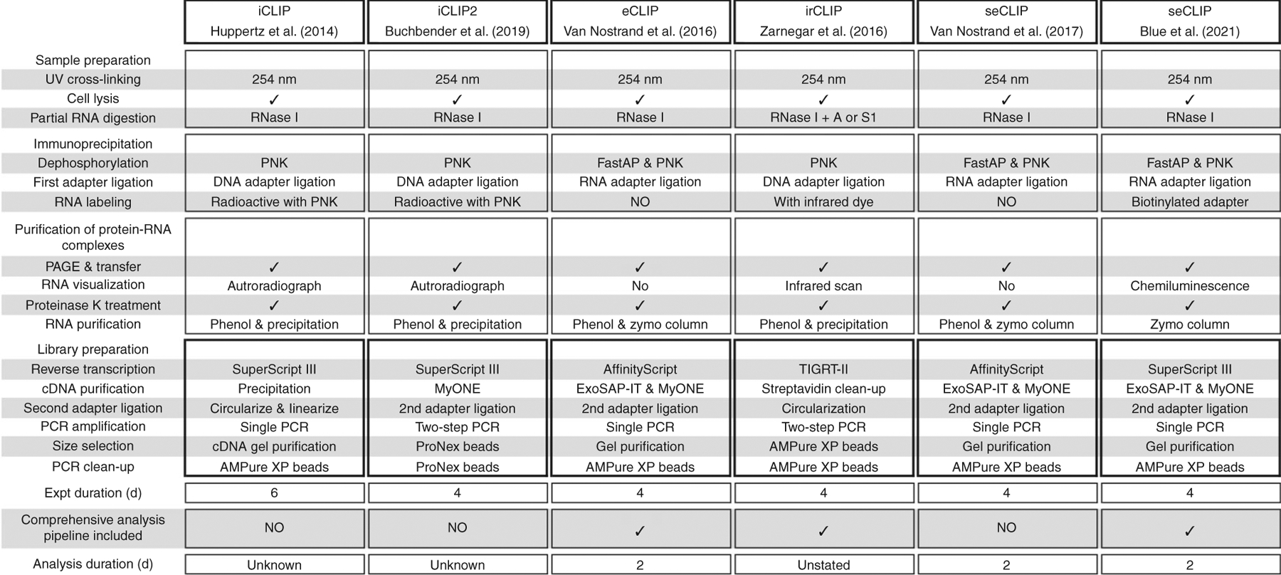
Discovery of interaction sites between RNA-binding proteins (RBPs) and their RNA targets plays a critical role in enabling our understanding of how these RBPs control RNA processing and regulation. Cross-linking and immunoprecipitation (CLIP) provides a generalizable, transcriptome-wide method by which RBP/RNA complexes are purified and sequenced to identify sites of intermolecular contact. By simplifying technical challenges in prior CLIP methods and incorporating the generation of and quantitative comparison against size-matched input controls, the single-end enhanced CLIP (seCLIP) protocol allows for the profiling of these interactions with high resolution, efficiency and scalability. Here, we present a step-by-step guide to the seCLIP method, detailing critical steps and offering insights regarding troubleshooting and expected results while carrying out the ~4-d protocol. Furthermore, we describe a comprehensive bioinformatics pipeline that offers users the tools necessary to process two replicate datasets and identify reproducible and significant peaks for an RBP of interest in ~2 d.


Conflict of interest statement

Competing interests

G.W.Y. is co-founder, member of the Board of Directors, on the Science Advisory Board, an equity holder and a paid consultant for Locanabio and Eclipse BioInnovations. G.W.Y. is a visiting professor at the National University of Singapore. G.W.Y.’s interest(s) have been reviewed and approved by the University of California, San Diego in accordance with its conflict-of-interest policies. A.A.S. is co-founder and Research & Development Director for Eclipse Bioinnovations. E.L.V.N. is co-founder, member of the Board of Directors, on the Science Advisory Board, an equity holder and a paid consultant for Eclipse BioInnovations. E.L.V.N.’s interest(s) have been reviewed and approved by Baylor College of Medicine in accordance with its conflict-of-interest policies. The authors declare no other competing interests.

Figures

Fig. 1 |
Fig. 1 |. Overview of CLIP methods.
Comparison of seCLIP experimental and analysis workflow to iCLIP, iCLIP2, eCLIP, irCLIP and a previously published version of seCLIP. Expt, experiment; iCLIP, individual-nucleotide resolution cross-linking and IP; irCLIP, infrared-CLIP. Figure adapted with permission from ref., Elsevier.
Fig. 2 |
Fig. 2 |. Overview of the seCLIP protocol.
a, UV cross-linking of cultured cells or tissue (Steps 1–4). b, Cells are lysed, and RNA is partially digested (Steps 8–11). c, The target protein is purified by using antibody-coupled magnetic beads (Step 12). d, RBP-RNA complexes are washed and dephosphorylated, enabling ligation of the 3′ linker (Steps 13–22). e, RBP-RNA complexes are denatured from beads and separated by SDS-PAGE, followed by transfer to nitrocellulose and PVDF membranes (Steps 23–27). IP success is visualized via western blot (Steps 28–31), and RBP-RNA complexes can be visualized via RNA biotinylation and streptavidin-HRP (Box 1). Western blot is used as a guide to isolate regions corresponding to RBP-bound RNA fragments. f, RNA is extracted from the membrane via proteinase digestion, SMInput RNA undergoes dephosphorylation and 3′ linker ligation and RNA is converted to cDNA by reverse transcription (Steps 32–56). g, RNA is degraded, and cDNA is purified (Steps 57–59). h, A UMI-containing linker is ligated to the 3′ end of cDNA molecules, followed by cleanup (Steps 60–65). i, cDNA libraries are quantified by qPCR, PCR-amplified and purified before quantification (Steps 66–82). j, Schematic of the final seCLIP library fragment. The unique molecular identifier is displayed in brown and labeled as UMI on the diagram.
Fig. 3 |
Fig. 3 |. Overview of the eCLIP bioinformatics workflow.
a, Outline of steps used to call significantly enriched peaks from fastq files as well as derive quality control metrics such as the number of usable reads and entropy total across peaks. b, Intermediates taken from the peak calling workflow may be used to discover bound repetitive elements. c, Irreproducibility discovery rate (IDR) may be used to merge two replicate sets of peaks and compute rescue and self-consistency ratios to be used to evaluate irreproducibility.
Fig. 4 |
Fig. 4 |. Overview of files required to assess irreproducibility statistics.
a and b, Beginning from PCR-duplicate-removed alignments from Step 91, files are merged and then split into pseudo-replicates (a), and peaks are called with CLIPper as described in Steps 107 and 108, respectively (b). c, Alignments from Step 8 are also individually shuffled and split to produce internal pseudo-replicate alignments and peaks as described in Steps 110 and 111, respectively. d, Depending on the experimental setup, PCR-duplicate-removed alignments for SMInputs may be merged if there are replicates available or left alone if there is only one. e, Outputs from ad are color-coded to match pairs of replicates and pseudo-replicates. IDR is performed as described in Steps 100–105 on each pair to obtain the number of peaks (N) required to compute the rescue ratio max(Np,Nt)min(Np,Nt) and self-consistency ratio max(N1,N2)min(N1,N2) described in Steps 106–109 and Steps 110–112, respectively.
Fig. 5 |
Fig. 5 |. Comparative visualization of biotin-labeled RNA detected by streptavidin-HRP and radiolabeled RNA.
Biotin- and 32P-based RNA labeling after TIAL1-specific IP (using RN059PW antibody) in HepG2 cells. Three samples underwent IP: UV cross-linked cells (XL) with standard (40 U) RNase (+), cross-linked cells with high (333 U) levels of RNase (++) and non-cross-linked (NXL) cells with either standard or high RNase. RNA was then either labeled with T4 RNA ligase and pCp-biotin followed by chemiluminescent imaging with streptavidin-bound HRP or radiolabeled with T4 PNK and [γ−32P]-ATP followed by autoradiographic imaging. Markers represent molecular weight in kilodaltons. Figure adapted with permission from ref., BioMed Central.
Fig. 6 |
Fig. 6 |. Representative expected results from an seCLIP experiment and analysis.
a, Example of PCR-amplified seCLIP cDNA libraries (PRPF39 IP in HepG2) from SMInput and IP samples after gel electrophoresis. Red dotted rectangles indicate excision window for sample purification, the blue arrow indicates unwanted adapter dimer and green arrows indicate unincorporated PCR primers. b, Example of TapeStation output traces corresponding to samples. The region selected in light green (150–700 bp) is used to calculate sample concentration, and samples are free of adapter dimer and unincorporated PCR primers after gel extraction. c, Example of a ‘skyscraper’-shaped peak (left), which should be flagged as a potential artefact and filtered. Examples of true binding signals (right) exhibit a more gradual increase of reads and are shaped like peaks rather than skyscrapers. chr, chromosome; FU, fluorescence units.

References

    1. Gerstberger S, Hafner M & Tuschl T A census of human RNA-binding proteins. Nat. Rev. Genet 15, 829–845 (2014). - PMC - PubMed
    1. Hentze MW, Castello A, Schwarzl T & Preiss T A brave new world of RNA-binding proteins. Nat. Rev. Mol. Cell Biol 19, 327–341 (2018). - PubMed
    1. Lukong KE, Chang K, Khandjian EW & Richard S RNA-binding proteins in human genetic disease. Trends Genet. 24, 416–425 (2008). - PubMed
    1. Nussbacher JK, Batra R, Lagier-Tourenne C & Yeo GW RNA-binding proteins in neurodegeneration: Seq and you shall receive. Trends Neurosci. 38, 226–236 (2015). - PMC - PubMed
    1. Brinegar AE & Cooper TA Roles for RNA-binding proteins in development and disease. Brain Res. 1647, 1–8 (2016). - PMC - PubMed

Publication types