Review
. 2022 May;17(5):1266-1305.
doi: 10.1038/s41596-022-00681-y. Epub 2022 Mar 23.

Generation and characterization of hair-bearing skin organoids from human pluripotent stem cells

Affiliations
Review

Generation and characterization of hair-bearing skin organoids from human pluripotent stem cells

Jiyoon Lee et al. Nat Protoc. 2022 May.

Erratum in

Abstract

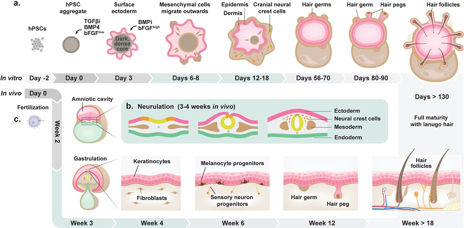
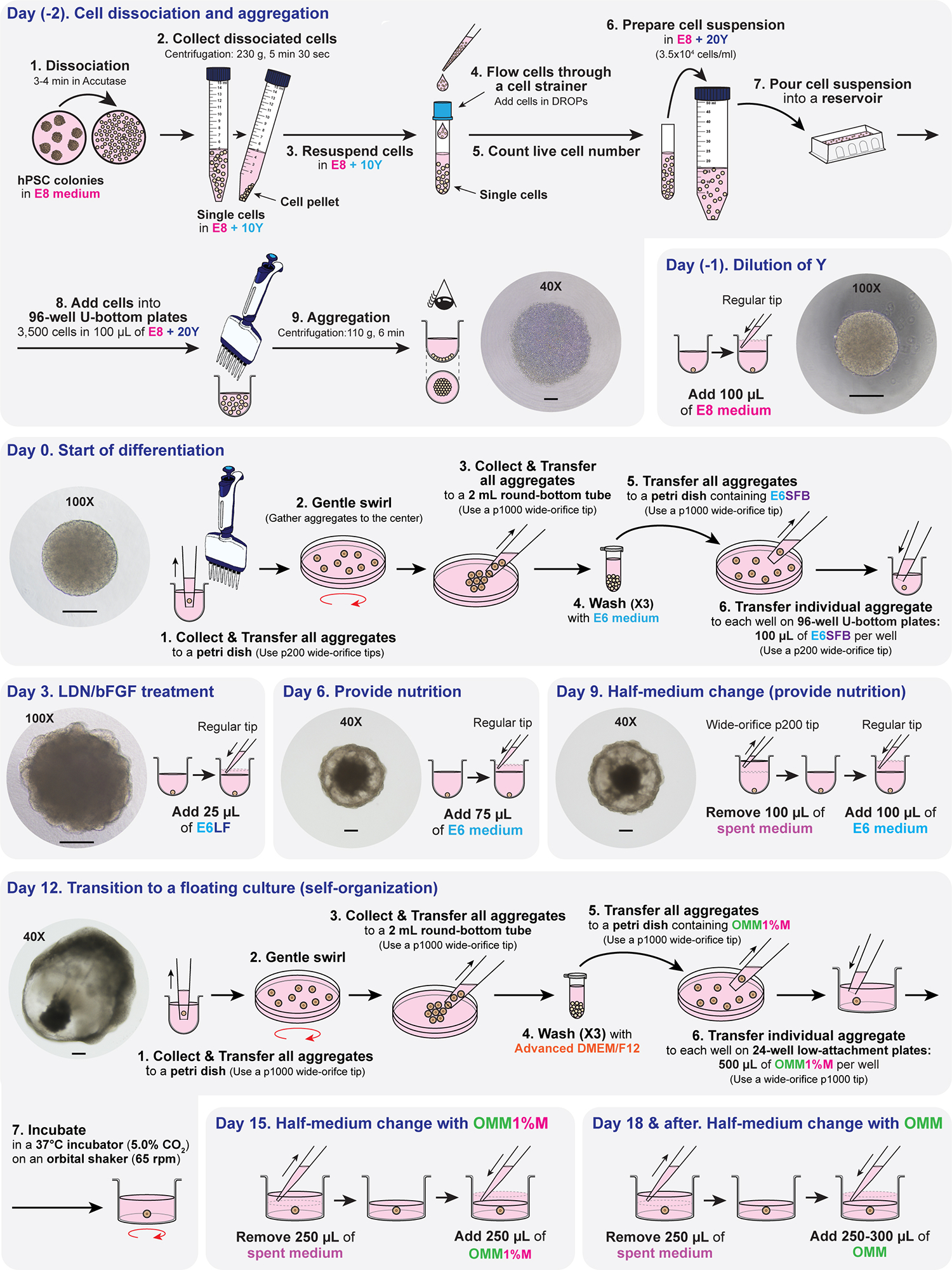
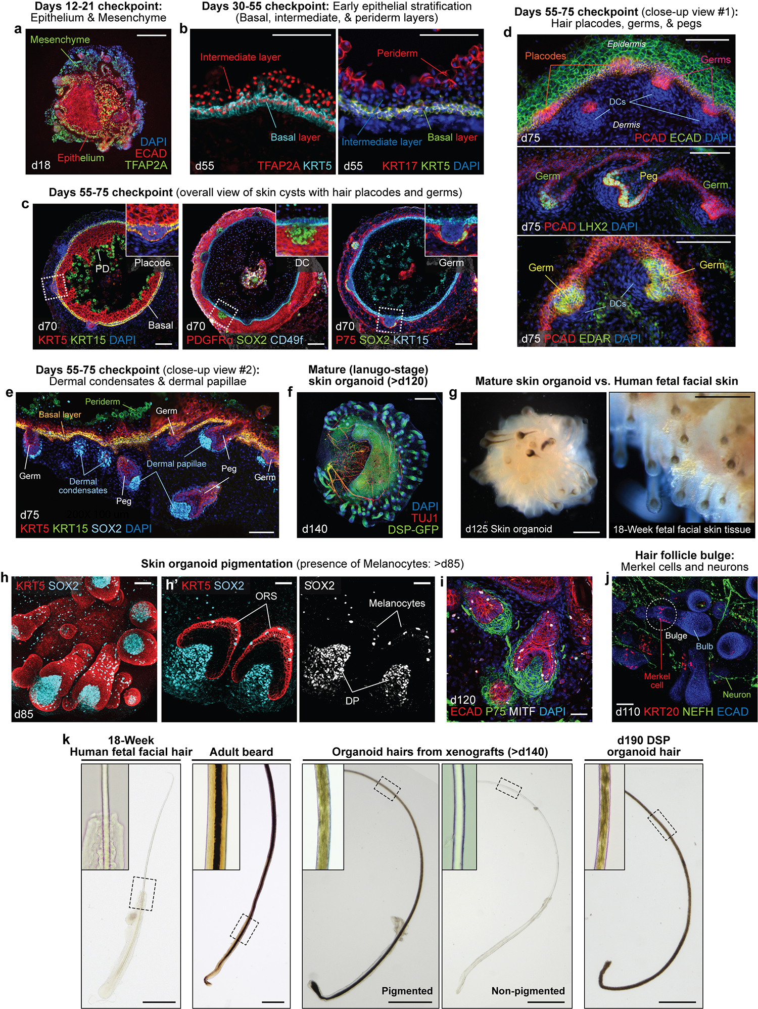
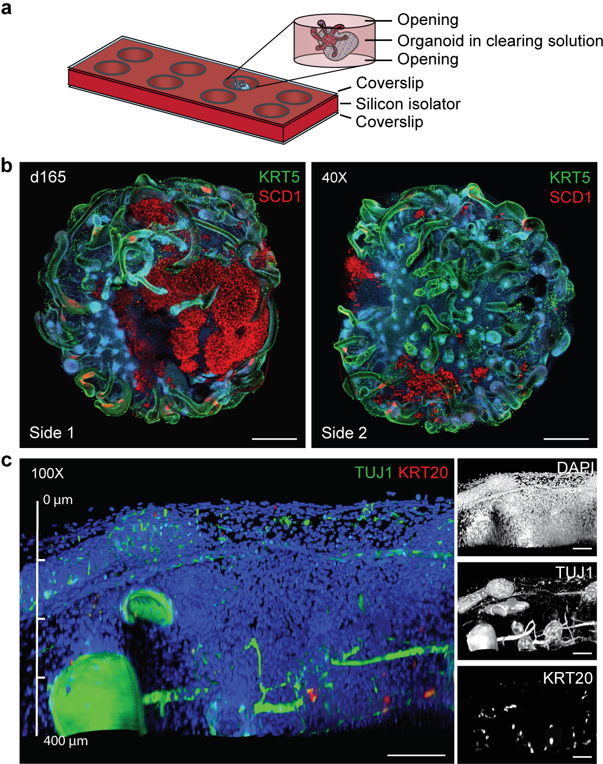
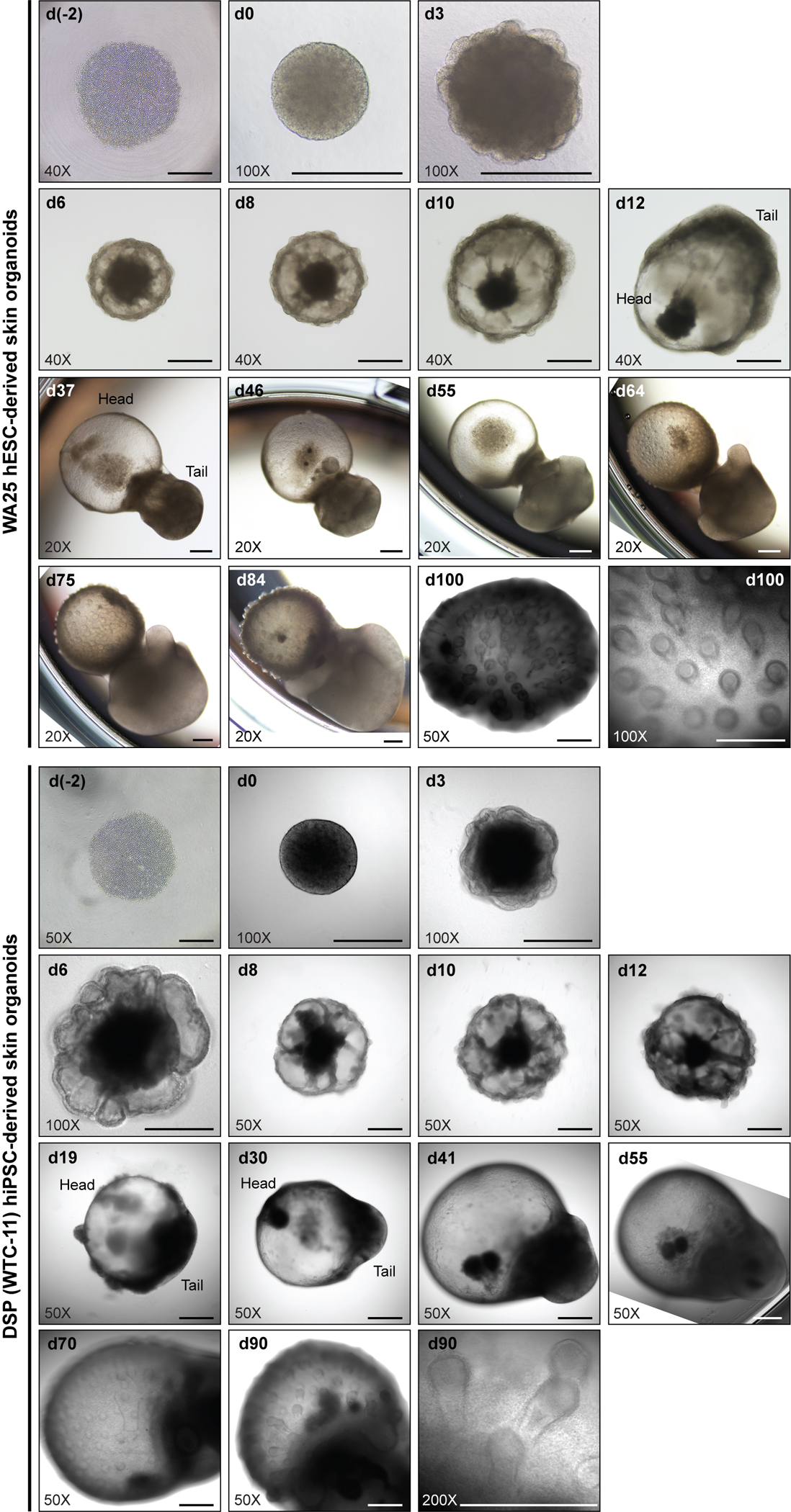
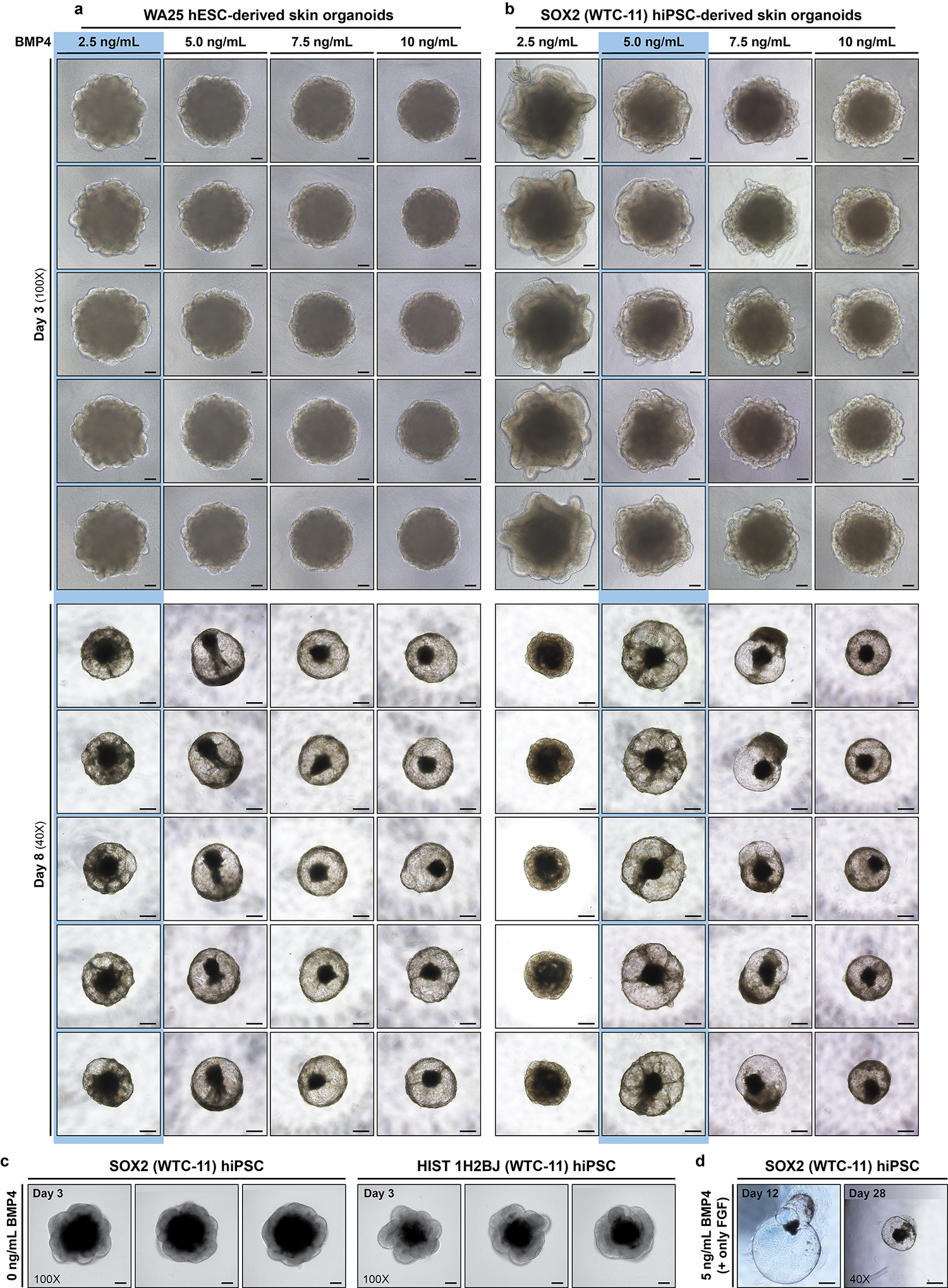
Human skin uses millions of hairs and glands distributed across the body surface to function as an external barrier, thermoregulator and stimuli sensor. The large-scale generation of human skin with these appendages would be beneficial, but is challenging. Here, we describe a detailed protocol for generating hair-bearing skin tissue entirely from a homogeneous population of human pluripotent stem cells in a three-dimensional in vitro culture system. Defined culture conditions are used over a 2-week period to induce differentiation of pluripotent stem cells to surface ectoderm and cranial neural crest cells, which give rise to the epidermis and dermis, respectively, in each organoid unit. After 60 d of incubation, the skin organoids produce hair follicles. By day ~130, the skin organoids reach full complexity and contain stratified skin layers, pigmented hair follicles, sebaceous glands, Merkel cells and sensory neurons, recapitulating the cell composition and architecture of fetal skin tissue at week 18 of gestation. Skin organoids can be maintained in culture using this protocol for up to 150 d, enabling the organoids to be used to investigate basic skin biology, model disease and, further, reconstruct or regenerate skin tissue.


Conflict of interest statement

COMPETING INTERESTS

J.L. and K.R.K, with the Indiana University Research and Technology Corporation, have a patent covering the entire skin organoid induction method (US11021688B2), which is licensed to STEMCELL Technologies, Inc. for research use. The other authors declare no competing interests.

Figures

Fig. 1
Fig. 1. Schematics comparing in vivo and in vitro skin organogenesis.
a, A timeline of in vitro skin organogenesis in the skin organoid model. hPSCs form aggregates on day 0 of differentiation. These aggregates are treated with TGF-β inhibitor (SB), BMP4, and a low concentration of bFGF, giving rise to surface ectoderm by day 3. The aggregates are then treated with BMP inhibitor (LDN) and a high concentration of bFGF. This day 3 treatment induces the development of non-epithelial cell populations consisting, in part, of cranial neural crest (CNC) cells, which further differentiate into diverse mesenchymal and neuro-glial cell populations contributing to the dermis layer of the organoid. Although rare, hair germs can be seen as early as day 56 of differentiation, with more mature hair pegs and hair follicles arising around day 70 through day 130 of differentiation. The fully mature skin organoid includes appendages, such as hair follicles, adipocytes, melanocytes, sebaceous glands, and sensory neurons. b, A schematic of neurulation in vivo. Neurulation occurs around 3–4 weeks of development, corresponding to days 6–18 of organoid culture. The ectoderm folds and pinches inward, resulting in the formation of the neural tube and, in the cranial region, delaminating CNC cells. CNC cells give rise to diverse cell lineages, such as chondrocytes, myocytes, fibroblasts, neurons, and Schwann cells. c, A timeline of in vivo skin development. Following fertilization, the zygote undergoes many rounds of rapid division. By day 12, the epiblast (amniotic cavity) and hypoblast (yolk sac) compose the bilaminar disk. The epiblast gives rise to the definitive germ layers, the ectoderm, mesoderm, and endoderm. In relation to the skin, the ectoderm gives rise to keratinocyte precursors (epidermis layer of skin), while the dermal fibroblasts (dermis layer) are derived primarily from the mesoderm in the body and CNCs in the face. By 6 weeks, melanocyte precursors, sensory neuron progenitors, and other diverse cell progenitors appear. Hair germs, which develop into hair pegs and finally into hair follicles, start to form around 12 weeks of gestation. Fully stratified skin with erupted lanugo hair is reached around week 18 of gestation. The skin further matures and develops diverse appendages, such as blood vessels, sweat glands, sebaceous glands, and a network of sensory neurons (thermoreceptors, mechanoreceptors, and nociceptors).
Fig. 2
Fig. 2. Illustration of day-by-day differentiation protocol and representative checkpoint images.
Briefly, on day (−2) of differentiation, the hPSCs cultured as colonies are dissociated into single cells and aggregated at a concentration of 3,500 cells in 100 μL of E8 + Y per well in a 96-well U-bottom plate. The E8 contains Y to inhibit apoptosis of cells when they become single-celled. After centrifugation, the single cells concentrate at the bottom of the 96-well U-bottom plate, forming a circular cell cluster layer. The single cells migrate and tightly bind each other, becoming a small sphere aggregate in the center of each well by day (−1). On day (−1), additional E8 is added to dilute Y and provide a better proliferative environment. During the process of introducing additional E8, any dead cells adhered to the aggregates get released so that the surface of the aggregates becomes clean. Starting on day 0, the differentiation initiates. All aggregates from the 96-well U-bottom plates are collected and washed thoroughly to remove any residues of E8 that may interfere with differentiation. The aggregates are individually transferred to each well of 96-well U-bottom plates in E6 medium containing SB, bFGF, and BMP4. SB is a TGF-β inhibitor that promotes ectoderm induction. BMP4 induces surface ectoderm formation. The combination of SB, BMP4, and bFGF (low concentration) at an optimal concentration and timing guides the outer layer of cells on the aggregate to differentiate purely into the surface ectoderm. By day 3 of differentiation, the aggregate will form a thin, bright, transparent-like epithelium surrounding the aggregate. Depending on the cell lines, the epithelium may appear wavy or linear and will be visible between days 3 and 5. Inhibition of the BMP signaling pathways with LDN and activation of FGF signaling with a high concentration of bFGF on day 3 induces the formation of NC cells. By day 6 of differentiation, the aggregate becomes more cystic, containing dark core and radial traces of migrating mesenchymal cells from the core to the epithelium. Fresh E6 is added to provide nutrition. On day 9 of differentiation, half the volume of spent medium is replenished with fresh E6, providing nutrition. The aggregate continues to grow larger, and the mesenchymal cells populate more on the surface of the aggregate. Around day 12 of differentiation, the mesenchymal cells typically start to concentrate on one pole of the aggregate, leaving the other pole more cystic. All aggregates are collected and washed thoroughly on day 12 of differentiation to remove any residual E6 differentiation medium. The individual aggregates are transferred to each well of the 24-well plate in OMM1%M and placed on an orbital shaker to provide a floating environment, where the aggregates self-organize further. The OMM is regularly replenished to provide sufficient nutrition during development. See Extended Data Fig. 1 and Supplementary information in ref. for additional optimization details. Representative images are taken at 40X (4X microscope objective × 10X eyepiece), or 100X (10X microscope objective × 10X eyepiece) magnifications as noted in each image. Scale bars, 200 μm.
Fig. 3
Fig. 3. Images from optimal time points to check differentiation and characterization of the resulting skin organoid structure.
a-e, See Table 1 for further descriptions. Representative IHC images showing key checkpoints throughout the differentiation procedure of WA25 hESC-derived skin organoids. a, Day 18 image representing the ECAD+TFAP2A+ epithelium and TFAP2A+ CNC or mesenchymal cells surrounding the aggregate; the presence of these cell populations should be checked between days 6–20 of differentiation. b, Day 55 images showing TFAP2A+KRT5+ and KRT17+KRT5+ basal layer, TFAP2A+ intermediate layer, and KRT17+ periderm layer; the periderm layer is detectable only at early stages of differentiation, prior to formation of the granular and cornified layers; the periderm layer is visible in organoids around days 40–75 of differentiation. c, Overall-view image of day 70 skin cysts; the images show the major layers of skin that are required to form the skin, the epidermal and the dermal layers, and the initiating hair germs; the basal layer of skin is highlighted by KRT5+KRT15+ and CD49f+ fluorescence signals; the periderm layer is visualized by KRT15; the dermal layer (fibroblasts) is visualized by PDGFRα, and NC cell-derived mesenchymal cells within the population express P75; the SOX2+ cells represent dermal condensates at the tip of the hair germs; the initial hair placode and germ formations can be observed starting around day 55 of differentiation. PD, periderm; DC, dermal condensate. d,e, Day 75 high-magnification images representing PCAD+ hair placodes, PCAD+EDAR+LHX2+ hair germs, and PCAD+LHX2+ hair pegs; SOX2+ cells represent dermal condensates of hair germs and dermal papillae of hair pegs. f, A representative IHC image of a day 140 skin organoid. The endogenous green fluorescence from the DSP-GFP cell line visualizes epithelium of the skin cyst in the center and the hair follicles protruding from the surface of the cyst. TUJ1+ neurons are wrapping around and innervating the epithelium and the hair follicles. The skin organoids reach the lanugo-like mature stage around day 120 of differentiation. g, Representative darkfield images of a day 125 WA25 hESC-derived skin organoid (left) and dermal view of 18-week human fetal facial skin tissue (right). Skin organoids at days 120–140 resemble the mid-second trimester fetal skin tissue with (pigmented) hair follicles and adipocytes. h,i, Representative whole-mount immunostaining images of hair follicles with dermal papillae and melanocytes in a day 85 (h, left and middle) and a day 120 (i) WA25 hESC-derived skin organoids. KRT5 visualizes epithelium, outer root sheath (ORS) of hair follicles and newly forming hair germs (h, middle). SOX2 marks for melanocytes or Merkel cells present in the ORS of hair follicles and on the epithelium (h, right). MITF also specifies melanocytes in the ORS and on the epithelium. Dermal papillae of the hair follicles are also visualized by P75 (i). j, A representative whole-mount immunostaining image of a day 110 skin organoid hair follicles. The hair follicles contain a bulge region where KRT20+ touch-sensing Merkel cells are present. NEFH+ sensory neurons innervate the upper bulge region near Merkel cells. k, Representative brightfield images of plucked hairs from human fetal facial tissue at 18 weeks of gestation, adult male’s cheek (beard), skin organoid xenograft, and DSP skin organoid at day 190 of differentiation. Insets present a magnified area indicated with dash boxes. The medulla is only present in the adult beard. The medulla layer is not visible in xenograft hairs, either pigmented or non-pigmented. Darker hairs from a xenograft and a DSP skin organoid appear to contain pigmented cells that are scattered throughout the cortex, but no sign of medulla is detectable in the center of the hair shaft. See ref. for additional images. The images are taken at the magnifications as follows: 200X (20X microscope objective × 10X eyepiece; b, d, e, h’); 100X (10X microscope objective × 10X eyepiece; a, c, h (left), i, and j); 50X (5X microscope objective × 10X eyepiece; g); 40X (4X microscope objective × 10X eyepiece; f and k (xenografts and organoid hairs)); 20X (2X microscope objective × 10X eyepiece; k (fetal hair and adult beard)). Scale bars, 500 μm (f, g, and k); 200 μm (a); 100 μm (b-e, and j); 50 μm (h (left) and i); 30 μm (h’).
Fig. 4
Fig. 4. qCe3D whole-mount immunostaining.
a, Schematic of mounting an immunostained organoid in a silicon isolator cassette after qCe3D clearing. A day 165 skin organoid is mounted with the qCe3D clearing solution in a well of a silicon isolator cassette. The silicone isolator is sealed with coverslips on both the top and the bottom, allowing the organoid to be imaged from both sides. b, Example of organoid imaging showing one side (left) and the other side (right) of the same organoid. KRT5 visualizes the epithelial layer of skin and ORS of hair follicles. SCD1 staining represents lipid-rich adipocytes and sebaceous glands. c, The application of the silicon isolator cassette allows for imaging up to 400 μm in depth of an organoid without loss of fluorescence signals. The panels on the right show extended depth of focus (EDF) images of individual channels of the image on the left. TUJ1 stains for neurons and KRT20 labels Merkel cells. Nuclei are visualized by DAPI. The images are taken at the magnification of 40X (4X microscope objective × 10X eyepiece; b) and 100X (10X microscope objective × 10X eyepiece; c). Scale bars, 500 μm (b); 100 μm (c).
Fig. 5
Fig. 5. Morphologies of developing skin organoids during differentiation.
Representative phase-contrast (WA25; day (−2)-day 3), brightfield (WA25; day 6-day 84), and differential interference contrast (DIC) (WA25; day 100, DSP; day (−2)-day 90) images of skin organoids derived from WA25 hESCs and DSP-GFP (WTC-11) hiPSCs. By day 35 of differentiation, the skin organoids typically form two poles where one end is a skin cyst (referred to as ‘head’) and the other end is composed of mesenchymal cells (referred to as ‘tail’). The initiation of hair follicle formation (hair germs) is visible between days 55 and 75 of differentiation, depending on cell lines. The cartilage formation on the tail is apparent under a microscope as the skin organoids mature. See ref. for additional images. Representative images are taken at 40X (4X microscope objective × 10X eyepiece), 50X (5X microscope objective × 10X eyepiece), 100X (10X microscope objective × 10X eyepiece), or 200X (20X microscope objective × 10X eyepiece) magnifications as noted in each image. Scale bars, 500 μm.
Fig. 6
Fig. 6. Morphological differences of skin organoids incubated with differing BMP4 concentrations.
a,b, Representative brightfield images of skin organoids derived from WA25 hESCs (a) and SOX2-GFP (WTC-11) hiPSCs (b) on (upper) day 3 and (lower) day 8 of differentiation. The aggregates are treated with BMP4 in a dose-dependent manner on day 0 of differentiation. This figure shows the importance of BMP4 concentration optimization depending on cell lines before initiating an experiment. The columns of optimal concentrations for each cell line are highlighted in blue. 2.5 ng/mL of BMP4 is sufficient for the WA25 cell line to produce a thin epithelium (surface ectoderm) by day 3 and further differentiate and form a transparent cystic morphology by day 8 of differentiation. On the other hand, 2.5 ng/mL of BMP4 has led the SOX2 cell line to form a thicker layer (reminiscent of neuroepithelium), which would develop into a cerebral organoid. By day 8 of differentiation, the 2.5 ng/mL BMP4 treated SOX2 cell line becomes an opaque, denser aggregate. For the SOX2 cell line and most of the WTC-11 background cell lines being used in the Koehler laboratory, 5 ng/mL of BMP4 is required to induce thin epithelium by day 3 and cystic aggregate by day 8 of differentiation. The day 8 aggregates also contain cells migrating from the core to the epithelium of the cystic aggregate. When evaluating BMP4 concentration, users should select the minimal concentration that produces this ‘spoked wheel’ morphology (highlighted in blue). The aggregates treated with slightly higher BMP4 concentrations (up to 10 ng/mL) than the minimally required concentrations ultimately mature into multi-layered skin generating hair follicles. However, be aware that the sizes of the cysts decrease, and morphological variabilities increase as higher BMP4 concentrations are introduced compared with the optimal BMP4 concentration. c, Representative day 3 DIC images of neuroepithelial formation (failed to differentiate into surface ectoderm) when BMP4 treatment is not applied on day 0 of differentiation. Thick neuroepithelium is visible surrounding the aggregates derived from SOX2 (left) and HIST 1H2BJ (right) (WTC-11 background) hiPSC lines. d, Representative brightfield images of aggregates that would fail to develop skin organoids. The images show SOX2 hiPSC-derived aggregate on day 12 (left) and day 28 (right) when treated with 5 ng/ml BMP4 (with SB and bFGF) on day 0 and only FGF (without LDN) on day 3. The aggregate develops into a thin-layered large cyst as shown in the day 12 image (left), which does not produce either skin layers or mesenchymal cells. Then, the cyst continues to shrink as shown in the day 28 image (right) and ultimately dies off. See Extended Data Fig. 1 and Supplementary information in ref. for additional optimization details. The representative images are taken at 40X (4X microscope objective × 10X eyepiece) and 100X (10X microscope objective × 10X eyepiece) magnifications as noted in the figure. Scale bars, 500 μm (a and b (day 8), d); 200 μm (c); 100 μm (a and b (day 3)).
Fig. 7
Fig. 7. Representative phase-contrast images of recommended cell seeding density and cell confluency.
a, A representative image showing ~50% of cell seeding density per well in a six-well plate, recommended for thawing and passaging procedures. Note that the cells are in tiny clusters; average six cells per cluster. Note that, when thawing a new vial of cells, seeding at a slightly higher cell density than what is shown in a is recommended. This is because the viability of cells in a vial could have decreased during the freezing-thawing cycle. b, A representative image of cell confluency and morphology, 1 day after thawing or passaging procedures. c, A representative image of the cell colonies that have reached 75–80% cell confluency. The cells shown in c are ready to be passaged or used for differentiation. Typically, the cells reach the 75–80% confluency after 4–5 days of passaging or thawing. All images are taken at 40X (4X microscope objective × 10X eyepiece) magnification as noted in each image. Scale bars, 500 μm.

Similar articles

Cited by

References

    1. Dąbrowska AK et al. The relationship between skin function, barrier properties, and body-dependent factors. Ski. Res. Technol. 24, 165–174 (2018). - PubMed
    1. Kolarsick PAJ, Kolarsick MA & Goodwin C Anatomy and physiology of the skin. J. Dermatol. Nurs. Assoc. 3, 203–213 (2011).
    1. Nose H, Kamijo Y & Masuki S Chapter 25—interactions between body fluid homeostasis and thermoregulation in humans. Handb. Clin. Neurol. 156, 417–429 (2018). - PubMed
    1. Woo S-H, Lumpkin EA & Patapoutian A Merkel cells and neurons keep in touch. Trends Cell Biol. 25, 74–81 (2015). - PMC - PubMed
    1. Zimmerman A, Bai L & Ginty DD The gentle touch receptors of mammalian skin. Science 346, 950–954 (2014). - PMC - PubMed

Publication types