. 2022 May;111(5):1107-1121.
doi: 10.1002/JLB.5COVA1121-626R. Epub 2022 Mar 24.

VIP plasma levels associate with survival in severe COVID-19 patients, correlating with protective effects in SARS-CoV-2-infected cells

Affiliations

VIP plasma levels associate with survival in severe COVID-19 patients, correlating with protective effects in SARS-CoV-2-infected cells

Jairo R Temerozo et al. J Leukoc Biol. 2022 May.

Abstract

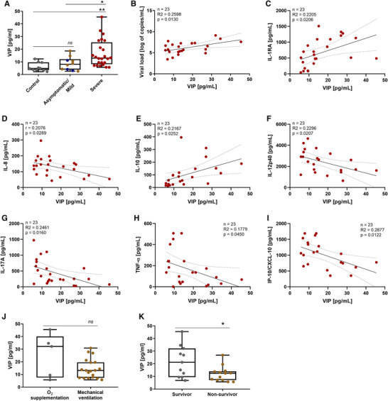
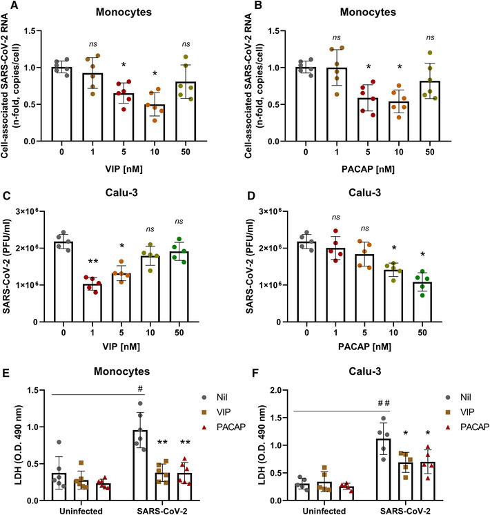
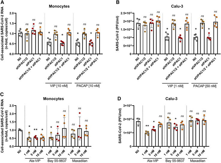
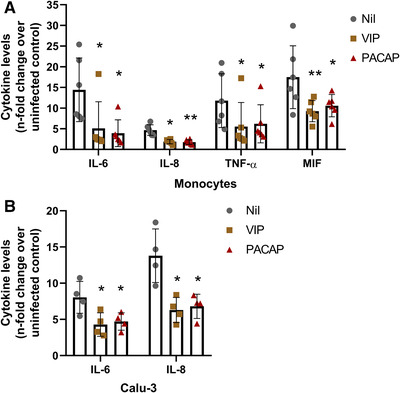
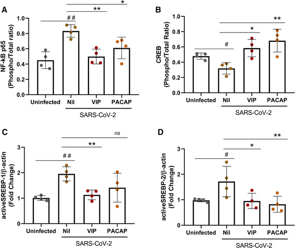
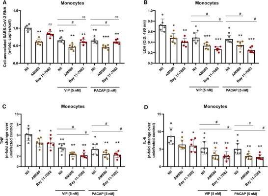
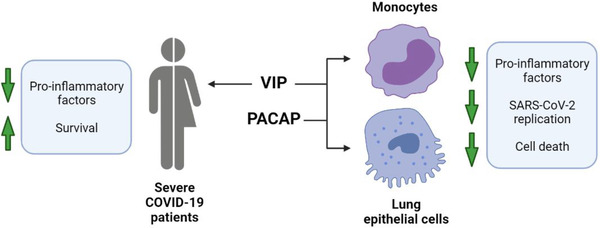
Infection by SARS-CoV-2 may elicit uncontrolled and damaging inflammatory responses. Thus, it is critical to identify compounds able to inhibit virus replication and thwart the inflammatory reaction. Here, we show that the plasma levels of the immunoregulatory neuropeptide VIP are elevated in patients with severe COVID-19, correlating with reduced inflammatory mediators and with survival on those patients. In vitro, vasoactive intestinal peptide (VIP) and pituitary adenylate cyclase-activating polypeptide (PACAP), highly similar neuropeptides, decreased the SARS-CoV-2 RNA content in human monocytes and viral production in lung epithelial cells, also reducing cell death. Both neuropeptides inhibited the production of proinflammatory mediators in lung epithelial cells and in monocytes. VIP and PACAP prevented in monocytes the SARS-CoV-2-induced activation of NF-kB and SREBP1 and SREBP2, transcriptions factors involved in proinflammatory reactions and lipid metabolism, respectively. They also promoted CREB activation, a transcription factor with antiapoptotic activity and negative regulator of NF-kB. Specific inhibition of NF-kB and SREBP1/2 reproduced the anti-inflammatory, antiviral, and cell death protection effects of VIP and PACAP. Our results support further clinical investigations of these neuropeptides against COVID-19.

Keywords: COVID-19; PACAP; SARS-CoV-2; VIP; neuropeptides.


Conflict of interest statement

The authors declare no competing financial interests.

Figures

FIGURE 1
FIGURE 1
Plasma levels of VIP are elevated in patients with severe forms of COVID‐19 and associates with reduced levels of inflammatory markers and with survival. The levels of VIP (A) in the plasma of SARS‐CoV‐2‐negative control participants, SARS‐CoV‐2‐positive asymptomatic subjects, or symptomatic patients presenting mild to severe COVID‐19 were quantified by ELISA. Correlation between levels of VIP and viral load (B) or inflammation markers (C–I). Severe COVID‐19 patients admitted to the ICU were subdivided between those requiring invasive mechanical ventilation or noninvasive O2 supplementation (J) and according to the 28‐day mortality outcome as survivors or non survivors (K). Linear regression (with the 95% confidence interval) and Spearman's correlation were calculated according to the distribution of the data. Dots represent: controls, grey; asymptomatic, blue; mild, brown; severe, red. The horizontal lines in the box plots represent the median, the box edges represent the interquartile ranges, and the whiskers indicate the minimal and maximal value in each group. *p ≤ 0.05; **p ≤ 0.01; ns, not significant
FIGURE 2
FIGURE 2
VIP and PACAP reduce SARS‐CoV‐2 RNA synthesis in human primary monocytes and viral replication in pulmonary cells, protecting them from virus‐mediated cytopathic effects. Monocytes (A and B) and Calu‐3 cells (C and D) were exposed (overnight) or not to the indicated concentrations of VIP (A and C) or PACAP (B and D). Culture medium was removed and then cells were infected with SARS‐CoV‐2 for 1 h, as described in Material and Methods. After infection, viral input was removed and cells were washed, then reexposed to the neuropeptides. Viral RNA synthesis was evaluated by qPCR in monocytes 24 h after infection. In Calu‐3 cells, supernatants were collected at 48 h after infection, and viral replication was evaluated by quantifying PFUs in Vero E6 plaque assays. Cellular viability was analyzed by measuring LDH release in the supernatants of uninfected or SARS‐CoV‐2‐infected monocytes (E) treated or not with VIP or PACAP (10 nM), and Calu‐3 cells (F) treated or not with VIP (1 nM) or PACAP (50 nM). Data in (A and B) are shown normalized to infected cells kept only with culture medium, and in (C, D, E, and F) represent means ± sd of absolute values. */#, p ≤ 0.05; **/##, p ≤ 0.01; ns, not significant; (A, B, and E) n = 6; (C, D, and F) n = 5. Each dot represents an independent assay with 3 replicates
FIGURE 3
FIGURE 3
Receptor contribution for the VIP‐ and PACAP‐mediated inhibition of SARS‐CoV‐2 replication. Monocytes (A) and Calu‐3 cells (B) were exposed (overnight) or not to antagonists for VPAC1/2 (atVPAC1/2; 100 nM for monocytes; 50 nM for Calu‐3 cells) and PAC1 receptors (atPAC1; 150 nM for monocytes; 300 nM for Calu‐3 cells) and to VIP or PACAP (antagonists were added 10 min before exposure to neuropeptides, and were kept in the culture medium). Also, monocytes (C) and Calu‐3 cells (D) were treated (overnight) or not with agonists for VIP and PACAP receptors, as indicated, at different concentrations. Upon overnight exposure (A–D), culture medium was removed and then cells were infected with SARS‐CoV‐2 for 1 h, as described in Material and Methods. After infection, viral input was removed, and cells were washed, and then reexposed to receptor antagonists and VIP or PACAP (A and B) or to receptor agonists, as before (C and D). Viral RNA synthesis was evaluated by qPCR in monocytes 24 h after infection. In Calu‐3 cells, supernatants were collected at 48 h after infection, and viral replication was evaluated by quantifying PFUs in Vero E6 plaque assays. Data in (A and C) are shown normalized to infected cells kept only with culture medium, and in (B and D) represents means ± sd of absolute values. *p ≤ 0.05; **p ≤ 0.01; ns, not significant; (A and B) n = 5; (C and D) n = 4. Each dot represents an independent assay with 3 replicates
FIGURE 4
FIGURE 4
VIP and PACAP reduce the production of proinflammatory mediators by SARS‐CoV‐2‐infected monocytes and Calu‐3 cells. Monocytes (A) and Calu‐3 cells (B) were treated (overnight) or not with VIP or PACAP (10 nM each for monocytes, 1 nM of VIP or 50 nM of PACAP for Calu‐3 cells). Culture medium was removed and then cells were infected with SARS‐CoV‐2 for 1 h, as described in Material and Methods. After infection, viral input was removed and cells were washed, and then re‐exposed to the neuropeptides. The levels of IL‐6, IL‐8, TNF‐α and MIF were measured in culture supernatants of monocytes after 24 h (A), and of IL‐6 and IL‐8 after 48 h for Calu‐3 cells (B), by ELISA. Data represent means ± sd. *p ≤ 0.05; **p ≤ 0.01; (A) n = 6; (B) n = 4. Each dot represents an independent assay with 3 replicates
FIGURE 5
FIGURE 5
VIP and PACAP regulate the activation of transcription factors in SARS‐CoV‐2‐infected monocytes. Monocytes were treated (overnight) or not with to VIP or PACAP (10 nM), culture medium was removed and then cells were infected with SARS‐CoV‐2 for 1 h, as described in Material and Methods. After infection, viral input was removed, and cells were washed and then reexposed to the neuropeptides. After 24 h, cells were lysed and the ratios between phosphoNF‐kBp65 and total NF‐kBp65 (A), phosphoCREB and total CREB (B), active SREBP‐1 and β‐actin (C), and active SREBP‐2 and β‐actin (D) were quantified by ELISA (A and B) or by Western blot (C and D) in the cell lysates. Data represent means ± sd. */#, p ≤ 0.05; **/##, p ≤ 0.01; ***/###, p ≤ 0.001; ns, not significant; (A, B, C, and D) n = 4. For A and B, each dot represents an independent assay with 2 replicates
FIGURE 6
FIGURE 6
Inhibition of NF‐kB and SREBP in monocytes reduces SARS‐CoV‐2 RNA synthesis, production of proinflammatory mediators and protects the cells from virus‐mediated cytopathic effects. Monocytes were treated (overnight) or not with to VIP or PACAP (5 nM), culture medium was removed and then cells were infected with SARS‐CoV‐2 for 1 h, as described in Material and Methods. After infection, viral input was removed, and cells were washed and then reexposed to the neuropeptides associated or not with inhibitors of SREBP (AM580, 5 μM) or NF‐kB (Bay 11–7082, 5 μM). Viral RNA synthesis (A), cellular viability (B) and levels of TNF‐α and IL‐6 (C and D) were evaluated by qPCR, ELISA, and LDH release, respectively, in the culture supernatants 24 h after infection. Data in (A) are shown normalized to infected cells kept only with culture medium, and in (B, C, and D) represent means ± sd of absolute values. */#, p ≤ 0.05; **/##, p ≤ 0.01; ***/###, p ≤ 0.001; ns, not significant; (A–D) n = 6. Each dot represents an independent assay with 3 replicates
FIGURE 7
FIGURE 7
Graphical summary of study data. In severe COVID‐19 patients, VIP plasma levels correlated with decreased inflammatory markers and survival. In in vitro assays with monocytes and lung epithelial cells, VIP and PACAP were found to decrease SARS‐CoV‐2 RNA synthesis (monocytes) and viral replication (lung epithelial cells). Both neuropeptides also reduced inflammatory factors and cell death of infected cells. Created with BioRender.com

Similar articles

Cited by

  • Inhaled aviptadil for the possible treatment of COVID-19 in patients at high risk for ARDS: study protocol for a randomized, placebo-controlled, and multicenter trial.
    Boesing M, Abig K, Brändle M, Brutsche M, Burri E, Frye BC, Giezendanner S, Grutters JC, Haas P, Heisler J, Jaun F, Leuppi-Taegtmeyer AB, Lüthi-Corridori G, Müller-Quernheim J, Nüesch R, Pohl W, Rassouli F, Leuppi JD. Boesing M, et al. Trials. 2022 Sep 20;23(1):790. doi: 10.1186/s13063-022-06723-w. Trials. 2022. PMID: 36127739 Free PMC article.
  • Occurrence of SARS-CoV-2 viremia is associated with genetic variants of genes related to COVID-19 pathogenesis.
    Roy-Vallejo E, Fernández De Córdoba-Oñate S, Delgado-Wicke P, Triguero-Martínez A, Montes N, Carracedo-Rodríguez R, Zurita-Cruz N, Marcos-Jiménez A, Lamana A, Galván-Román JM, Villapalos García G, Zubiaur P, Ciudad M, Rabes L, Sanz M, Rodríguez C, Villa A, Rodríguez JÁ, Marcos C, Hernando J, Díaz-Fernández P, Abad F, de Los Santos I, Rodríguez Serrano DA, García-Vicuña R, Suárez Fernández C, P Gomariz R, Muñoz-Calleja C, Fernández-Ruiz E, González-Álvaro I, Cardeñoso L; PREDINMUN-COVID Group; Barrios A, Sanz J, Casado P, Gutiérrez Á, Bautista A, Hernández P, Ruiz Giménez N, Moyano B, Gil P, Jesús Delgado M, Parra P, Sánchez B, Sáez C, Fernández Rico M, Arévalo Román C, Castañeda S, Llorente I, G Tomero E, García Castañeda N, Uriarte M, Fontán García-Rodrigo L, Domingo García D, Alarcón Cavero T, Auxiliadora Semiglia Chong M, Gutiérrez Cobos A, Sánchez-Madrid F, Martín Gayo E, Sánchez-Cerrillo I, Martínez-Fleta P, López-Sanz C, Gabrie L, Del Campo Guerola L, Tejedor R, Ancochea J, García Castillo E, Ávalos E, Sánchez-Azofra A, Alonso T, Cisneros C, Valenzuela C, Javier García Pérez F, María Girón R, Aspa J, Marcos C, Del Perpetuo Socorro Churruca M, Zamora E, Martínez A, Barrio May… See abstract for full author list ➔ Roy-Vallejo E, et al. Front Med (Lausanne). 2023 Sep 22;10:1215246. doi: 10.3389/fmed.2023.1215246. eCollection 2023. Front Med (Lausanne). 2023. PMID: 37809329 Free PMC article.
  • Evaluation of serum VIP and aCGRP during pulmonary exacerbation in cystic fibrosis: A longitudinal pilot study of patients undergoing antibiotic therapy.
    Al-Keilani MS, Awad S, Hammouri HM, Al Shalakhti T, Almomani BA, Dahabreh MM, Ajlony MJ. Al-Keilani MS, et al. PLoS One. 2023 May 5;18(5):e0284511. doi: 10.1371/journal.pone.0284511. eCollection 2023. PLoS One. 2023. PMID: 37146001 Free PMC article.
  • Role of endocrine PACAP in age-related diseases.
    Toth D, Reglodi D, Schwieters L, Tamas A. Toth D, et al. Front Endocrinol (Lausanne). 2023 Mar 9;14:1118927. doi: 10.3389/fendo.2023.1118927. eCollection 2023. Front Endocrinol (Lausanne). 2023. PMID: 36967746 Free PMC article. Review.
  • VIP/VPAC Axis Expression in Immune-Mediated Inflammatory Disorders: Associated miRNA Signatures.
    Lamana A, Castro-Vázquez D, de la Fuente H, Triguero-Martínez A, Martínez-Hernández R, Revenga M, Villanueva-Romero R, Llamas-Velasco M, Chicharro P, Juarranz Y, Marazuela M, Sales-Sanz M, García-Vicuña R, Tomero E, González-Álvaro I, Martínez C, Gomariz RP. Lamana A, et al. Int J Mol Sci. 2022 Aug 2;23(15):8578. doi: 10.3390/ijms23158578. Int J Mol Sci. 2022. PMID: 35955723 Free PMC article.

References

    1. García LF. Immune response, inflammation, and the clinical spectrum of COVID‐19. Front Immunol. 2020;11:1441. - PMC - PubMed
    1. Huang C, Wang Y, Li X, et al. Clinical features of patients infected with 2019 novel coronavirus in Wuhan, China. Lancet. 2020;395:497–506. - PMC - PubMed
    1. Chen G, Wu D, Guo W, et al. Clinical and immunological features of severe and moderate coronavirus disease 2019. J Clin Invest. 2020;130:2620–2629. - PMC - PubMed
    1. Gupta A, Madhavan M V., Sehgal K, et al. Extrapulmonary manifestations of COVID‐19. Nat Med. 2020;26:1017–1032. - PMC - PubMed
    1. Tang D, Comish P, Kang R. The hallmarks of COVID‐19 disease. PLoS Pathog. 2020;16:e1008536. - PMC - PubMed

Publication types

MeSH terms

Substances