. 2022 Apr 5;11(7):e024536.
doi: 10.1161/JAHA.121.024536. Epub 2022 Mar 24.

PSG7 and 9 (Pregnancy-Specific β-1 Glycoproteins 7 and 9): Novel Biomarkers for Preeclampsia

Affiliations

PSG7 and 9 (Pregnancy-Specific β-1 Glycoproteins 7 and 9): Novel Biomarkers for Preeclampsia

Manju Kandel et al. J Am Heart Assoc. .

Abstract

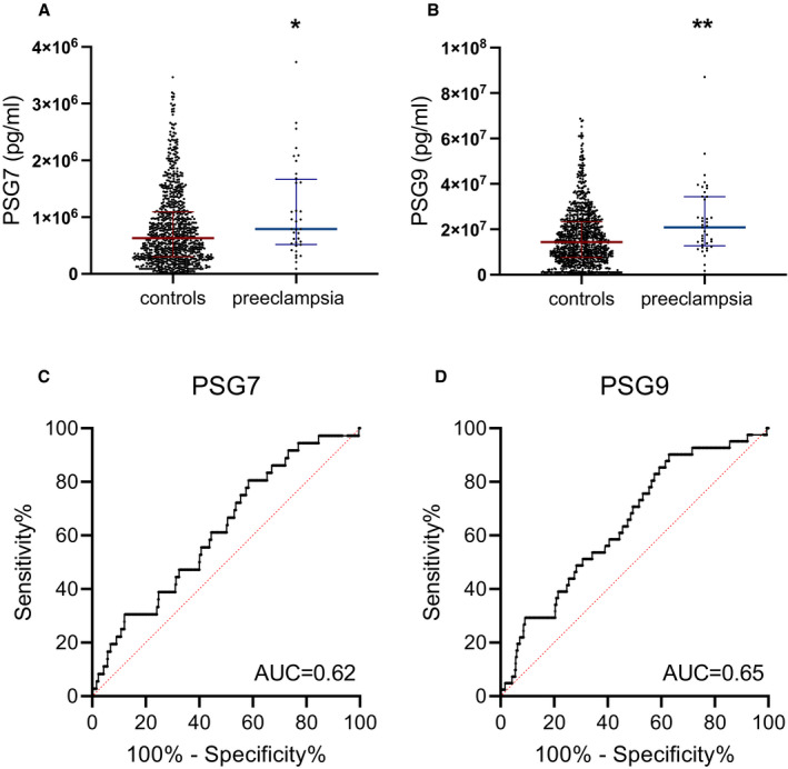
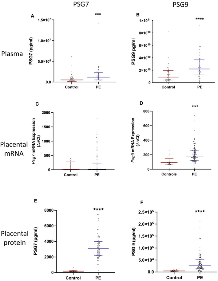
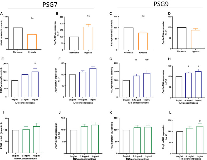
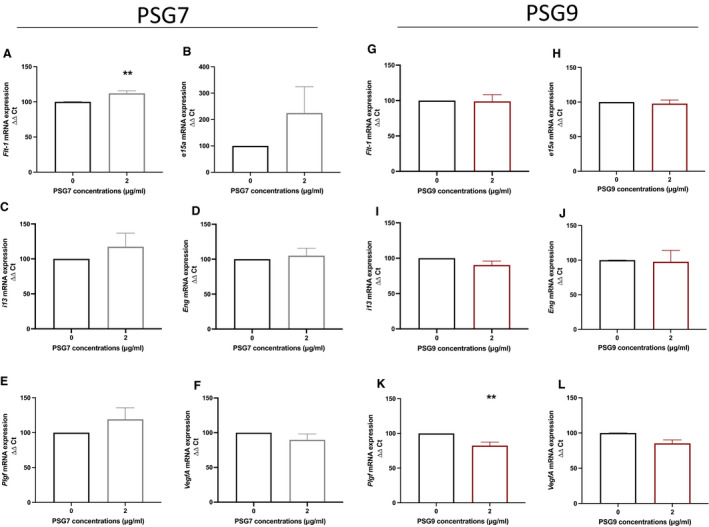
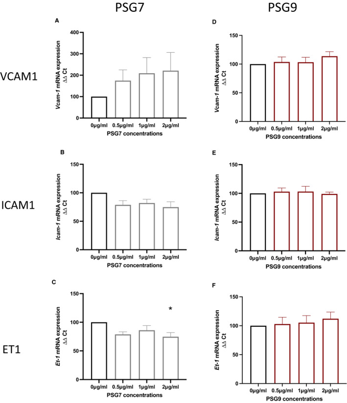
Background Preeclampsia is pregnancy specific, involving significant maternal endothelial dysfunction. Predictive biomarkers are lacking. We evaluated the biomarker potential, expression, and function of PSG7 (pregnancy-specific β-1 glycoprotein 7) and PSG9 (pregnancy-specific β-1 glycoprotein 9) in preeclampsia. Methods and Results At 36 weeks gestation preceding term preeclampsia diagnosis, PSG7 and PSG9 (in Australian cohorts of n=918 and n=979, respectively) were significantly increased before the onset of term preeclampsia (PSG7, P=0.013; PSG9, P=0.0011). In samples collected at 28 to 32 weeks from those with preexisting cardiovascular disease and at high risk of preeclampsia (Manchester Antenatal Vascular Service, UK cohort, n=235), both PSG7 and PSG9 were also significantly increased preceding preeclampsia onset (PSG7, P<0.0001; PSG9, P=0.0003) relative to controls. These changes were validated in the plasma and placentas of patients with established preeclampsia who delivered at <34 weeks gestation (PSG7, P=0.0008; PSG9, P<0.0001). To examine whether PSG7 and PSG9 are associated with increasing disease severity, we measured them in a cohort from South Africa stratified for this outcome, the PROVE (Preeclampsia Obstetric Adverse Events) cohort (n=72). PSG7 (P=0.0027) and PSG9 (P=0.0028) were elevated among patients who were preeclamptic with severe features (PROVE cohort), but not significantly changed in those without severe features or with eclampsia. In syncytialized first trimester cytotrophoblast stem cells, exposure to TNFα (tumor necrosis factor α) or IL-6 (interleukin 6) significantly increased the expression and secretion of PSG7 and PSG9. In contrast, when we treated primary endothelial cells with recombinant PSG7 and PSG9, we only observed modest changes in Flt-1 (FMS-like tyrosine kinase-1) expression and Plgf (placental growth factor) expression, and no other effects on proangiogenic/antiangiogenic or endothelial dysfunction markers were observed. Conclusions Circulating PSG7 and PSG9 are increased before preeclampsia onset and among those with established disease with their production and release potentially driven by placental inflammation.

Keywords: biomarkers; placenta; preeclampsia; pregnancy; pregnancy specific beta‐1 glycoproteins.


Figures

Figure 1
Figure 1. Circulating PSG7 and PSG9 are increased before diagnosis of preeclampsia.
Circulating PSG7 and PSG9 were measured in a large prospective study, the FLAG (Fetal Longitudinal Assessment of Growth) study. These proteins were measured in the plasma of patients preceding their diagnosis with preeclampsia at 36 weeks gestation relative to controls. PSG7 and PSG9 were found to be significantly elevated in the plasma of patients who developed preeclampsia after blood sampling (A and B) relative to controls; individual symbols represent individual patients. The discriminatory power of each biomarker is shown as a receiver operating characteristic curve with the AUC annotated (C and D). Data are expressed as median (interquartile range), and the significance levels determined using a Mann–Whitney U test were *P<0.05 and **P<0.01. AUC indicates area under the curve; PSG7, pregnancy‐specific β‐1 glycoprotein 7; and PSG9, pregnancy‐specific β‐1 glycoprotein 9.
Figure 2
Figure 2. Circulating and placental PSG7 and PSG9 are increased in patients diagnosed with preeclampsia.
PSG7 and PSG9 proteins were measured in patients with preeclampsia who delivered at <34 weeks gestation. Plasma (A) PSG7 (n=28 control, n=46 preeclampsia) and (B) PSG9 (n=28 control, n=46 preeclampsia) protein concentrations were significantly increased in plasma from women with early‐onset preeclampsia relative to gestation‐matched controls. In placental samples, (C) Psg7 mRNA expression (n=19 control, n=81 preeclampsia) was not significantly different between preeclampsia and control groups, whereas (D) Psg9 mRNA expression (n=19 control, n=81 preeclampsia) was significantly increased in the preeclamptic cohort. Both (E) PSG7 (n=20 control, n=82 preeclampsia) and (F) PSG9 (n=20 control, n=82 preeclampsia) protein concentrations were significantly increased in preeclamptic placental lysates relative to controls. Placental mRNA expression for both PSG7 and PSG9 were normalized to the geometric mean of the reference housekeeping genes, topoisomerase‐1 and cyclin‐1. Individual symbols represent individual patients. Data are expressed as median (interquartile range), and the significance levels determined using a Mann–Whitney U test were ***P<0.001 and ****P<0.0001. PE indicates preeclampsia; PSG7, pregnancy‐specific β‐1 glycoprotein 7; PSG9, pregnancy‐specific β‐1 glycoprotein 9; and ΔΔCT, delta delta CT analysis method.
Figure 3
Figure 3. The effect of hypoxia and inflammation on PSG7 and PSG9 mRNA expression and secretion in syncytialized cytotrophoblast stem cells.
Syncytiotrophoblast cells were exposed to either (A through D) hypoxic conditions or (E through L) inflammatory stimuli. When cells were exposed to hypoxia (1% oxygen; orange), both (A) PSG7 and (C) PSG9 protein secretion were significantly reduced compared with the normoxic (8% oxygen; black) group. Psg7 mRNA expression was significantly increased in syncytiotrophoblast cells cultured under hypoxic conditions (B); however, there was no significant difference in Psg9 mRNA expression (D). Next, syncytiotrophoblast cells were treated with increasing doses of either IL‐6 (E through H; blue) or TNFα (I through L; green; 0 ng/mL, 0.1 ng/mL, 1 ng/mL). Treatment with IL‐6 significantly increased PSG7 secretion from syncytiotrophoblast cells with the 1 ng/mL dose (E), whereas there was no significant change in Psg7 mRNA expression (F). Treatment with 0.1 ng/mL and 1 ng/mL IL‐6 significantly increased both PSG9 secretion (G) and mRNA expression (H) in syncytiotrophoblast cells. In TNFα‐treated syncytiotrophoblast cells, there was no significant change in PSG7 (I) and PSG9 (K) protein secretion and Psg7 mRNA expression (J), whereas treatment with 1 ng/mL TNFα significantly increased Psg9 mRNA expression (L). Protein levels were normalized as percentage of controls within the experiment, and mRNA expression was normalized to the geometric mean of housekeeping genes. All experiments were repeated 5 times with triplicate repeats. Data are expressed as mean±SEM, and the significance levels determined using a Mann–Whitney U test for 2 groups or a Kruskal–Wallis test with multiple comparisons for ≥3 groups were *P<0.05 and **P<0.01. IL‐6 indicates interleukin‐6; PSG7, pregnancy‐specific β‐1 glycoprotein 7; PSG9, pregnancy‐specific β‐1 glycoprotein 9; TNFα, tumor necrosis factor α; and ΔΔCT, delta delta CT analysis method.
Figure 4
Figure 4. The effect of PSG7 and PSG9 on proangiogenic and antiangiogenic factors in primary HUVECs.
Primary HUVECs were treated with a 2‐µg/mL dose of either recombinant PSG7 (gray) or recombinant PSG9 (maroon), and the effect on Flt‐1, sFlt‐1 e15a, sFlt‐1 i13, Eng, Plgf, and VegfA mRNA expression was assessed. Treatment with PSG7 significantly increased (A) Flt‐1 mRNA expression, whereas PSG7 had no significant effect on (B) sFlt‐1 e15a, (C) sFlt‐1 i13, (D) Eng, (E) Plgf, or (F) VegfA mRNA expression. Furthermore, there was no significant change in (G) Flt‐1, (H) sFlt‐1 e15a, (I) sflt‐1 i13, (J) Eng, or (L) VegfA expression in PSG9‐treated HUVECs. However, (K) Plgf was significantly reduced in PSG9‐treated HUVECs. mRNA expression was normalized to the reference housekeeper Ywhaz. All experiments were repeated 5 times, and each treatment was run in triplicate. Data are expressed as mean±SEM, and the significance level determined using a Mann–Whitney U test was **P<0.01. Eng indicates endoglin; Flt‐1, FMS‐like tyrosine kinase‐1; HUVEC, human umbilical vein endothelial cell; Plgf, placental growth factor; PSG7, pregnancy‐specific β‐1 glycoprotein 7; PSG9, pregnancy‐specific β‐1 glycoprotein 9; sFlt‐1, soluble fms‐like tyrosine kinase‐1; VegfA, vascular endothelial growth factor a; Ywhaz, Tyrosine 3‐Monooxygenase/Tryptophan 5‐Monooxygenase Activation Protein Zeta; and ΔΔCT, delta delta CT analysis method.
Figure 5
Figure 5. The effect of PSG7 and PSG9 on markers of endothelial dysfunction in primary HUVECs.
Primary HUVECs were treated with recombinant PSG7 (gray) or recombinant PSG9 (maroon; 0 µg/mL [control], 0.5, 1, or 2 µg/mL) and the effect on endothelial dysfunction markers was assessed. In PSG7‐treated HUVECs, no significant effect on Vcam‐1 and Icam‐1 expression in HUVECS was observed (A and B), whereas PSG7 significantly decreased Et‐1 mRNA expression (C). In PSG9‐treated HUVECs, no significant effects of PSG9 on Vcam‐1, Icam‐1, or Et‐1 mRNA expression was observed (D through F). mRNA expression was normalized to the housekeeper Ywhaz. All experiments were repeated 5 times, and each treatment was run in triplicate. Data are expressed as mean±SEM, and the significance level determined using a Kruskal–Wallis test with multiple comparisons was *P<0.05. Et‐1 indicates endothelin‐1; HUVEC, human umbilical vein endothelial cell; Icam‐1, intracellular cell adhesion molecule‐1; PSG7, pregnancy‐specific β‐1 glycoprotein 7; PSG9, pregnancy‐specific β‐1 glycoprotein 9; Vcam‐1, vascular cell adhesion molecule‐1; Ywhaz, Tyrosine 3‐Monooxygenase/Tryptophan 5‐Monooxygenase Activation Protein Zeta; and ΔΔCT, delta delta CT analysis method.

Similar articles

Cited by

References

    1. Ghulmiyyah L, Sibai B. Maternal mortality from preeclampsia/eclampsia. Semin Perinatol. 2012;36:56–59. doi: 10.1053/j.semperi.2011.09.011 - DOI - PubMed
    1. Maynard SE, Karumanchi SA. Angiogenic factors and preeclampsia. Semin Nephrol. 2011;31:33–46. doi: 10.1016/j.semnephrol.2010.10.004 - DOI - PMC - PubMed
    1. Gathiram P, Moodley J. Pre‐eclampsia: its pathogenesis and pathophysiolgy. Cardiovasc J Afr. 2016;27:71. doi: 10.5830/CVJA-2016-009 - DOI - PMC - PubMed
    1. Redman C, Sargent I. Placental stress and pre‐eclampsia: a revised view. Placenta. 2009;30:38–42. doi: 10.1016/j.placenta.2008.11.021 - DOI - PubMed
    1. Brümmendorf T, Rathjen FG. Cell adhesion molecules. 1: immunoglobulin superfamily. Protein Profile. 1994;1:951–1058. - PubMed

Substances

LinkOut - more resources