

Carboxymethyl chitosan-grafted polyvinylpyrrolidone-iodine microspheres for promoting the healing of chronic wounds
- PMID: 35322745
- PMCID: PMC9161872
- DOI: 10.1080/21655979.2022.2054911
Carboxymethyl chitosan-grafted polyvinylpyrrolidone-iodine microspheres for promoting the healing of chronic wounds
Abstract
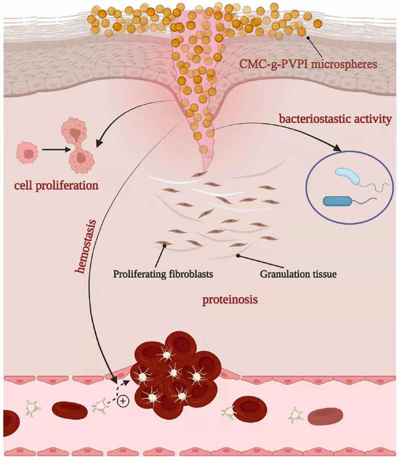
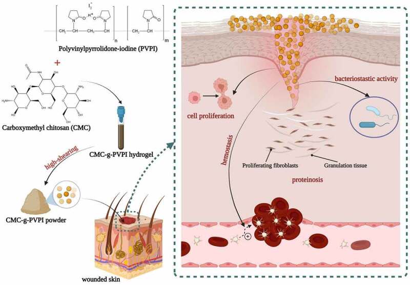
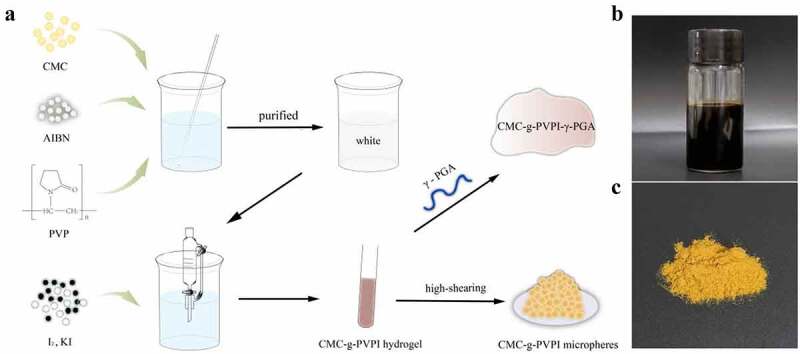
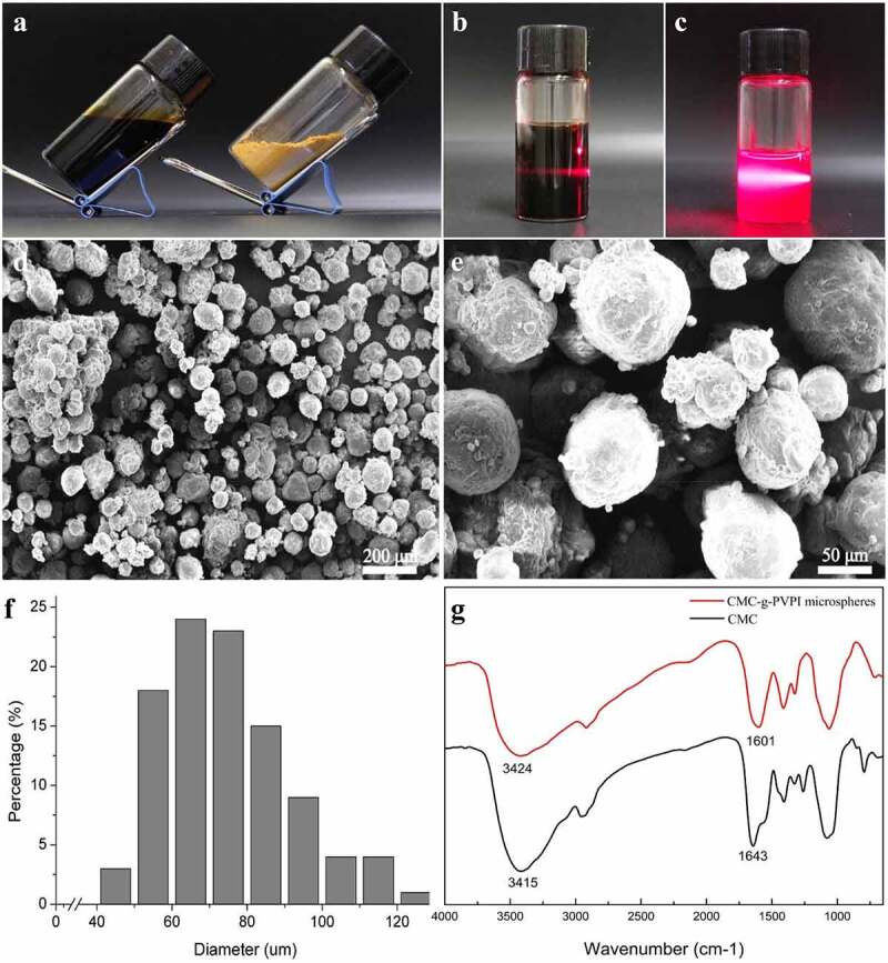
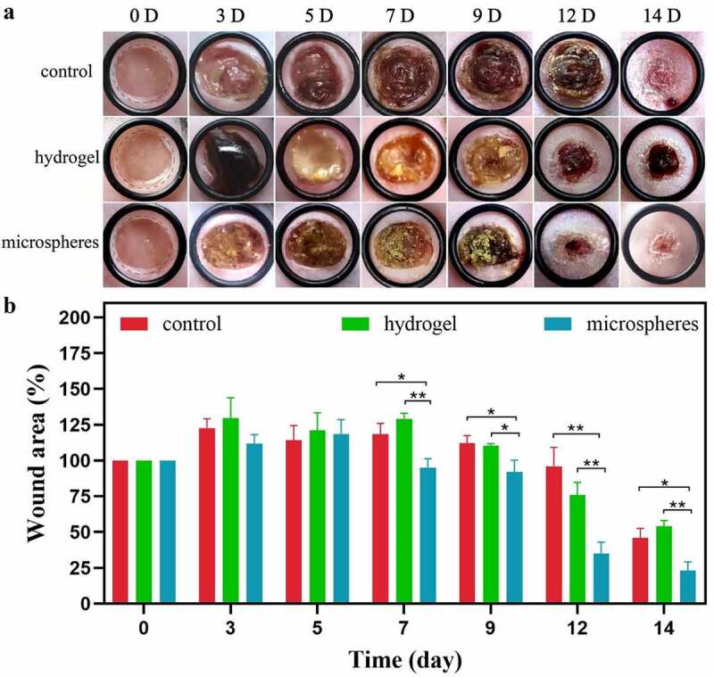
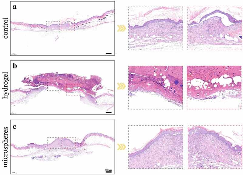
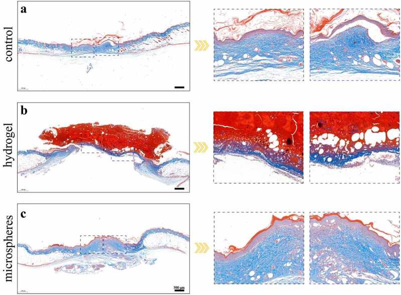
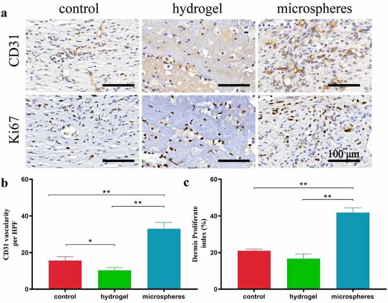
Chronic wounds that fail to heal are the most common complications experienced by diabetic patients, and current treatment remains unsatisfactory, mainly due to the vulnerability of diabetic wounds to bacterial infections. Chitosan derivatives are widely used to treat chronic wounds due to their excellent hydrophilicity, biodegradability, and antimicrobial activity and substantial contribution to tissue regeneration. However, the antimicrobial effect of chitosan is not sufficient due to the complicated pathological mechanism of diabetes mellitus. Here, we prepared carboxymethyl chitosan-grafted polyvinylpyrrolidone-iodine (CMC-g-PVPI) microspheres and used them to treat chronic wounds. Carboxymethyl chitosan (CMC) was used as the skeleton and was grafted with polyvinylpyrrolidone-iodine (PVPI) to form a CMC-g-PVPI complex hydrogel and CMC-g-PVPI microspheres, which formed as a result of the high shearing dispersion of the complex hydrogel. In vivo experiments on diabetic wounds revealed significantly accelerated wound closure in the presence of the microspheres, demonstrating the excellent potential of CMC-g-PVPI to promote skin wound regeneration under diabetic conditions.
Keywords: Carboxymethyl chitosan; chronic wounds; healing; microspheres; polyvinylpyrrolidone-iodine.
Conflict of interest statement
No potential conflict of interest was reported by the author(s).
Figures
Similar articles
-
Polyvinyl pyrrolidone/carboxymethyl chitosan hydrogel loaded with Paris polyphylla var. yunnanensis extracellular vesicles promotes wound healing.Int J Biol Macromol. 2025 May;306(Pt 4):141782. doi: 10.1016/j.ijbiomac.2025.141782. Epub 2025 Mar 5. Int J Biol Macromol. 2025. PMID: 40054821
-
Comparative Analysis of Antibacterial and Wound Healing Activities of Chitosan and Povidone-Iodine-Based Hydrogels.Ann Plast Surg. 2024 Feb 1;92(2):240-244. doi: 10.1097/SAP.0000000000003755. Ann Plast Surg. 2024. PMID: 38198629
-
Preparation, property of the complex of carboxymethyl chitosan grafted copolymer with iodine and application of it in cervical antibacterial biomembrane.Mater Sci Eng C Mater Biol Appl. 2016 Oct 1;67:247-258. doi: 10.1016/j.msec.2016.05.027. Epub 2016 May 14. Mater Sci Eng C Mater Biol Appl. 2016. PMID: 27287120 Clinical Trial.
-
Povidone-iodine solution in wound treatment.Phys Ther. 1998 Feb;78(2):212-8. doi: 10.1093/ptj/78.2.212. Phys Ther. 1998. PMID: 9474112 Review.
-
An appraisal of povidone-iodine and wound healing.Postgrad Med J. 1993;69 Suppl 3:S97-105. Postgrad Med J. 1993. PMID: 8290466 Review.
Cited by
-
Functional microspheres for tissue regeneration.Bioact Mater. 2022 Aug 9;25:485-499. doi: 10.1016/j.bioactmat.2022.07.025. eCollection 2023 Jul. Bioact Mater. 2022. PMID: 37056261 Free PMC article. Review.
-
Innovative Functional Biomaterials as Therapeutic Wound Dressings for Chronic Diabetic Foot Ulcers.Int J Mol Sci. 2023 Jun 8;24(12):9900. doi: 10.3390/ijms24129900. Int J Mol Sci. 2023. PMID: 37373045 Free PMC article. Review.
-
Silk Fibroin Nanofibers: Advancements in Bioactive Dressings through Electrospinning Technology for Diabetic Wound Healing.Pharmaceuticals (Basel). 2024 Sep 30;17(10):1305. doi: 10.3390/ph17101305. Pharmaceuticals (Basel). 2024. PMID: 39458946 Free PMC article. Review.
-
Mannose-modified erythrocyte membrane-encapsulated chitovanic nanoparticles as a DNA vaccine carrier against reticuloendothelial tissue hyperplasia virus.Front Immunol. 2023 Jan 4;13:1066268. doi: 10.3389/fimmu.2022.1066268. eCollection 2022. Front Immunol. 2023. PMID: 36776397 Free PMC article.
References
-
- Saeedi P, Petersohn I, Salpea P, et al. Global and regional diabetes prevalence estimates for 2019 and projections for 2030 and 2045: results from the International diabetes federation diabetes atlas, 9(th) edition. Diabetes Res Clin Pract. 2019;157:107843. - PubMed
-
- Armstrong DG, Boulton AJM, Bus SA.. Diabetic foot ulcers and their recurrence. N Engl J Med. 2017;376(24):2367–2375. - PubMed
-
- Yin M, Wu J, Deng M, et al. Multifunctional magnesium organic framework-based microneedle patch for accelerating diabetic wound healing. ACS Nano. 2010;4(4):2021. - PubMed
-
- Maranda EL, Rodriguez-Menocal L, Badiavas EV.. Role of Mesenchymal stem cells in dermal repair in burns and diabetic wounds. Curr Stem Cell Res Ther. 2017;12(1):61–70. - PubMed
Publication types
MeSH terms
Substances
LinkOut - more resources
Full Text Sources
Other Literature Sources