. 2022 Jul;235(1):188-203.
doi: 10.1111/nph.18115. Epub 2022 Apr 12.

GENOMES UNCOUPLED1 plays a key role during the de-etiolation process in Arabidopsis

Affiliations

GENOMES UNCOUPLED1 plays a key role during the de-etiolation process in Arabidopsis

Tamara Hernández-Verdeja et al. New Phytol. 2022 Jul.

Abstract

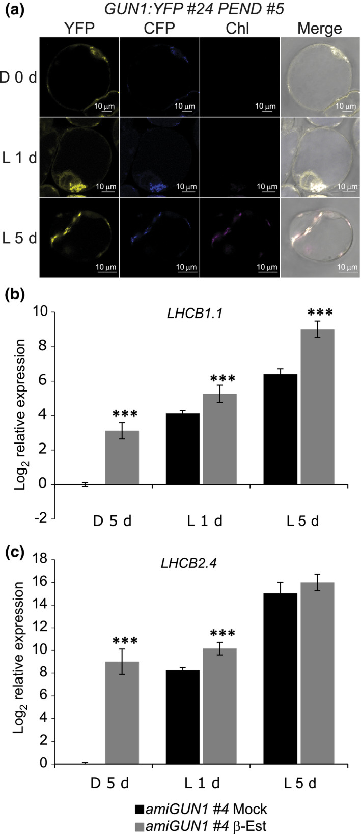
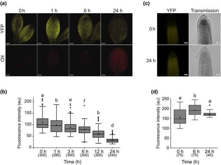
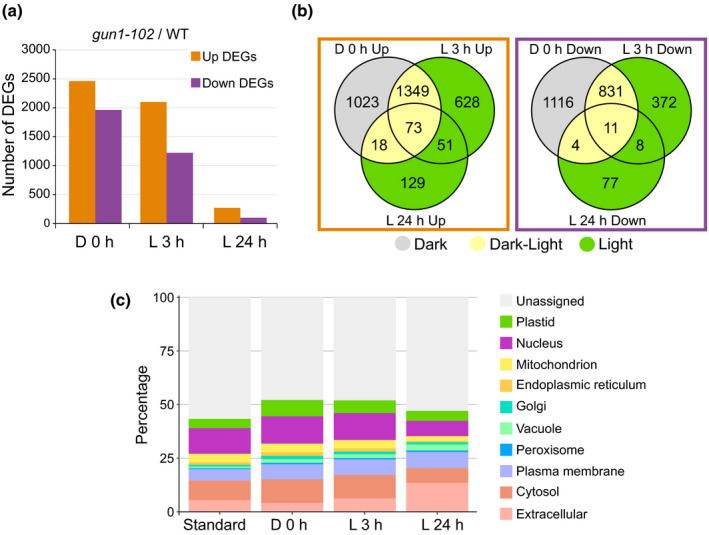
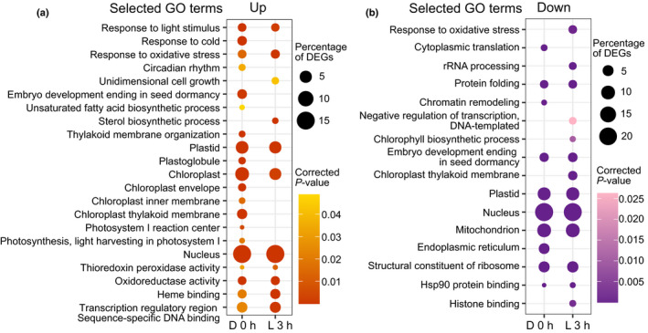
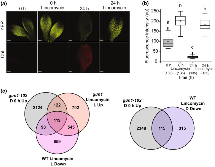
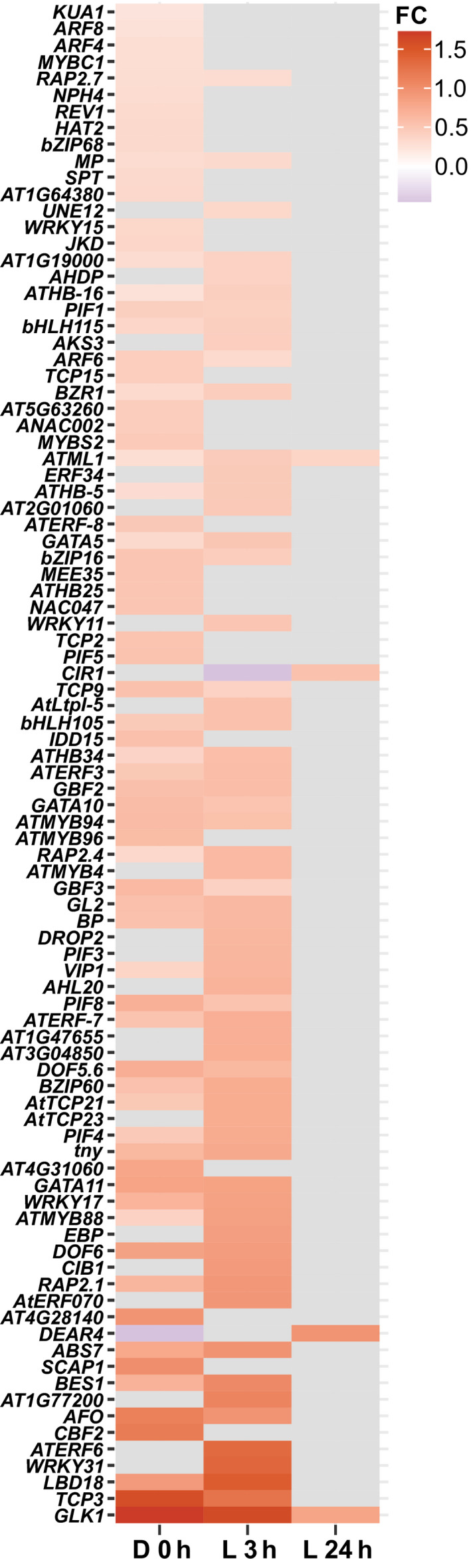
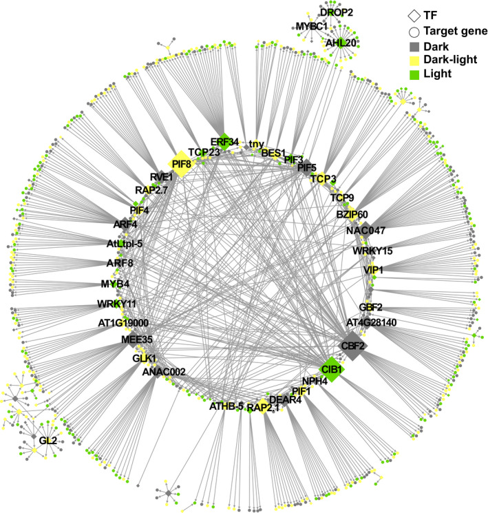
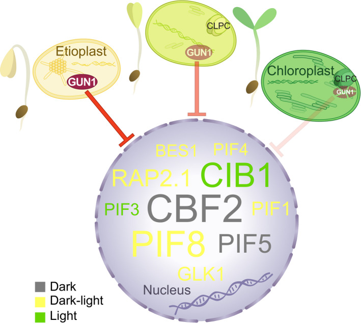
One of the most dramatic challenges in the life of a plant occurs when the seedling emerges from the soil and exposure to light triggers expression of genes required for establishment of photosynthesis. This process needs to be tightly regulated, as premature accumulation of light-harvesting proteins and photoreactive Chl precursors causes oxidative damage when the seedling is first exposed to light. Photosynthesis genes are encoded by both nuclear and plastid genomes, and to establish the required level of control, plastid-to-nucleus (retrograde) signalling is necessary to ensure correct gene expression. We herein show that a negative GENOMES UNCOUPLED1 (GUN1)-mediated retrograde signal restricts chloroplast development in darkness and during early light response by regulating the transcription of several critical transcription factors linked to light response, photomorphogenesis, and chloroplast development, and consequently their downstream target genes in Arabidopsis. Thus, the plastids play an essential role during skotomorphogenesis and the early light response, and GUN1 acts as a safeguard during the critical step of seedling emergence from darkness.

Keywords: GUN1; chloroplast; greening; light signalling; plastid retrograde signalling; transcriptional regulation.


Figures

Fig. 1
Fig. 1
GUN1:YFP and GUN1 silencing in Arabidopsis cell culture. (a) Representative images of GUN1:YFP fluorescence in 7‐d‐old dark grown (0 d) and 1 d and 5 d of constant white light (150 μmol m−2 s−1) exposed GUN1:YFP #24 PEND #5 Arabidopsis cells transformed with PGUN1:GUN1:YFP construct. PEND:CFP was used as a marker for plastids. All the images were taken using the same confocal microscope settings. Bar, 10 μm. (b, c) LHCB1.1 and LHCB2.4 log2 relative expression in amiGUN1 #4 transgenic cells treated with ethanol (mock) or β‐oestradiol (β‐Est) to silence GUN1 and grown in dark for 5 d (D 5 d) and 1 or 5 d (L 1 d, L 5 d) following constant light exposure (150 μmol photons m−2 s−1). Gene expression was normalized to AT1G13320 and calibrated to 1 relative to mock treated cells D 5 d. Data represents the mean ± SE of the mean. Significance of the differences were determined by Wilcoxon test; ***, P < 0.001. ami, artificial microRNA; GUN1, GENOMES UNCOUPLED1; CFP, cyan fluorescent protein; LHCB, LIGHT HARVESTING CHLOROPHYLL A/B; PEND, PLASTID ENVELOPE DNA‐BINDING; YFP, yellow fluorescent protein.
Fig. 2
Fig. 2
GUN1:YFP in dark‐grown and light‐exposed Arabidopsis seedlings. (a) Fluorescence of GUN1:YFP in 5‐d‐old etiolated cotyledons of gun1‐102 GUN1:YFP #6.1.1 seedlings exposed 0, 1, 6, or 24 h to constant light (150 μmol m−2 s−1). Bar, 100 μm. (b) Quantification of GUN1:YFP based on fluorescence intensity (arbitrary units (au)) in plastids of seedlings grown as in (a). Results from three independent replicates, with four or five cotyledons per replicate and time‐point, and 20 chloroplasts per cotyledon (total n in parentheses). (c) Fluorescence of GUN1:YFP in 5‐d‐old etiolated roots of gun1‐102 GUN1:YFP #6.1.1 seedlings exposed 0 or 24 h to constant light (150 μmol m−2 s−1). Bar, 20 μm. (d) Quantification of GUN1:YFP based on fluorescence intensity in plastids of roots grown as in (c). Results from three or five roots and 15 chloroplasts per root (total n in parentheses). (a, c) All the images were taken using the same confocal microscopy settings. (b, d) Boxplot centre lines show the medians and plus signs show the mean of all the fluorescence intensity measurements; box limits indicate the 25th and 75th percentiles; whiskers extend 1.5 times the interquartile range from the 25th and 75th percentiles; potential outliers are plotted as circles. Significance of the differences and differences between groups were determined with a Kruskal–Wallis test, (b) P < 2.2e−16 and (d) P < 1.7e−7, and a post hoc Dunn test with Bonferroni correction. Groups sharing a letter do not differ significantly (α = 0.05). GUN1, GENOMES UNCOUPLED1; YFP, yellow fluorescent protein.
Fig. 3
Fig. 3
Transcriptomic analysis of dark‐grown and light‐exposed wild‐type (WT) and genomes uncoupled1 (gun1)‐102 Arabidopsis seedlings. (a) Number of differentially expressed genes (DEGs) in the gun1‐102 seedlings compared with the WT. Up and downregulated genes are shown for etiolated (D 0 h) and de‐etiolated for 3 h (L 3 h) or 24 h (L 24 h). (b) Venn diagrams showing the overlaps between conditions for the up and downregulated genes in gun1‐102. The DEGs exclusively deregulated in dark, dark and light, or only in light are highlighted in grey, yellow, or green, respectively. (c) Relative subcellular localization of the protein products in gun1‐102 DEGs. Estimation of the subcellular abundance was done with SUBA4 based on published experimental datasets. Standard represents all available TAIR10 Arabidopsis Genome Initiative identifiers with assigned high‐confidence subcellular localization.
Fig. 4
Fig. 4
Gene Ontology (GO) enrichment analysis of deregulated genes in genomes uncoupled1 (gun1)‐102. (a) Selected GO terms enriched in upregulated genes in etiolated (D 0 h) and de‐etiolated (L 3 h) gun1‐102 Arabidopsis seedlings. (b) Selected GO terms enriched in downregulated genes in etiolated (D 0 h) and de‐etiolated (L 3 h) gun1‐102 seedlings. (a, b) The size of the circles indicates the percentage of the deregulated genes, and the colour intensity indicates the significance (P‐value Bonferroni correction). The complete tables of enriched GO terms are in Supporting Information Fig. S5.
Fig. 5
Fig. 5
GUN1:YFP response to lincomycin and comparison of etiolated gun1‐102 differentially expressed genes (DEGs) with gun1 or wild‐type (WT) Arabidopsis seedlings treated with lincomycin. (a) Fluorescence of GUN1:YFP in 5‐d‐old etiolated cotyledons of gun1‐103 GUN1:YFP #6.3.6 seedlings grown with or without lincomycin and exposed 0 or 24 h to light. Bar, 100 μm. (b) Quantification of GUN1:YFP based on fluorescence intensity (arbitrary units (au)) in plastids’ seedlings grown as in (a). Results from three independent replicates, with three cotyledons per replicate and time‐point, and 15 chloroplasts per cotyledon (total n in parentheses). Boxplot centre lines show the medians and plus signs show the mean of all the fluorescence intensity measurements; box limits indicate the 25th and 75th percentiles; whiskers extend 1.5 times the interquartile range from the 25th and 75th percentiles; potential outliers are plotted as circles. Significance of the differences and differences between groups were determined with a Kruskal–Wallis test, P < 2.2e−16, and a post hoc Dunn test with Bonferroni correction. Groups sharing a letter do not differ significantly (α = 0.05). (c) Venn diagrams show the overlaps between gun1‐102 upregulated DEGs in etiolated seedlings (D 0 h Up) and gun1 upregulated DEGs in seedlings grown with lincomycin in light (Lincomycin L Up) or WT downregulated DEGs in seedlings grown with lincomycin in light (Lincomycin L Down) or dark (Lincomycin D Down). GUN1, GENOMES UNCOUPLED1; YFP, yellow fluorescent protein.
Fig. 6
Fig. 6
Upregulated transcription factors in Arabidopsis gun1‐102. Heatmap of the 92 upregulated transcription factors in gun1‐102 with potential targets among the differentially expressed genes according to TF2Network. FC, fold change.
Fig. 7
Fig. 7
Gene regulatory network representation of predicted regulatory interactions of 92 upregulated Arabidopsis transcription factors (TFs) and their respective differentially expressed gene (DEG) targets are represented. Positive coexpression values and motifs overrepresented on promoters of target DEGs were considered to create regulatory interactions. Only the main connected component is represented. Sizes of TF nodes are proportional to the number of connections. A file with the network information is available in Supporting Information Table S7.
Fig. 8
Fig. 8
Working model for GENOMES UNCOUPLED1 (GUN1) during chloroplast biogenesis in Arabidopsis. GUN1 is present in proplastids and etioplasts giving rise to a retrograde signal that regulates a large number of critical transcription factors (TFs) linked to light response, photomorphogenesis, and chloroplast development, and consequently their downstream genes. As the light response proceeds, GUN1 levels decrease through degradation by the chloroplast chaperone CLPC, the regulation of TFs and their downstream genes is released, transcription of photosynthesis‐associated nuclear genes is boosted and the establishment of fully functional chloroplasts is achieved. In the nucleus are depicted representative GUN1‐repressed TFs only in etiolated seedlings (grey), in etiolated and de‐etiolated seedlings (yellow), or only in de‐etiolated seedlings (green). Sizes of TFs are relative to the amount of connections in the gene regulatory network (Fig. 7; Supporting Information Table S7). BES1, BRI1‐EMS‐SUPPRESSOR 1; CBF2, C‐REPEAT BINDING FACTOR 2; CIB1, CRYPTOCHROME‐INTERACTING BASIC‐HELIX‐LOOP‐HELIX 1; GLK1, GOLDEN2‐LIKE 1; PIF, PHYTOCHROME‐INTERACTING FACTOR; RAP2.1, RELATED TO AP2 1.

Similar articles

Cited by

References

    1. Alabadí D, Gallego‐Bartolomé J, Orlando L, García‐Cárcel L, Rubio V, Martínez C, Frigerio M, Iglesias‐Pedraz JM, Espinosa A, Deng XW et al. 2008. Gibberellins modulate light signaling pathways to prevent Arabidopsis seedling de‐etiolation in darkness. The Plant Journal 53: 324–335. - PubMed
    1. Armarego‐Marriott T, Kowalewska Ł, Burgos A, Fischer A, Thiele W, Erban A, Strand D, Kahlau S, Hertle A, Kopka J et al. 2019. Highly resolved systems biology to dissect the etioplast‐to‐chloroplast transition in tobacco leaves. Plant Physiology 180: 654–681. - PMC - PubMed
    1. Armarego‐Marriott T, Sandoval‐Ibañez O, Kowalewska Ł. 2020. Beyond the darkness: recent lessons from etiolation and de‐etiolation studies. Journal of Experimental Botany 71: 1215–1225. - PMC - PubMed
    1. Castillo‐Davis CI, Hartl DL. 2003. GeneMerge – post‐genomic analysis, data mining, and hypothesis testing. Bioinformatics 19: 891–892. - PubMed
    1. Cheminant S, Wild M, Bouvier F, Pelletier S, Renou J, Erhardt M, Hayes S, Terry MJ, Genschik P, Achard P. 2011. DELLAs regulate chlorophyll and carotenoid biosynthesis to prevent photooxidative damage during seedling deetiolation in Arabidopsis. Plant Cell 23: 1849–1860. - PMC - PubMed

Publication types

LinkOut - more resources