. 2022 Jun;82(8):933-941.
doi: 10.1002/pros.24339. Epub 2022 Mar 24.

Tumorigenic transformation of human prostatic epithelial cell line RWPE-1 by growth hormone-releasing hormone (GHRH)

Affiliations

Tumorigenic transformation of human prostatic epithelial cell line RWPE-1 by growth hormone-releasing hormone (GHRH)

Laura Muñoz-Moreno et al. Prostate. 2022 Jun.

Abstract

Background: Growth hormone-releasing hormone (GHRH) and its receptors have been implicated in the progression of various tumors. In this study, we analyzed the carcinogenetic potential of exposure to GHRH of a nontumor human prostate epithelial cell line (RWPE-1) as well as its transforming effect in a xenograft model.

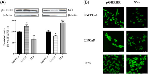
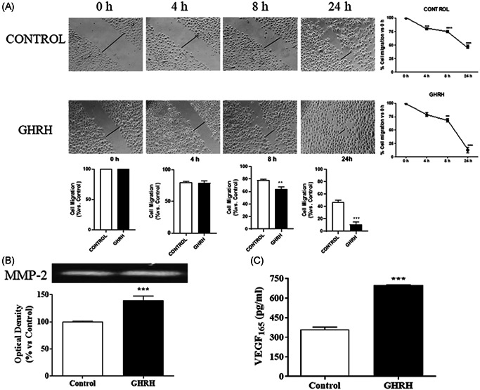
Methods: We performed cell viability, cell proliferation, adhesion and migration assays. In addition, metalloprotease (MMP)-2 activity by means gelatin zymography, GHRH-R subcellular location using confocal immunofluorescence microscopy and vascular endothelial growth factor (VEGF) levels by enzyme-linked immunoassay were assessed. Besides, we developed an in vivo model in order vivo model to determine the role of GHRH on tumorigenic transformation of RWPE-1 cells.

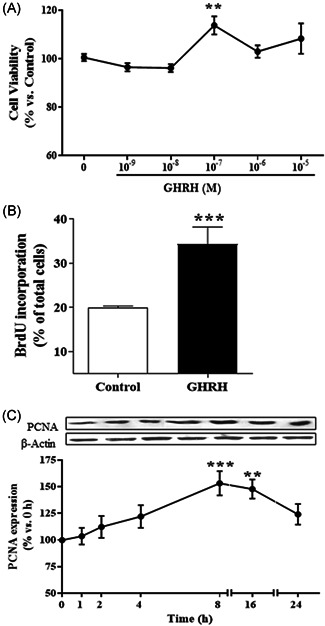
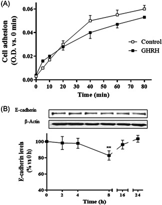
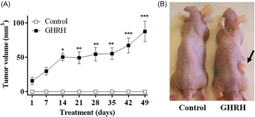
Results: In cell cultures, we observed development of a migratory phenotype consistent with the gelatinolytic activity of MMP-2, expression of VEGF, as well as E-cadherin-mediated cell-cell adhesion and increased cell motility. Treatment with 0.1 µM GHRH for 24 h significantly increased cell viability and cell proliferation. Similar effects of GHRH were seen in RWPE-1 tumors developed by subcutaneous injection of GHRH-treated cells in athymic nude mice, 49 days after inoculation.

Conclusions: Thus, GHRH appears to act as a cytokine in the transformation of RWPE-1 cells by mechanisms that likely involve epithelial-mesenchymal transition, thus reinforcing the role of GHRH in tumorigenesis of prostate.

Keywords: GHRH; RWPE-1 cells; epithelial-mesenchymal transition; prostate cancer; tumorigenesis.


Conflict of interest statement

The authors declare no conflicts of interest.

Figures

Figure 1
Figure 1
Expression and subcellular location of GHRH receptors in RWPE‐1, LNCaP and PC3 cells. (A) Expression levels of GHRH receptors were evaluated by Western blotting using specific antibodies for pGHRHR and SVs. The diagram represents the mean ± SEM of four experiments. *p < 0.05; **p < 0.01; ***p < 0.001 compared with those obtained in RWPE‐1 cells. (B) Immunofluorescence detection of pGHRHR and SVs. Cells were fixed and incubated with anti‐pGHRHR and anti‐SVs as described in Section 2.2. After an examination by confocal microscopy, the intensity of the emitted fluorescence was measured using image performing software LCS‐SL. Results are representative of four independent experiments. Cells were observed at ×200 magnification, scale bar = 20 μm. GHRH, growth hormone‐releasing hormone; SV, splice variant [Color figure can be viewed at wileyonlinelibrary.com]
Figure 2
Figure 2
Effect of GHRH on cell viability and cell proliferation in RWPE‐1 cells. (A) RWPE‐1 cells were incubated in the absence or presence of 0.1 μM GHRH at different concentration (10−9–10−5 M) for 24 h. GHRH provoked a significant increase of cell viability at 10−7 M (n = 4). (B) RWPE‐1 cells were incubated in the absence or presence of 0.1 μM GHRH and 10 μM bromodeoxyuridine (BrdU). GHRH increased BrdU incorporation after 24 h‐treatment (n = 4). (C) Expression of PCNA and β‐actin levels were evaluated by Western blotting at indicated times (1–24 h). GHRH (1–44) (0.1 μM) augmented the cell proliferation marker at 8 and 16 h. A representative experiment is shown. The bar diagrams represent the mean ± SEM of four experiments. **p < 0.01; ***p < 0.001 compared with control. GHRH, growth hormone‐releasing hormone
Figure 3
Figure 3
Effect of GHRH on cell adhesion in RWPE‐1 cells. (A) Cells were plated onto coated culture wells in the presence or absence of 0.1 µM GHRH; after the indicated times, MTT (1 mg/ml) was added for 4 h followed by aspiration and addition of 0.1 ml DMSO; the absorbance was measured at 530/640 nm (n = 4). (B) Expression of E‐cadherin and β‐actin levels was evaluated by Western blotting at indicated times (2–24 h). GHRH (0.1 μM) significantly decreased the cell adhesion marker at 8 h. A representative experiment is shown. The bar diagrams represent the mean ± SEM of four experiments. **p < 0.01 compared with control. DMSO, dimethyl sulfoxide; GHRH, growth hormone‐releasing hormone
Figure 4
Figure 4
Effect of GHRH on cell migration in RWPE‐1 cells. (A) Cells were damaged by mechanical scrapping and incubated in the presence or absence of 0.1 µM GHRH for the indicated times (4–24 h). Recovery of cell monolayer wounds was followed by microscopy. Representative images from four experiments are shown. (B) Cells were incubated with the peptide for 24 h. Total protein from cell secretion was subjected to zymography to detect the gelatinases. The expression of the latent isoform of metalloproteinase (MMP)‐2 was increased by 0.1 μM GHRH. A representative experiment is shown (n = 4). (C) Secretion of the vascular endothelial growth factor (VEGF) was evaluated by ELISA after incubation with GHRH (0.1 μM). The neuropeptide augmented the secreted VEGF165 by 90% at 24 h of the treatment. The diagrams represent the mean ± SEM of four experiments. **p < 0.01; ***p < 0.001 compared with control. ELISA, enzyme‐linked immunoassay; GHRH, growth hormone‐releasing hormone
Figure 5
Figure 5
Effect of GHRH on the promotion of tumorigenesis. (A) RWPE‐1 cells were incubated in the absence or the presence of 0.1 µM GHRH for 24 h. The cell suspension was mixed with Matrigel synthetic basement membrane and then injected under the skin in the right flank (1 × 107 cells/mouse). Seven animals were used per group. Experiment lasted for 49 days. Data in each bar are the means ± SEM. *p < 0.05; **p < 0.01; ***p < 0.001 versus control. (B) Image from a representative animal of each group. Tumor mass is marked with a black arrow. GHRH, growth hormone‐releasing hormone; GHRH, growth hormone‐releasing hormone [Color figure can be viewed at wileyonlinelibrary.com]

Similar articles

Cited by

References

    1. Ferlay J, Colombet M, Soerjomataram I, et al. Estimating the global cancer incidence and mortality in 2018: GLOBOCAN sources and methods. Int J Cancer. 2019;144(8):1941‐1953. 10.1002/ijc.31937 - DOI - PubMed
    1. Nowacka‐Zawisza M, Wiśnik E. DNA methylation and histone modifications as epigenetic regulation in prostate cancer (Review). Oncol Rep. 2017;38(5):2587‐2596. 10.3892/or.2017.5972 - DOI - PubMed
    1. Junejo NN, AlKhateeb SS. BRCA2 gene mutation and prostate cancer risk. Comprehensive review and update. Saudi Med J. 2020;41(1):9‐17. 10.15537/smj.2020.1.24759 - DOI - PMC - PubMed
    1. Ramanand SG, Mani RS. Genetic, environmental, and nuclear factors governing genomic rearrangements. Adv Exp Med Biol. 2019;1210:57‐66. 10.1007/978-3-030-32656-2-3 - DOI - PubMed
    1. Singh VK, Pal R, Srivastava P, Misra G, Shukla Y, Sharma PK. Exposure of androgen mimicking environmental chemicals enhances proliferation of prostate cancer (LNCaP) cells by inducing AR expression and epigenetic modifications. Environ Pollut. 2021;272:116397. 10.1016/j.envpol.2020.116397 - DOI - PubMed

Publication types

Substances