Review
. 2022 Mar 4;29(3):1645-1671.
doi: 10.3390/curroncol29030138.

Current Advances in the Management of Adult Craniopharyngiomas

Affiliations
Review

Current Advances in the Management of Adult Craniopharyngiomas

Montserrat Lara-Velazquez et al. Curr Oncol. .

Abstract

Craniopharyngiomas (CPs) are slow growing, histologically benign intracranial tumors located in the sellar-suprasellar region. Although known to have low mortality, their location and relationship to the adjacent neural structures results in patients having significant neurologic, endocrine, and visual comorbidities. The invasive nature of this tumor makes complete resection a challenge and contributes to its recurrence. Additionally, these tumors are bimodally distributed, being treated with surgery, and are followed by other adjuncts, such as focused radiation therapy, e.g., Gamma knife. Advances in surgical techniques, imaging tools, and instrumentations have resulted in the evolution of surgery using endoscopic techniques, with residual components being treated by radiotherapy to target the residual tumor. Advances in molecular biology have elucidated the main pathways involved in tumor development and recurrence, but presently, no other treatments are offered to patients, besides surgery, radiation, and endocrine management, as the disease and tumor evolve. We review the contemporary management of these tumors, from the evolution of surgical treatments, utilizing standard open microscopic approaches to the more recent endoscopic surgery, and discuss the current recommendations for care of these patients. We discuss the developments in radiation therapy, such as radiosurgery, being used as treatment strategies for craniopharyngioma, highlighting their beneficial effects on tumor resections while decreasing the rates of adverse outcomes. We also outline the recent chemotherapy modalities, which help control tumor growth, and the immune landscape on craniopharyngiomas that allow the development of novel immunotherapies.

Keywords: chemotherapy; craniopharyngiomas; gross total resection; minimally invasive; molecular biology; neurological complications; radiation therapy; sellar tumors; surgical resection.


Conflict of interest statement

The authors declare no conflict of interest.

Figures

Figure 1
Figure 1
Twenty-nine year old patient with recurrent adamantinomatous craniopharyngioma treated with salvage surgery and post-operative proton radiotherapy. Row 1. Pre-op MRI shows suprasellar cystic tumor with enhancing wall involving the optic pathway. Pre- and post-operative tumor volumes inform the radiation target contours as overlayed on the images. GTV (red) = gross tumor volume (based on pre-op gross tumor), CTV (yellow) = clinical target volume (encompassing areas of tumor extent at initial presentation and areas at risk of subclinical disease), PTV (green) = planning target volume. Row 2. Post-op MRI shows residual cystic tumor involving the optic pathway. Row 3. Radiation planning CT with radiation dose overlay. Pencil beam scanning proton radiation was utilized to deliver a prescription dose of 54 GyRBE in 1.8 GyRBE fractions.
Figure 2
Figure 2
Craniopharyngioma with mixed papillary and adamantinomatous patterns with well differentiated squamous epithelium around fibrovascular cores. (A) Focal arears in the tumor show adamantinomatous pattern of craniopharyngioma displaying nodular and trabecular cellular growth with peripheral nuclear palisading ((B), arrow head) and looser plumper stellate reticulum cells ((B), arrows). HE = hematoxylin and eosin.
Figure 3
Figure 3
Tissue sections showing papillary configuration with cauliflower-like morphology of solid sheets of well-differentiated nonkeratinizing squamous epithelium around fibrovascular cores (A,B). Mitotic figures are rare to absent and no necrosis or significant nuclear atypia are seen. BRAF V600E immunostain is positive in tumor cells (C), supporting the diagnosis of papillary craniopharyngioma. HE = hematoxylin and eosin.
Figure 4
Figure 4
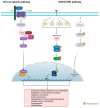
Representation of the molecular pathways involved in tumor formation and progression in CPs. The wingless (Wnt)/β-catenin and the mitogen-activated protein kinases/extracellular signal-regulated kinase (MAPK/ERK) pathways, are the main biological cascades involved in the development of the two histological variants of CPs: ACPs and PCPs. Created with Biorender.com (accessed on 28 November 2021).
Figure 5
Figure 5
Algorithm by Gazanfar and Gene for the treatment of CPs with minimally invasive techniques combined with radiation. Taken without modifications from [58]. Abbreviations: KPS, Karnofsky Performance Status Scale; ASA, American Society of Anesthesiologists; MRI, magnetic resonance imaging; IMR, intensity modulated radiation; SRT, stereotactic radiation therapy; SRS, stereotactic radiosurgery. (published under Creative Commons Attribution License, which permits unrestricted use, distribution, and reproduction in any medium, provided the original author and source are credited). * Patient choice for minimally invasive produce. ** MRI scans every 3 months/1 year then 6 months/2 years, earlier imaging if symptomatic, clinical history, and examination + neuro-endocrine review.
Figure 6
Figure 6
Timeline that includes the preceding events that lead the development of the surgical and radiation treatments of CPs [115,116,117,118,119].
Figure 7
Figure 7
The study of the immune landscape of CPs allowed the development of novel immunotherapies for the treatment of these tumors. By targeting immunological checkpoints (CTLA-4, PD-L1, B7-H3, VISTA) the action of the immune system can be enhanced to specifically target cancer cells. Created with Biorender.com (accessed on 28 November 2021).

References

    1. Bunin G.R., Surawicz T.S., Witman P.A., Preston-Martin S., Davis F., Bruner J.M. The descriptive epidemiology of craniopharyngioma. J. Neurosurg. 1998;89:547–551. doi: 10.3171/jns.1998.89.4.0547. - DOI - PubMed
    1. Karavitaki N., Cudlip S., Adams C.B., Wass J.A. Craniopharyngiomas. Endocr. Rev. 2006;27:371–397. doi: 10.1210/er.2006-0002. - DOI - PubMed
    1. Ostrom Q.T., Gittleman H., Liao P., Vecchione-Koval T., Wolinsky Y., Kruchko C., Barnholtz-Sloan J.S. CBTRUS Statistical Report: Primary brain and other central nervous system tumors diagnosed in the United States in 2010–2014. Neur. Oncol. 2017;19:v1–v88. doi: 10.1093/neuonc/nox158. - DOI - PMC - PubMed
    1. Yamini B., Narayanan M. Craniopharyngiomas: An update. Expert Rev. Anticancer Ther. 2006;6:S85–S92. doi: 10.1586/14737140.6.9s.S85. - DOI - PubMed
    1. Muller H.L., Merchant T.E., Warmuth-Metz M., Martinez Barbera J.P., Puget S. Craniopharyngioma. Nat. Rev. Dis Primers. 2019;5:75. doi: 10.1038/s41572-019-0125-9. - DOI - PubMed

Publication types