

Echocardiographic Assessment of Atrial Function: From Basic Mechanics to Specific Cardiac Diseases
- PMID: 35323616
- PMCID: PMC8955277
- DOI: 10.3390/jcdd9030068
Echocardiographic Assessment of Atrial Function: From Basic Mechanics to Specific Cardiac Diseases
Abstract
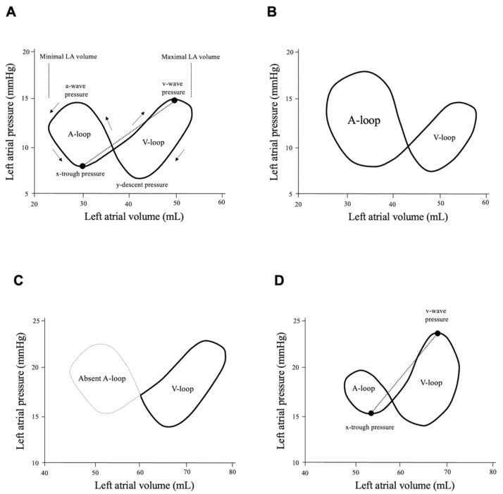
The left and right atria serve as buffer chambers to control the flow of venous blood for ventricular filling. If an atrium is absent, blood does not flow effectively into the ventricle, leading to venous blood retention and low cardiac output. The importance of atrial function has become increasingly recognized, because left atrial (LA) function contributes to cardiac performance, and loss of LA function is associated with heart failure. LA volume change has been used for LA function assessment in experimental and clinical studies. In conjunction with LA pressure, the LA pressure-volume relationship provides a better understanding of LA mechanics. LA strain measurement by speckle tracking echocardiography was introduced to evaluate three components of LA function as a (booster) pump, reservoir and conduit. Furthermore, increasing evidence supports the theory that LA reservoir strain has prognostic utility in various cardiac diseases. In this review, we summarize LA contribution to maintain cardiac performance by evaluating LA function with echocardiography according to our experiences and previous reports. Furthermore, we discuss LA dysfunction in challenging cardiac diseases of cardiac amyloidosis and adult congenital heart disease.
Keywords: adult congenital heart disease; atrial fibrillation; cardiac amyloidosis; heart failure; left atrial function; left atrial strain; pressure–volume loop.
Conflict of interest statement
The authors declare no conflict of interest regarding to this review article.
Figures


Similar articles
-
Prognostic Utility and Clinical Significance of Cardiac Mechanics in Heart Failure With Preserved Ejection Fraction: Importance of Left Atrial Strain.Circ Cardiovasc Imaging. 2016 Mar;9(3):10.1161/CIRCIMAGING.115.003754 e003754. doi: 10.1161/CIRCIMAGING.115.003754. Circ Cardiovasc Imaging. 2016. PMID: 26941415 Free PMC article.
-
Right and Left Atrial Dissimilarities in Normal Subjects Explored by Speckle Tracking Echocardiography.Echocardiography. 2015 Sep;32(9):1392-9. doi: 10.1111/echo.12880. Epub 2015 Jan 21. Echocardiography. 2015. PMID: 25611312
-
Left Atrial Mechanics Assessed Early during Hospitalization for Cryptogenic Stroke Are Associated with Occult Atrial Fibrillation: A Speckle-Tracking Strain Echocardiography Study.J Am Soc Echocardiogr. 2021 Feb;34(2):156-165. doi: 10.1016/j.echo.2020.09.009. Epub 2020 Oct 29. J Am Soc Echocardiogr. 2021. PMID: 33132019
-
Unveiling the Hidden Chamber: Exploring the Importance of Left Atrial Function and Filling Pressure in Cardiovascular Health.J Cardiovasc Echogr. 2023 Jul-Sep;33(3):117-124. doi: 10.4103/jcecho.jcecho_44_23. Epub 2023 Nov 20. J Cardiovasc Echogr. 2023. PMID: 38161774 Free PMC article. Review.
-
Assessment of Left Atrial Function by Echocardiography: Novel Insights.Curr Cardiol Rep. 2018 Aug 27;20(10):96. doi: 10.1007/s11886-018-1044-1. Curr Cardiol Rep. 2018. PMID: 30151628 Review.
Cited by
-
Right heart and left atrial strain to differentiate cardiac amyloidosis and Fabry disease.Sci Rep. 2024 Jan 30;14(1):2445. doi: 10.1038/s41598-024-52890-y. Sci Rep. 2024. PMID: 38291191 Free PMC article.
-
Current and Clinically Relevant Echocardiographic Parameters to Analyze Left Atrial Function.J Cardiovasc Dev Dis. 2024 Aug 5;11(8):241. doi: 10.3390/jcdd11080241. J Cardiovasc Dev Dis. 2024. PMID: 39195149 Free PMC article. Review.
-
Left Atrial Strain Imaging by Speckle Tracking Echocardiography: The Supportive Diagnostic Value in Cardiac Amyloidosis and Hypertrophic Cardiomyopathy.J Cardiovasc Dev Dis. 2023 Jun 15;10(6):261. doi: 10.3390/jcdd10060261. J Cardiovasc Dev Dis. 2023. PMID: 37367426 Free PMC article.
-
Echocardiographic Predictors of Postoperative Atrial Fibrillation After Cardiac Surgery: Assessing Atrial Mechanics for Risk Stratification.J Cardiovasc Dev Dis. 2025 Apr 17;12(4):160. doi: 10.3390/jcdd12040160. J Cardiovasc Dev Dis. 2025. PMID: 40278219 Free PMC article.
-
Atrial Dysfunction in Significant Atrial Functional Mitral Regurgitation: Phenotypes and Prognostic Implications.Circ Cardiovasc Imaging. 2023 May;16(5):e015089. doi: 10.1161/CIRCIMAGING.122.015089. Epub 2023 May 9. Circ Cardiovasc Imaging. 2023. PMID: 37158081 Free PMC article.
References
-
- Nagueh S.F., Smiseth O.A., Appleton C.P., Byrd B.F., 3rd, Dokainish H., Edvardsen T., Flachskampf F.A., Gillebert T.C., Klein A.L., Lancellotti P., et al. Recommendations for the Evaluation of Left Ventricular Diastolic Function by Echocardiography: An Update from the American Society of Echocardiography and the European Association of Cardiovascular Imaging. J. Am. Soc. Echocardiogr. 2016;29:277–314. doi: 10.1016/j.echo.2016.01.011. - DOI - PubMed
-
- Paulus W.J., Tschope C., Sanderson J.E., Rusconi C., Flachskampf F.A., Rademakers F.E., Marino P., Smiseth O.A., De Keulenaer G., Leite-Moreira A.F., et al. How to diagnose diastolic heart failure: A consensus statement on the diagnosis of heart failure with normal left ventricular ejection fraction by the Heart Failure and Echocardiography Associations of the European Society of Cardiology. Eur. Heart J. 2007;28:2539–2550. doi: 10.1093/eurheartj/ehm037. - DOI - PubMed
-
- Fukuda N., Oki T., Iuchi A., Tabata T., Yamada H., Ito S., Takeichi N., Shinohara H., Socki T., Shinomiya H., et al. Tricuspid inflow and regurgitant flow dynamics after mitral valve replacement: Differences relating to surgical repair of the tricuspid valve. J. Heart Valve Dis. 1997;6:184–188. - PubMed
Publication types
LinkOut - more resources
Full Text Sources
Medical
