. 2022 Apr 29;145(3):1124-1138.
doi: 10.1093/brain/awab408.

Sexual dimorphism in a neuronal mechanism of spinal hyperexcitability across rodent and human models of pathological pain

Affiliations

Sexual dimorphism in a neuronal mechanism of spinal hyperexcitability across rodent and human models of pathological pain

Annemarie Dedek et al. Brain. .

Abstract

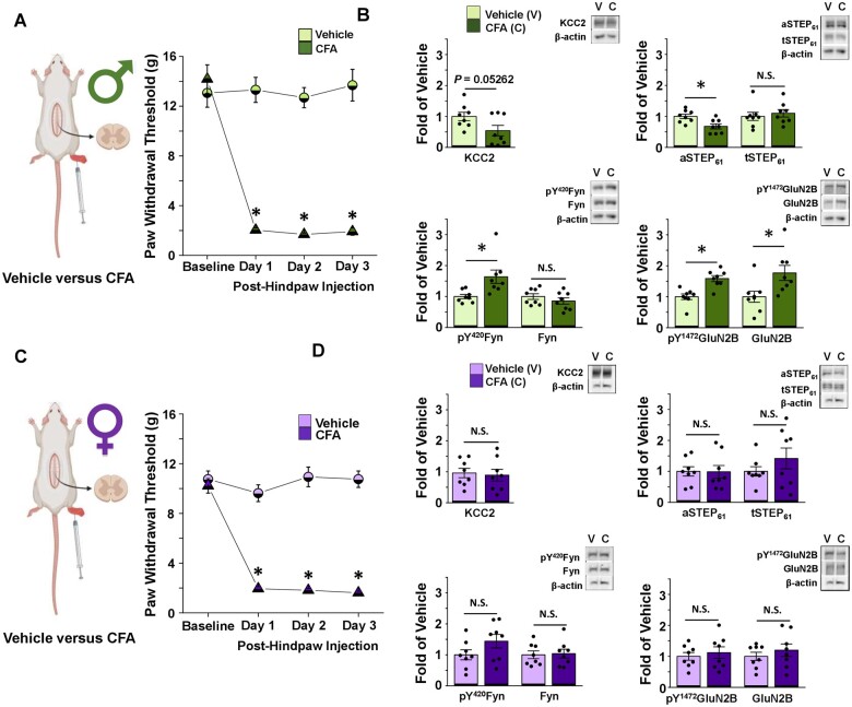
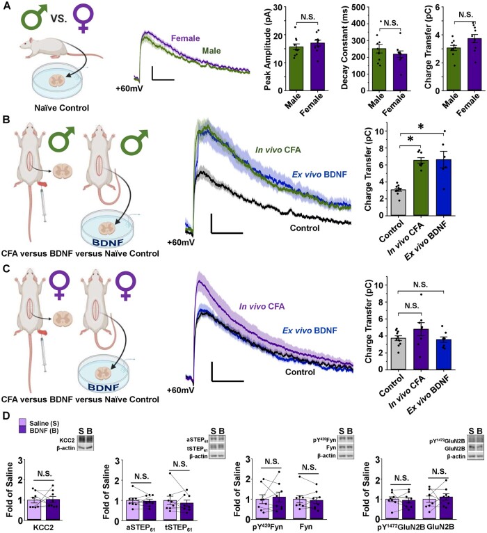
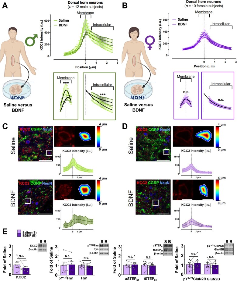
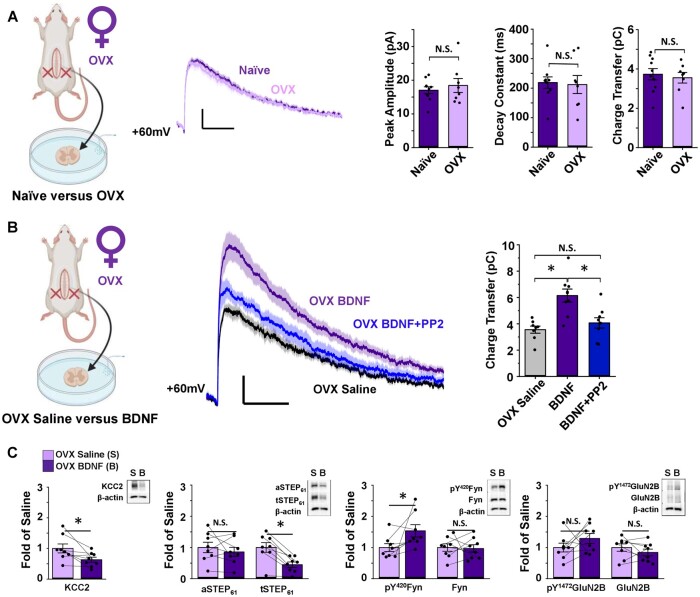
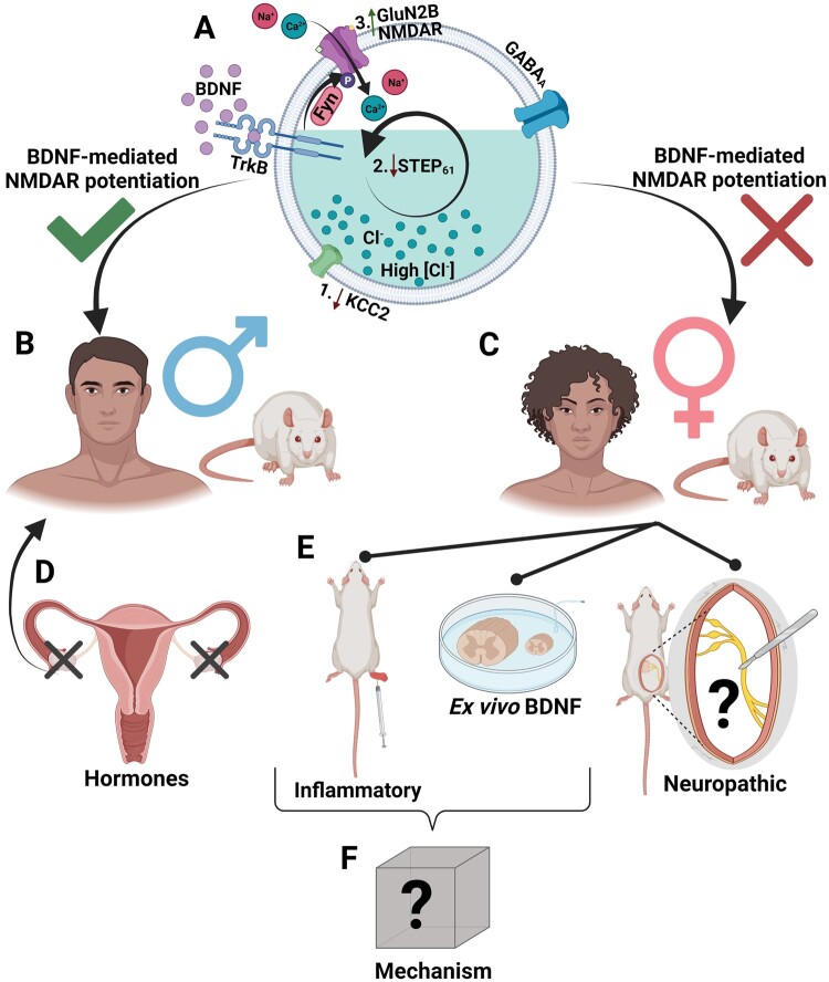
The prevalence and severity of many chronic pain syndromes differ across sex, and recent studies have identified differences in immune signalling within spinal nociceptive circuits as a potential mediator. Although it has been proposed that sex-specific pain mechanisms converge once they reach neurons within the superficial dorsal horn, direct investigations using rodent and human preclinical pain models have been lacking. Here, we discovered that in the Freund's adjuvant in vivo model of inflammatory pain, where both male and female rats display tactile allodynia, a pathological coupling between KCC2-dependent disinhibition and N-methyl-D-aspartate receptor (NMDAR) potentiation within superficial dorsal horn neurons was observed in male but not female rats. Unlike males, the neuroimmune mediator brain-derived neurotrophic factor (BDNF) failed to downregulate inhibitory signalling elements (KCC2 and STEP61) and upregulate excitatory elements (pFyn, GluN2B and pGluN2B) in female rats, resulting in no effect of ex vivo brain-derived neurotrophic factor on synaptic NMDAR responses in female lamina I neurons. Importantly, this sex difference in spinal pain processing was conserved from rodents to humans. As in rodents, ex vivo spinal treatment with BDNF downregulated markers of disinhibition and upregulated markers of facilitated excitation in superficial dorsal horn neurons from male but not female human organ donors. Ovariectomy in female rats recapitulated the male pathological pain neuronal phenotype, with BDNF driving a coupling between disinhibition and NMDAR potentiation in adult lamina I neurons following the prepubescent elimination of sex hormones in females. This discovery of sexual dimorphism in a central neuronal mechanism of chronic pain across species provides a foundational step towards a better understanding and treatment for pain in both sexes.

Keywords: NMDAR; inflammatory pain; pain; sex difference; spinal hyperexcitability.


Figures

Figure 1
Figure 1
CFA elicits tactile allodynia in male and female adult rats, but only activates the STEP61/pFyn/pGluN2B spinal hyperexcitability pathway at male SDH synapses. (A) Left: In vivo CFA inflammatory pain model, created using BioRender.com. Right: CFA elicits tactile allodynia in adult male rats. Von Frey behaviour testing shows that rats with a CFA injection (n = 14) to their right hindpaw have decreased PWT compared to vehicle-injected animals (n = 8; one-way repeated-measures ANOVA, followed by Bonferroni-adjusted pairwise comparisons when P < 0.05). Animal tissue was collected for use in either western blot analysis or electrophysiological recordings. (B) CFA inflammatory pain model drives upregulation of pFyn, pGluN2B, GluN2B and downregulation of aSTEP61 in male rat SDH synaptosomes. Plots (left) and representative western blots (right) from SDH synaptosomes of male rats 5 days after hindpaw injection of either vehicle (light green, n = 8 for all targets) or CFA (dark green, n = 8 for all targets; compared using independent samples t-test). (C) Left: In vivo CFA inflammatory pain model, created using Biorender.com. Right: Von Frey behaviour testing shows that rats with a CFA (n = 14) injection to their right hindpaw have decreased PWT compared to vehicle injected animals (n = 8; one-way repeated-measures ANOVA, followed by Bonferroni-adjusted pairwise comparisons when P < 0.05). Animal tissue was collected for use in either western blot analysis or electrophysiological recordings. (D) CFA inflammatory pain model elicits no effect in the STEP61/pFyn/pGluN2B spinal hyperexcitability pathway of female rat SDH synaptosomes. Plots (left) and representative western blots (right) from SDH synaptosomes of female rats 5 days after injection of either vehicle (lilac, n = 8) or CFA (purple, n = 8; compared using independent samples t-test). *P < 0.05.
Figure 2
Figure 2
NMDARs at female SDH synapses are not potentiated or upregulated by the ex vivo BDNF or in vivo CFA models of pathological pain. (A) Baseline lamina I mEPSCs do not differ between male and female adult SD rats. Left: Saline-treated male vs female lamina I neurons, created using BioRender.com. Middle: Average mEPSCs at +60 mV in lamina I neurons from male (green) and female (purple) adult rats. Right: Peak amplitude, decay constant and charge transfer of the NMDAR component of mEPSCs at +60 mV do not differ between male and female SD rats. n = 10 for males and n = 9 for females. Compared using independent samples t-test. (B) Male lamina I NMDAR mEPSCs are potentiated following CFA hindpaw injection and ex vivo BDNF treatment. Left: Experimental paradigm showing male in vivo CFA versus ex vivo BDNF models, created using BioRender.com. Middle: NMDAR mEPSCs from male rat lamina I neurons; control in black, CFA in green, BDNF in blue. Right: Charge transfer of NMDAR mEPSCs for groups shown to left. n = 10 for control, n = 8 for CFA and n = 6 for BDNF. Compared using Welch’s test followed by Games–Howell comparison. (C) Female lamina I NMDAR mEPSCs are not potentiated following CFA hindpaw injection or ex vivo BDNF treatment. Left: Experimental paradigm showing female in vivo CFA versus ex vivo BDNF models, created using BioRender.com. Middle: NMDAR mEPSCs from female rat lamina I neurons; control in black, CFA in purple, BDNF in blue. Right: Charge transfer of NMDAR mEPSCs shown to left. n = 10 for control, n = 8 for CFA and n = 8 for BDNF. Compared using the Kruskal–Wallis one-way ANOVA. (D) Ex vivo BDNF treatment model elicits no effect in female rat SDH synaptosomes. Plots (left) and representative western blots (right) from female rat SDH synaptosomes of tissue treated with either control saline (lilac, n = 8) or 50 ng/ml recombinant BDNF for 70 min (purple, n = 8; compared using paired-samples t-test). *P < 0.05.
Figure 3
Figure 3
In contrast to males, ex vivo BDNF treatment does not activate markers of disinhibition or facilitated excitation at SDH synapses of female human spinal tissue. (A) The ex vivo BDNF model elicits KCC2 internalization in adult male human SDH neurons. Left: Experimental paradigm showing treatment of human SDH tissue in either saline or BDNF, created using BioRender.com. Top right: Average KCC2 intensity values from SDH neurons of male human donor tissue (n = 12) incubated in saline (light green) versus BDNF (dark green). Bottom right: Comparisons of membrane and intracellular regions by the extra sum-of-squares F-test method; the membrane component is fitted to a Gaussian distribution, while the intracellular component is fitted to an exponential decay. (B) The ex vivo BDNF model has no effect on KCC2 internalization in adult female human SDH neurons. Left: Experimental paradigm showing treatment of human SDH tissue in either saline or BDNF, created using BioRender.com. Top right: Average KCC2 intensity values from SDH neurons of saline (lilac) versus BDNF-treated (purple) spinal segments of 10 female human donors. Bottom right: Comparisons of membrane and intracellular regions by the extra sum-of-squares F-test method; the membrane component is fitted to a Gaussian distribution, while the intracellular component is fitted to an exponential decay. (C and D) Representative confocal images of male (C) and female (D) human superficial dorsal horn incubated in saline or BDNF. KCC2 (red), CGRP (green) and NeuN (blue). A zoomed region (top right) shows a neuron expressing KCC2 together with the delineation of the membrane and the distance to the membrane of each pixel analysed in a colour-coded distance map. KCC2 intensity (i.u.) versus distance to the membrane profile (bottom). Scale bar = 50 µm; inset = 10 µm. (E) The ex vivo BDNF treatment model elicits no effect in human female SDH synaptosomes. Plots (left) and representative western blots (right) from SDH synaptosomes of human female spinal cord treated with either control saline (lilac, n = 6) or 100 ng/ml recombinant BDNF for 70 min (purple, n = 6). Comparisons were made using paired samples t-tests in all cases except for aSTEP61, where the Wilcoxon signed-rank test was used. *P < 0.05.
Figure 4
Figure 4
Ovariectomy triggers BDNF-mediated NMDAR potentiation by the KCC2/STEP61/SFK pathway in SDH neurons of female rats. (A) Ovariectomy (OVX) has no effect on baseline lamina I NMDAR mEPSCs of female SD rats. Left: Saline-treated naïve female versusOVX female lamina I neurons, created using BioRender.com. Middle: Average mEPSCs at +60 mV in lamina I neurons of naïve (purple) and ovariectomized (lilac) female rats. Right: Peak amplitude (compared using Mann–Whitney test), decay constant (compared using independent samples t-test) and charge transfer (compared using independent samples t-test) of the NMDAR component of mEPSCs do not differ between naïve (n = 10) and OVX (n = 8) female rats. (B) In OVX rats, NMDAR mEPSCs in lamina I neurons are potentiated following ex vivo BDNF treatment. This potentiation is blocked using co-treatment with the SFK inhibitor, PP2. Left: Recordings from lamina I neurons were compared for saline- versus BDNF-treated slices from OVX rats, created using BioRender.com. Middle: Average mEPSCs at +60 mV from OVX female rat lamina I neurons; control in black, BDNF in purple, BDNF+PP2 in blue. Right: Charge transfer of NMDAR mEPSCs shown on left. n = 8 for control, n = 9 for BDNF and BDNF + PP2. Comparisons were made using one-way ANOVA, followed by Tukey HSD when P < 0.05. (C) Ex vivo BDNF treatment in OVX rat SDH synaptosomes results in upregulation of pY420Fyn and downregulation of KCC2 and STEP61. Plots (left) and representative western blots (right) from OVX female rat SDH synaptosomes of tissue treated with either control saline (lilac, n = 8) or 50 ng/ml recombinant BDNF for 70 min (purple, n = 8, compared using paired samples t-tests). *P < 0.05.
Figure 5
Figure 5
Working model of sexual dimorphism in a superficial dorsal horn mechanism underlying pathological pain. (A) In lamina I superficial dorsal horn (SDH) neurons, BDNF drives (1) disinhibition through KCC2 downregulation that (2) is coupled to reduced STEP61 activity and subsequent (3) phosphorylation and potentiation of GluN2B NMDARs by Fyn kinase. (B) We have found that this molecular mechanism of BDNF-mediated NMDAR potentiation is conserved across species in males, (C) but is not observed in the SDH of female rats and humans. (D) Rodent ovariectomy before reaching sexual maturity results in the male-like BDNF-induced hyperexcitability pathway, indicating that this sex difference is hormonally mediated. (E) We have found that the SDH STEP61/pFyn/pGluN2B spinal hyperexcitability pathway is not triggered by the in vivo CFA inflammatory pain or the ex vivo BDNF treatment models of pathological pain; however, it remains unknown if this molecular mechanism is involved in neuropathic pain models in females. (F) Further investigations are needed to identify the molecular mechanisms underlying spinal hyperexcitability and pathological pain in the female SDH. Figure created using BioRender.com.

Similar articles

Cited by

References

    1. Pleis JR, Ward BW, Lucas JW. Summary health statistics for U.S. adults: National Health Interview Survey, 2009. Vital Health Stat 2010;249:1–207. - PubMed
    1. Chen G, Luo X, Qadri MY, Berta T, Ji R-R. Sex-dependent glial signaling in pathological pain: distinct roles of spinal microglia and astrocytes. Neurosci Bull. 2017;34(1):98–108. - PMC - PubMed
    1. Bouhassira D, Lantéri-Minet M, Attal N, Laurent B, Touboul C. Prevalence of chronic pain with neuropathic characteristics in the general population. Pain. 2008;136(3):380–387. - PubMed
    1. Bartley EJ, Fillingim RB. Sex differences in pain: A brief review of clinical and experimental findings. Br J Anaesth. 2013;111(1):52–58. - PMC - PubMed
    1. Mogil JS. Qualitative sex differences in pain processing: Emerging evidence of a biased literature. Nat Rev Neurosci. 2020;21(7):353–365. - PubMed

Publication types

Substances