. 2022 Jul 14;107(8):e3515-e3525.
doi: 10.1210/clinem/dgac166.

Kisspeptin Overcomes GnRH Neuronal Suppression Secondary to Hyperprolactinemia in Humans

Affiliations

Kisspeptin Overcomes GnRH Neuronal Suppression Secondary to Hyperprolactinemia in Humans

Katerina Hoskova et al. J Clin Endocrinol Metab. .

Erratum in

Abstract

Context: Hyperprolactinemia suppresses gonadotropin-releasing hormone (GnRH)-induced luteinizing hormone (LH) pulses. The hypothalamic neuropeptide kisspeptin potently stimulates the secretion of GnRH. The effects of exogenous kisspeptin administration on GnRH pulse generation in the setting of hyperprolactinemia have not previously been explored.

Objective: This work aimed to examine the effects of kisspeptin on GnRH secretion, as reflected by LH secretion, in women with hyperprolactinemia.

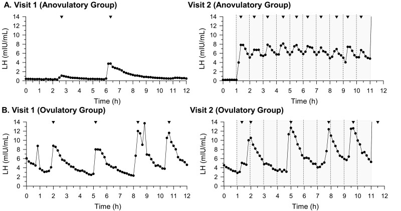
Methods: Women with hyperprolactinemia (n = 11) participated in two 12-hour visits. Before study visits, participants underwent washout of dopamine agonist and/or combined oral contraceptive. Frequent blood sampling was performed (1 sample was collected every 10 minutes). Visit 1 involved no intervention, to examine baseline LH pulsatility. During visit 2, kisspeptin 112-121 (0.24 nmol/kg) was administered every 1 hour, for 10 hours. At hour 11, one intravenous bolus of GnRH (75 ng/kg) was administered.

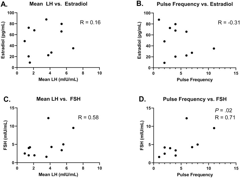
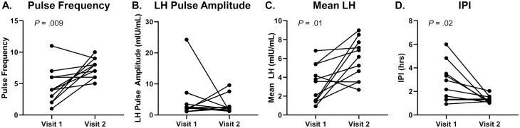
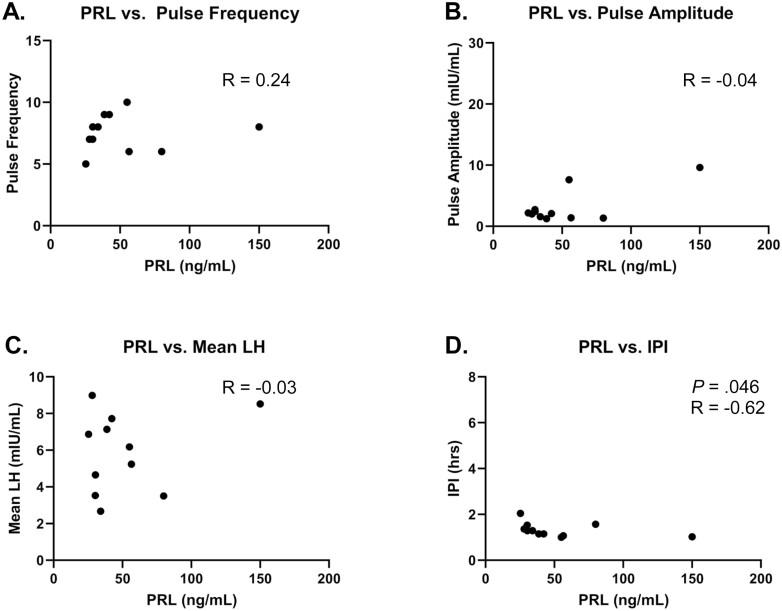
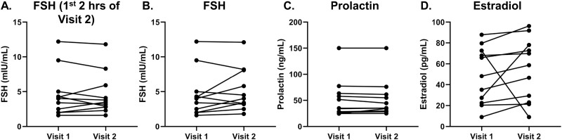
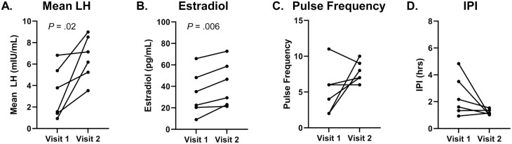
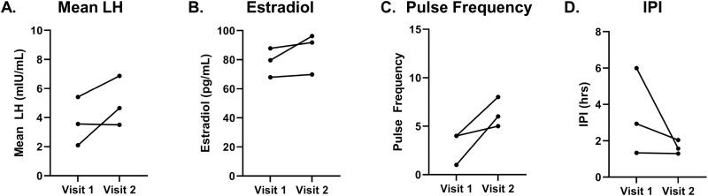
Results: Repetitive intravenous bolus kisspeptin administration increased the total number of LH pulses in the setting of hyperprolactinemia. The interpulse interval declined during the same time frames. LH pulse amplitude did not change, but the mean LH rose. In 6 participants with progesterone levels suggestive of an anovulatory state, mean LH and estradiol levels increased significantly at visit 2. In the entire cohort, follicle-stimulating hormone and prolactin levels did not change significantly across the 2 visits. A total of 73% of subjects exhibited an LH pulse within 30 minutes of first kisspeptin dose.

Conclusion: Kisspeptin is capable of stimulating hypothalamic GnRH-induced LH pulses in the setting of hyperprolactinemia.

Keywords: hyperprolactinemia; kisspeptin.


Figures

Figure 1.
Figure 1.
Relationships among gonadotropins (luteinizing hormone [LH] and follicle-stimulating hormone [FSH]) and estradiol of individuals at visit 1 (n = 11). Mean LH and pulse frequency are calculated across hour 1 to hour 11. Values for FSH and estradiol span the entire sampling period of visit 1 (hour 0-hour 12). A, Mean LH vs estradiol; B, pulse frequency vs estradiol; C, mean LH vs FSH; D, pulse frequency vs FSH. Statistically significant P and R values are included in the figure.
Figure 2.
Figure 2.
Measures of luteinizing hormone (LH) pulsatility profile at visits 1 and 2 (n = 11). Pulse frequency, mean LH, and interpulse interval (IPI) were calculated across the time frame of kisspeptin administration (hour 1-hour 11). LH pulse amplitude for visit 2 includes value from response to first kisspeptin bolus only. One participant experienced only one pulse during visit 1. For this individual, hour 11 was used as the equivalent to the nadir of subsequent pulse for IPI calculations. A, pulse frequency; B, LH pulse amplitude; C, mean LH levels; and D, IPI. Statistically significant P values are included in the figure.
Figure 3.
Figure 3.
Relationship between prolactin (PRL) levels of individuals at visit 2 and features of luteinizing hormone (LH) response to kisspeptin at visit 2 (n = 11). PRL level from visit 2 before kisspeptin administration (hour 0-hour 1) is included in this analysis. Pulse frequency, mean LH, and interpulse interval (IPI) were calculated across the time frame of kisspeptin administration (hour 1-hour 11). LH pulse amplitude from response to first kisspeptin bolus is included in this analysis. A, PRL vs pulse frequency; B, PRL vs LH pulse amplitude; C, PRL vs mean LH levels; and D, PRL vs IPI. Statistically significant P and R values are included in the figure.
Figure 4.
Figure 4.
Comparison of hormone levels between visits 1 and 2 (n = 11). Estradiol and prolactin for visit 1 were calculated as mean of all visit 1 time points (hour 0-hour 12). Follicle-stimulating hormone (FSH) values include all FSH levels during visit 1 baseline sampling (hour 0-hour 12) and FSH levels across kisspeptin administration during visit 2 (hour 1-hour 11). FSH First 2H of Kisspeptin administration includes all FSH levels during visit 1 baseline sampling and FSH across first 2 hours of kisspeptin administration during visit 2 (hour 1-hour 3). A, FSH visit 1 vs. visit 2 restricted to first 2Hrs of kisspeptin administration; B, FSH; C, prolactin; and D, estradiol.
Figure 5.
Figure 5.
Representative luteinizing hormone (LH) pulse profiles from participant subsets. A, This section shows results from a representative individual (participant 4) from subset with progesterone levels suggestive of anovulatory state (n = 6), with a suppressed hypothalamic-pituitary-gonadal (HPG) axis at visit 1, showing dampened LH pulsatility and hypogonadal state expected in the context of high prolactin levels; B, This section shows results from a representative individual (participant 8) from a subset with progesterone levels suggestive of ovulatory state (n = 3), with an HPG axis that was not fully suppressed. The shaded region represents the time frame of kisspeptin administration. Dashed lines represent kisspeptin bolus administration. Inverted triangles represent pulses as defined by modified Santen and Bardin method.
Figure 6.
Figure 6.
Hormone profiles of individuals with anovulatory progesterone levels (n = 6). For visit 1 and visit 2, analyses were restricted to time frame of kisspeptin administration (hour 1-hour 11), except for estradiol values from visit 1, for which all values were included (hour 0-hour 12). A, Mean LH; B, estradiol; C, pulse frequency; and D, inter-pulse interval (IPI). Statistically significant P-values are included in figure.
Figure 7.
Figure 7.
Hormone profiles of individuals with ovulatory progesterone levels (n = 3). These analyses were restricted to time frame of kisspeptin administration (hour 1-hour 11), except for estradiol values from visit 1, for which all values were included (hour 0-hour 12). A, Mean LH; B, estradiol; C, pulse frequency; and D, inter-pulse interval (IPI). Statistically significant P-values are included in figure.

Similar articles

Cited by

References

    1. Vroonen L, Jaffrain-Rea ML, Petrossians P, et al. . Prolactinomas resistant to standard doses of cabergoline: a multicenter study of 92 patients. Eur J Endocrinol. 2012;167(5):651-662. - PubMed
    1. Delgrange E, Daems T, Verhelst J, Abs R, Maiter D. Characterization of resistance to the prolactin-lowering effects of cabergoline in macroprolactinomas: a study in 122 patients. Eur J Endocrinol. 2009;160(5):747-752. - PubMed
    1. Vergès B, Boureille F, Goudet P, et al. . Pituitary disease in MEN type 1 (MEN1): data from the France-Belgium MEN1 multicenter study. J Clin Endocrinol Metab. 2002;87(2):457-465. - PubMed
    1. Molitch ME. Pharmacologic resistance in prolactinoma patients. Pituitary. 2005;8(1):43-52. - PubMed
    1. Weintraub D, David AS, Evans AH, Grant JE, Stacy M. Clinical spectrum of impulse control disorders in Parkinson’s disease. Mov Disord. 2015;30(2):121-127. - PubMed

Publication types