. 2022 Aug 23;31(16):2728-2737.
doi: 10.1093/hmg/ddac066.

Elongin C (ELOC/TCEB1)-associated von Hippel-Lindau disease

Affiliations

Elongin C (ELOC/TCEB1)-associated von Hippel-Lindau disease

Avgi Andreou et al. Hum Mol Genet. .

Abstract

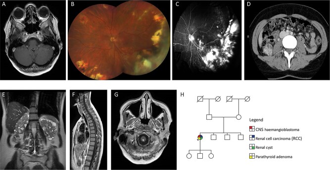
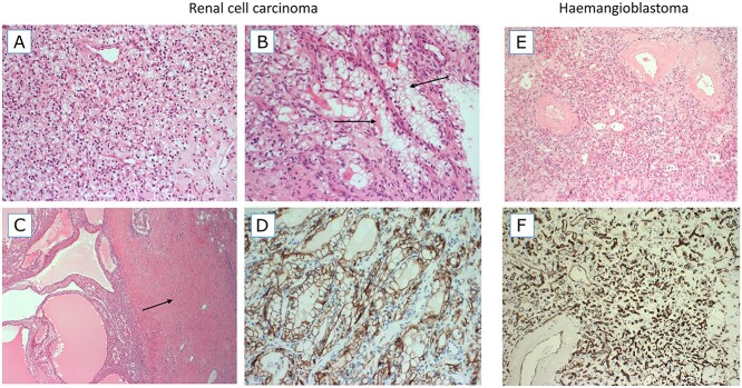
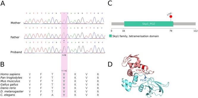
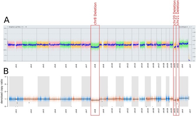
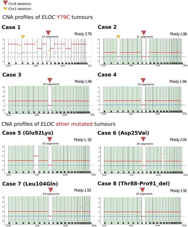
Around 95% of patients with clinical features that meet the diagnostic criteria for von Hippel-Lindau disease (VHL) have a detectable inactivating germline variant in VHL. The VHL protein (pVHL) functions as part of the E3 ubiquitin ligase complex comprising pVHL, elongin C, elongin B, cullin 2 and ring box 1 (VCB-CR complex), which plays a key role in oxygen sensing and degradation of hypoxia-inducible factors. To date, only variants in VHL have been shown to cause VHL disease. We undertook trio analysis by whole-exome sequencing in a proband with VHL disease but without a detectable VHL mutation. Molecular studies were also performed on paired DNA extracted from the proband's kidney tumour and blood and bioinformatics analysis of sporadic renal cell carcinoma (RCC) dataset was undertaken. A de novo pathogenic variant in ELOC NM_005648.4(ELOC):c.236A>G (p.Tyr79Cys) gene was identified in the proband. ELOC encodes elongin C, a key component [C] of the VCB-CR complex. The p.Tyr79Cys substitution is a mutational hotspot in sporadic VHL-competent RCC and has previously been shown to mimic the effects of pVHL deficiency on hypoxic signalling. Analysis of an RCC from the proband showed similar findings to that in somatically ELOC-mutated RCC (expression of hypoxia-responsive proteins, no somatic VHL variants and chromosome 8 loss). These findings are consistent with pathogenic ELOC variants being a novel cause for VHL disease and suggest that genetic testing for ELOC variants should be performed in individuals with suspected VHL disease with no detectable VHL variant.


Figures

Figure 1
Figure 1
(A) Axial T1-weighted post-contrast image through the orbits shows a retinal angioma in the left globe. (B) Colour fundus photograph of left eye at most recent clinic visit showing areas of previous laser and cryotherapy treatment with dragging of optic nerve vessels towards inferotemporal quadrant. Haemangiomas present in macula and nasal quadrant. Multiple peripheral chorioretinal scars related to previously treated haemangiomas. Superotemporal vessels with perivascular exudate. Right eye normal (not shown). Visual acuity Right 6/5 and Left 6/12. (C) Fluorescein angiogram performed 4 years previously showing areas of scarring and retinal detachment related to exudation and effect of treatment. Optic nerve leak related to the effect of traction and glial proliferation with areas of hyperfluorescence in the macula related to new small haemangiomas, visible anterior to the internal limiting membrane on OCT scans (not shown). Right eye normal (not shown). (D) CT scan showing 29 mm diameter RCC in right kidney. (E) Coronal T2-weighted image through the abdomen shows numerous small cysts in both kidneys. (F) Sagittal T1-weighted post-contrast image of the spinal cord shows a solid enhancing haemangioblastoma with associated hypertrophied vessels on the dorsal surface of the spinal cord. (G) Axial T1-weighted post-contrast image through the cervicomedullary junction shows a small solid haemangioblastoma. (H) Family pedigree of sporadic case of VHL.
Figure 2
Figure 2
Haematoxylin and eosin (H&E)-stained images (AC) and CA-IX staining image from the RCC. H&E and CD34-stained images from the haemangioblastoma. (A) An area with typical features of a ccRCC, composed of a sheet of small cells with clear cytoplasm and a delicate background vascular network. (B) Focus on branching tubules in which the tumour cells have more voluminous clear cytoplasm (arrows). (C) A cystic area of the RCC tumour (left) and a dense band of leiomyomatous (muscular) stroma (right, arrow). (D) The tumour was diffusely positive for CA-IX, a classic marker of HIF up-regulation. (E) The haemangioblastoma tumour is composed of very small cells with clear cytoplasm and a background vascular network. Larger blood vessels have thickened hyalinized walls. (F) The vascular network of haemangioblastoma is highlighted by CD34 immunohistochemistry.
Figure 3
Figure 3
(A) Direct (Sanger) sequencing in trio shows the presence of the ELOC c.236A>G (p.Tyr79Cys) variant in the proband and absence from the parents. (B) Evolutionary conservation. Tyrosine at codon 79 (Y79) is evolutionary conserved across vertebrates and invertebrates (22). (C) ELOC domains. The Y79C variant (p.Tyr79Cys) is in the tetramerization domain of the ELOC gene (23). (D) ELOC Y79C-VHL interaction. Tyr79 mediates a hydrogen bond with Pro154 of VHL via a water molecule; adapted from (25). The X-ray crystallographic structure of the ELOC/VHL complex was downloaded from the Protein Data Bank (PDB:4WQO) (26). Molecules other than ELOC and VHL were removed from the structure for clarity.
Figure 4
Figure 4
(A) aCGH (750k array) of germline/tumour pair showed monosomy for chromosomes 8, 21, 22 (in ~20% of cells). (B) Targeted WES of germline/tumour pair identified in terms of copy number profile. This shows broad losses involving the full chromosome 8 and the long arms of chromosomes 21 and 22 (in ~40% of cells, tumour fraction ~47.8%). Any of the known recurrent RCC-related copy number aberrations (i.e. 3p, 9p or 14p losses and 5q or chr7 amplification) were not found.
Figure 5
Figure 5
Copy number analysis profiles for the eight RCC tumours with somatic ELOC variants from the 100,000 Genomes Project using Battenberg caller (subclonal copy number caller) (56). The ELOC c.236A>G (p.Tyr79Cys) missense variant was identified in cases 1–4, cases 5–7 had a non-codon 79 missense ELOC variant [NM_005648.4:c.274G>A (p.Glu92Lys), NM_005648.4:c.311T>A (p.Leu104Gln), NM_005648.4:c.74A>T (p.Asp25Val)] and case 8 harboured anin-frame deletion [NM_005648.4:c.261_272del (p.Thr88_Pro91del)].

References

    1. Gossage, L., Eisen, T. and Maher, E.R. (2015) VHL, the story of a tumour suppressor gene. Nat. Rev. Cancer, 15, 55–64. - PubMed
    1. Maher, E.R., Yates, J.R., Harries, R., Benjamin, C., Harris, R., Moore, A.T. and Ferguson-Smith, M.A. (1990) Clinical features and natural history of von Hippel-Lindau disease. Q. J. Med., 77, 1151–1163. - PubMed
    1. Melmon, K.L. and Rosen, S.W. (1964) Lindau’s disease. Review of the literature and study of a large kindred. Am. J. Med., 36, 595–617. - PubMed
    1. Maher, E.R., Iselius, L., Yates, J.R., Littler, M., Benjamin, C., Harris, R., Sampson, J., Williams, A., Ferguson-Smith, M.A. and Morton, N. (1991) Von Hippel-Lindau disease: a genetic study. J. Med. Genet., 28, 443–447. - PMC - PubMed
    1. Seizinger, B.R., Rouleau, G.A., Ozelius, L.J., Lane, A.H., Farmer, G.E., Lamiell, J.M., Haines, J., Yuen, J.W., Collins, D., Majoor-Krakauer, D.et al. (1988) Von Hippel-Lindau disease maps to the region of chromosome 3 associated with renal cell carcinoma. Nature, 332, 268–269. - PubMed

Publication types

MeSH terms

Substances