

Clinical and laboratory characterization of patients with localized scleroderma and response to UVA-1 phototherapy: In vivo and in vitro skin models
- PMID: 35324032
- PMCID: PMC9790552
- DOI: 10.1111/phpp.12786
Clinical and laboratory characterization of patients with localized scleroderma and response to UVA-1 phototherapy: In vivo and in vitro skin models
Abstract
Background/purpose: Localized scleroderma (LS) is a rare disease leading to progressive hardening and induration of the skin and subcutaneous tissues. LS is responsive to UVA-1 phototherapy, though its exact mechanism of action dermal fibrosis is yet to be fully elucidated. We aimed to investigate the molecular changes induced by UVA-1 rays in human primary fibroblasts cultures.
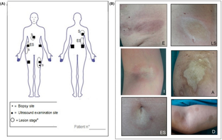
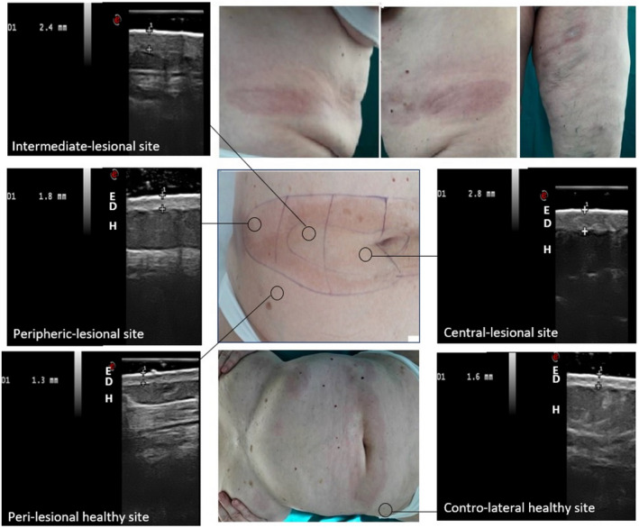
Methods: A total of 16 LS patients were treated with medium-dose UVA-1 phototherapy. At baseline, during and after therapy, Localized Scleroderma Assessment Tool, Dermatology Life Quality Index and lesions' staging and mapping were performed along with high-frequency ultrasound (HFUS) examination for dermal thickness assessment. Gene expression analysis for 23 mRNA transcripts, in vitro UVA-1 irradiation and viability tests were realized on lesional fibroblasts' primary cultures, before and 3 months after therapy.
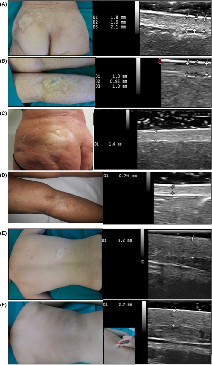
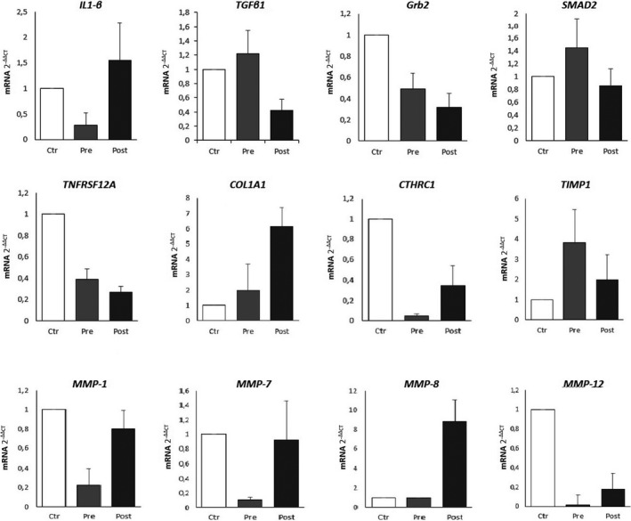
Results: The dermal thickness, the LoSCAT and the DLQI progressively decreased starting from the last phototherapy session up to the 6 and 9 month follow-ups (-57% and -60%, respectively). Molecular gene analysis (rt-PCR) revealed that UVA-1 phototherapy exerts multiple effects: the activation of specific anti-fibrotic pathways (e.g., overexpression of CTHRC1 and metalloproteases 1, 2, 7, 8, 9, 12, suppression of TIMP-1), the downregulation of peculiar pro-fibrotic pathways (e.g., downregulation of TGF-ß, TGF-ßrII, Grb2, SMAD 2/3, TNRSF12A, CTGF) through a significant overexpression of IL-1ß; the stabilization of collagen synthesis acting on genes COL1A1, COL3A1, COL8A1, COL10A1, COL12A1.
Conclusion: UVA-1 phototherapy adds significant benefits in local tissue remodeling, rebalancing the alteration between pro-fibrotic and anti-fibrotic pathways; these changes can be well monitored by HFUS.
Keywords: UVA-1 phototherapy; high-frequency ultrasound; in vitro UVA-1 irradiation; localized scleroderma; primary fibroblast cultures.
© 2022 The Authors. Photodermatology, Photoimmunology & Photomedicine published by John Wiley & Sons Ltd.
Conflict of interest statement
None.
Figures
Similar articles
-
UVA-1 phototherapy as adjuvant treatment for eosinophilic fasciitis: in vitro and in vivo functional characterization.Int J Dermatol. 2022 Jun;61(6):718-726. doi: 10.1111/ijd.16003. Epub 2021 Dec 8. Int J Dermatol. 2022. PMID: 34881449 Free PMC article.
-
Ultraviolet A1 phototherapy decreases inhibitory SMAD7 gene expression in localized scleroderma.Arch Dermatol Res. 2006 Nov;298(6):265-72. doi: 10.1007/s00403-006-0695-8. Epub 2006 Sep 19. Arch Dermatol Res. 2006. PMID: 17009056 Clinical Trial.
-
Successful ultraviolet A1 phototherapy in the treatment of localized scleroderma: a retrospective and prospective study.Br J Dermatol. 2010 Feb 1;162(2):445-7. doi: 10.1111/j.1365-2133.2009.09438.x. Epub 2009 Aug 8. Br J Dermatol. 2010. PMID: 19785603
-
UVA irradiation induced heme oxygenase-1: a novel phototherapy for morphea.Photochem Photobiol. 2015 Jan-Feb;91(1):210-20. doi: 10.1111/php.12342. Epub 2014 Nov 8. Photochem Photobiol. 2015. PMID: 25207998 Review.
-
Phototherapy for scleroderma: biologic rationale, results, and promise.Curr Opin Rheumatol. 2002 Nov;14(6):723-6. doi: 10.1097/00002281-200211000-00016. Curr Opin Rheumatol. 2002. PMID: 12410098 Review.
Cited by
-
Efficacy and Satisfaction of Low Doses UVA1 Phototherapy: A Spanish Experience from a Single Centre.Life (Basel). 2023 Feb 28;13(3):669. doi: 10.3390/life13030669. Life (Basel). 2023. PMID: 36983825 Free PMC article.
References
-
- Asano Y, Fujimoto M, Ishikawa O, et al. Diagnostic criteria, severity classification and guidelines of localized scleroderma. J Dermatol. 2018;45(7):755‐780. - PubMed
-
- Florez‐Pollack S, Kunzler E, Jacobe HT. Morphea: current concepts. Clin Dermatol. 2018;36(4):475‐486. - PubMed
-
- Knobler R, Moinzadeh P, Hunzelmann N, et al. European dermatology forum S1‐guideline on the diagnosis and treatment of sclerosing diseases of the skin, part 1: localized scleroderma, systemic sclerosis and overlap syndromes. J Eur Acad Dermatol Venereol. 2017;31(9):1401‐1424. - PubMed
MeSH terms
Substances
LinkOut - more resources
Full Text Sources
Medical
Research Materials
Miscellaneous
