. 2022 Apr 5;94(13):5335-5343.
doi: 10.1021/acs.analchem.1c05279. Epub 2022 Mar 24.

Enhancing the Throughput of FT Mass Spectrometry Imaging Using Joint Compressed Sensing and Subspace Modeling

Enhancing the Throughput of FT Mass Spectrometry Imaging Using Joint Compressed Sensing and Subspace Modeling

Yuxuan Richard Xie et al. Anal Chem. .

Abstract

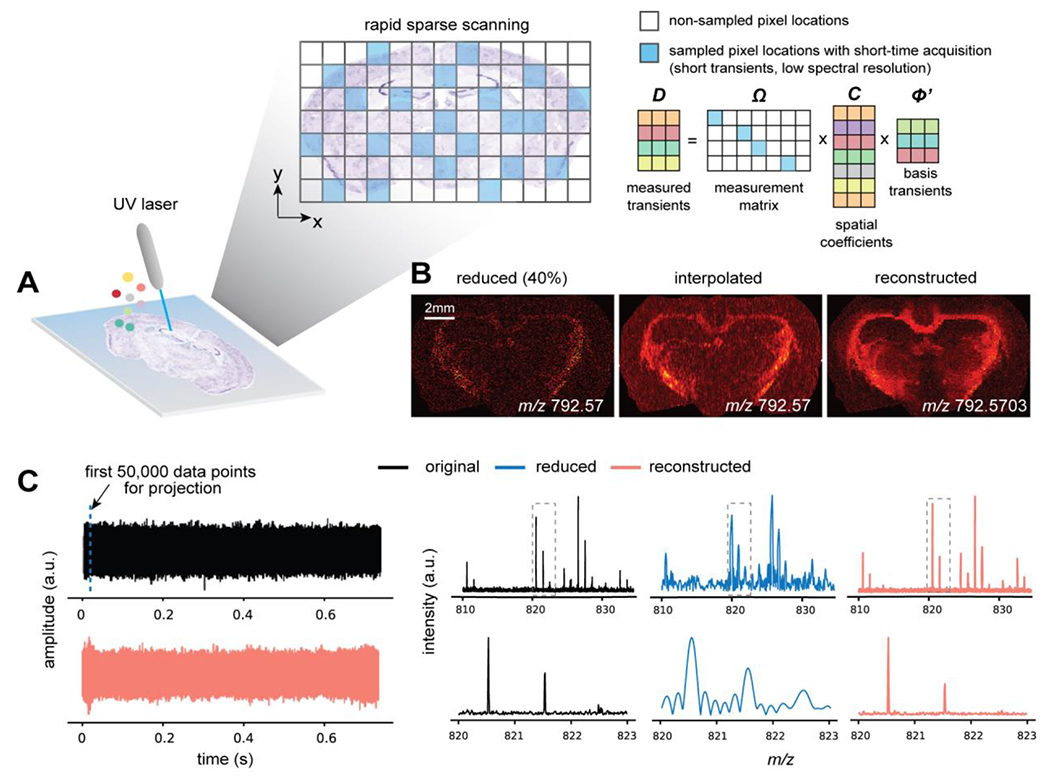
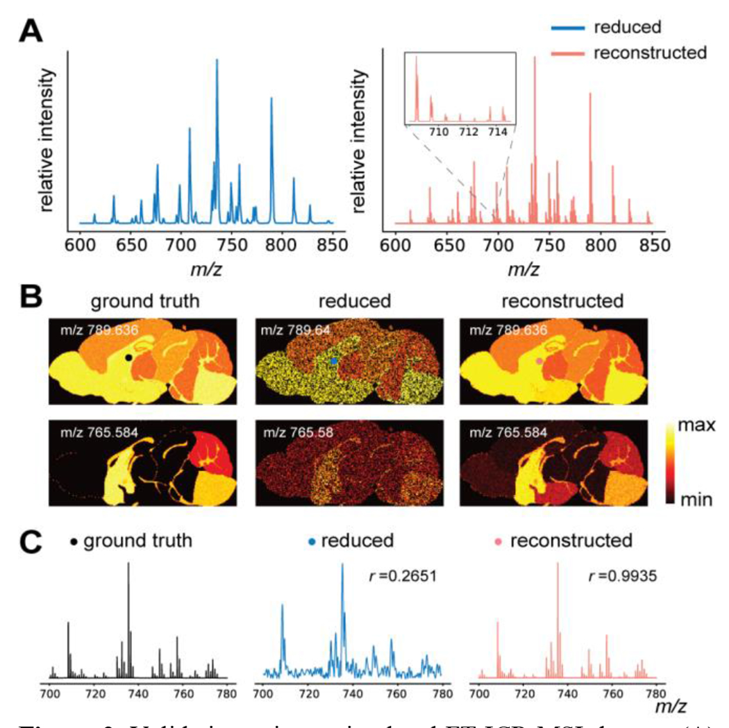
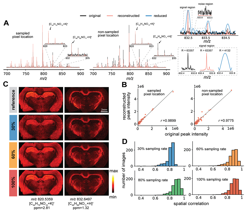
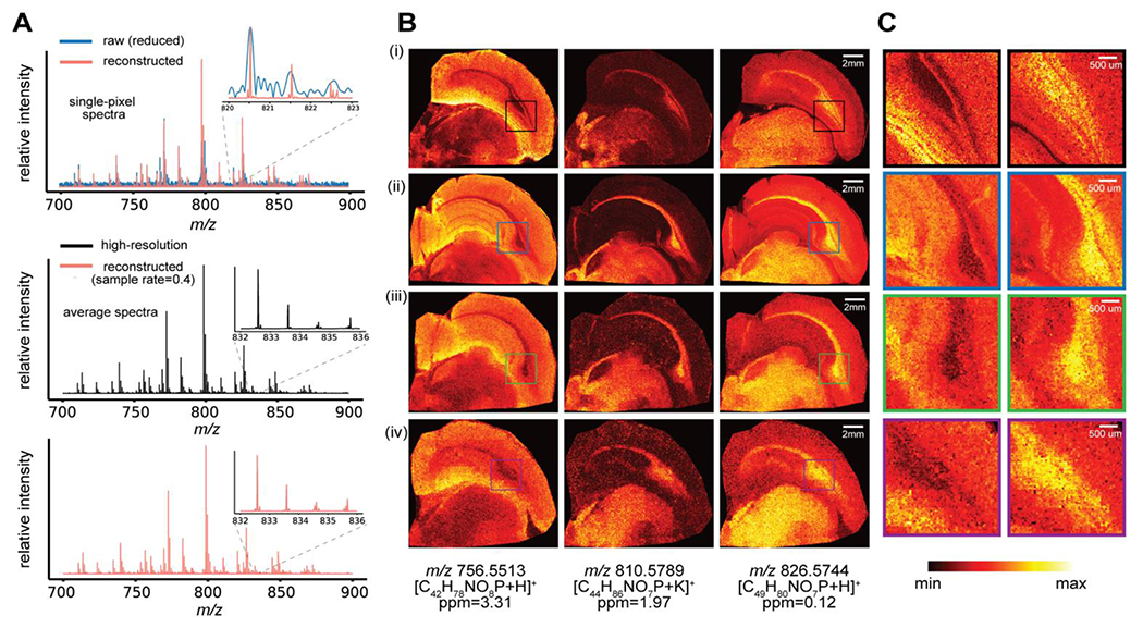
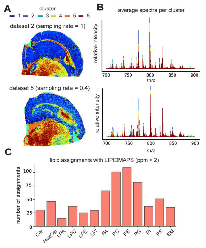
Mass spectrometry imaging (MSI) allows for untargeted mapping of the chemical composition of tissues with attomole detection limits. MSI using Fourier transform (FT)-based mass spectrometers, such as FT-ion cyclotron resonance (FT-ICR), grants the ability to examine the chemical space with unmatched mass resolution and mass accuracy. However, direct imaging of large tissue samples using FT-ICR is slow. In this work, we present an approach that combines the subspace modeling of ICR temporal signals with compressed sensing to accelerate high-resolution FT-ICR MSI. A joint subspace and spatial sparsity constrained model computationally reconstructs high-resolution MSI data from the sparsely sampled transients with reduced duration, allowing a significant reduction in imaging time. Simulation studies and experimental implementation of the proposed method in investigation of brain tissues demonstrate a 10-fold enhancement in throughput of FT-ICR MSI, without the need for instrumental or hardware modifications.


Conflict of interest statement

Conflict of Interest

The authors declare no competing financial interest.

Figures

Figure 1.
Figure 1.
Overview of the proposed approach for enhanced throughput FT-ICR MSI. (A) An illustration of the rapid scanning strategy integrating both the compressed sensing based spatial sparse sampling and the subspace model based short-time acquisition. The data matrix, D, contains collected transients with much fewer temporal points per transient than standard acquisition, at randomly sampled pixel locations (corresponding to different color shadings) defined by the measurement matrix (blue grids are 1 and white grids are 0). With the predetermined basis transient in Φ, the reconstruction of entire high-resolution MSI dataset is defined as estimating the much lower-dimensional spatial coefficients given the sparsely measured data in D. (B) An ion image (left) directly formed from a sparsely sampled dataset (40% pixels) provides limited interpretation about the tissue morphological features and the spatial distributions of molecules (unsampled pixels filled with zeros). The reconstructed ion image (right) effectively recovers tissue features with a much higher mass accuracy for the corresponding ion signal than the simple linear interpolation of the sparse ion image (middle). (C) An original transient (top left) and its reconstructed transient (bottom left) by fitting the first 50,000 data points to the subspace spanned by a set of basis transients. The original and reconstructed mass spectra exhibit highly similar mass spectral profiles and consistent intensity levels, while the mass spectra obtained from a direct Fourier transform of the first 50,000 data points alone with zero-filling have poor spectral quality and strong artifact.
Figure 2.
Figure 2.
Validation using a simulated FT-ICR MSI dataset. (A) Average mass spectra of the reduced noisy data (left, 60% pixels sampled at a 5% transient duration) and the reconstruction (right) from the same reduced data. Spectral features with close m/z values that cannot be resolved by direct Fourier-transform were successfully resolved after the proposed reconstruction (inset). (B) Representative ion images from the ground truth, the reduced noisy data, and the proposed reconstruction for two simulated ions. (C) Individual mass spectra of the ground truth, the reduced noisy data, and the reconstructed data from a selected pixel location (marked in the ion images). Pearson correlation coefficients were calculated to quantitatively evaluate the fidelity of the different methods using a ground truth mass spectrum as the standard for comparison. Spectra shown were not from transients used to estimate the basis.
Figure 3.
Figure 3.
Reconstruction of a high-resolution FT-ICR MSI dataset from a rat brain coronal section. (A) Mass spectra from the reference dataset (all pixels fully sampled with long transients) and reconstruction from the sparsely sampled reduced data (5% transient duration and 30% sampling rate) by the proposed approach exhibit strong consistency for both sampled and non-sampled pixel locations. The insets show the isotopic distributions for two ion signals. The mass spectra obtained from the reconstructed data have a lower noise level than the reference data and achieved significantly improved mass resolution over the standard Fourier transform of the reduced data. Mass spectra were obtained from apodized transients with magnitude mode. (B) High-level similarity between the reference and reconstructed mass spectra is supported by quantitative analysis of peak intensity profiles using the Pearson correlation. (C) Selected ion images are shown at m/z 820.5359 and m/z 832.6497 from the reference dataset (row 1) as well as the reconstructed dataset with 30, 60 and 100% pixels sampled, all with 5% transient duration (rows 2 to 4 respectively). (D) The histograms of the spatial correlation measures of ion images from reference and reconstructed datasets suggest that the proposed method can produce accurate molecular distributions from sparsely sampled data.
Figure 4.
Figure 4.
Representative results from different datasets listed in Table 1 generated by experimental implementation of the proposed sparse sampling strategy and reconstruction using our algorithm. (A) Reduced and reconstructed single-pixel mass spectra (sampled location) from dataset 5 with 40% sampling rate (top), average mass spectra in the range of m/z 700-900 from the fully sampled high-resolution dataset 1 (middle), and the average mass spectra from the reconstruction of dataset 5 (bottom). Insets show the isotopic distribution for a selected ion in a small m/z window. Individual mass spectra were obtained from apodized transients with magnitude mode FT processing. (B) Three representative ion images for m/z 756.5513, 810.5789, and 826.5744 are shown for the the fully sampled high-resolution (HR) dataset 1 (i), short-time acquisition without spatial sparse sampling (dataset 2) (ii), short-time acquisition with 60% pixels sampled (iii), and short-time acquisition with 40% pixels sampled (iv). (C) Zoomed in ion images at m/z 756.5513 and 826.5744 for certain brain regions, which include part of the corpus callosum, display significant recovery of the spatial information and structural details while using a relatively low sampling rate.
Figure 5.
Figure 5.
Downstream data analysis of the reconstructed datasets listed in Table 1 demonstrates the preservation of chemical insights of the tissue by the proposed method with enhanced throughput. (A) Spatial segmentation using the chemical profiles from the reconstructed data through k-means clustering (k=6) provides structurally intuitive cluster assignments, which are consistent between datasets acquired without (top) and with spatially sparse sampling at a 40% sampling rate (bottom). (B) Average mass spectra of 6 clusters for the two datasets, respectively. (C) Putative lipid assignments of the major lipid classes obtained by searching against the LIPIDMAPS with a <2ppm threshold for dataset 5

Similar articles

Cited by

References

    1. Norris JL; Caprioli RM Analysis of Tissue Specimens by Matrix-Assisted Laser Desorption/Ionization Imaging Mass Spectrometry in Biological and Clinical Research. Chem. Rev 2013, 113 (4), 2309–2342. - PMC - PubMed
    1. Rae Buchberger A; DeLaney K; Johnson J; Li L Mass Spectrometry Imaging: A Review of Emerging Advancements and Future Insights. Anal Chem 2018, 90 (1), 240–265. - PMC - PubMed
    1. Wang T; Cheng X; Xu H; Meng Y; Yin Z; Li X; Hang W Perspective on Advances in Laser-Based High-Resolution Mass Spectrometry Imaging. Anal. Chem 2020, 92 (1), 543–553. - PubMed
    1. Scigelova M; Hornshaw M; Giannakopulos A; Makarov A Fourier Transform Mass Spectrometry. Mol Cell Proteomics 2011, 10 (7). - PMC - PubMed
    1. Xian F; Hendrickson CL; Marshall AG High Resolution Mass Spectrometry. Anal. Chem 2012, 84 (2), 708–719. - PubMed

Publication types