. 2022 Apr 6;144(13):5956-5964.
doi: 10.1021/jacs.2c00178. Epub 2022 Mar 24.

Ruthenium Polypyridyl Complex Bound to a Unimolecular Chair-Form G-Quadruplex

Affiliations

Ruthenium Polypyridyl Complex Bound to a Unimolecular Chair-Form G-Quadruplex

Kane T McQuaid et al. J Am Chem Soc. .

Abstract

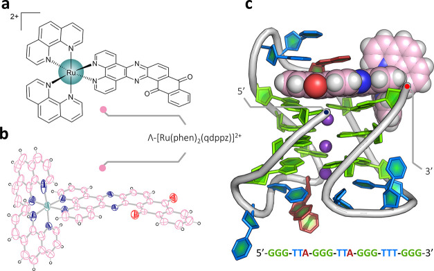
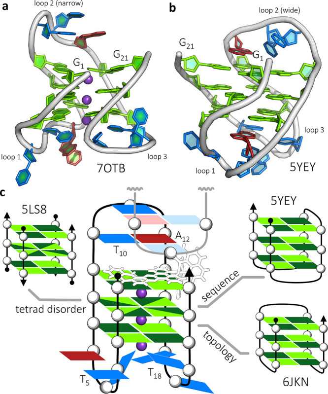
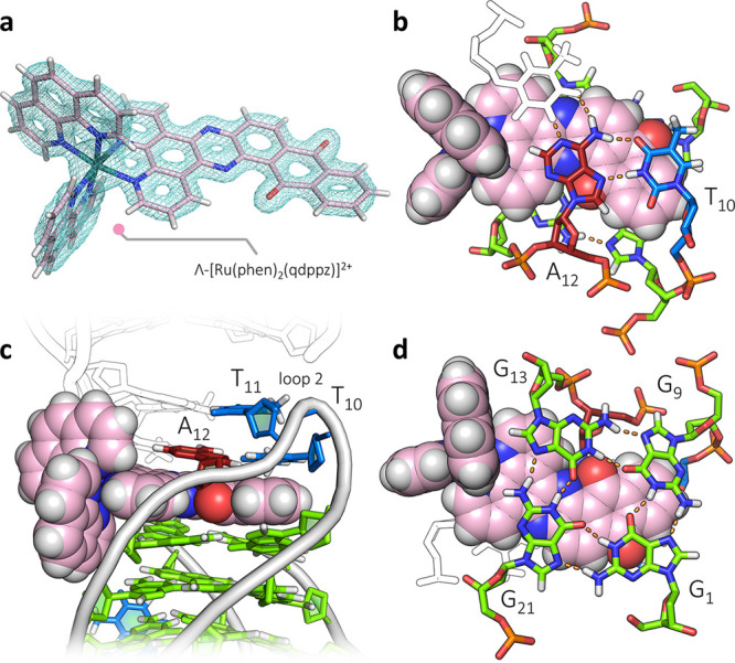
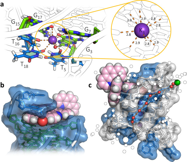
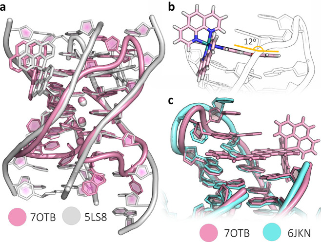
The DNA G-quadruplex is known for forming a range of topologies and for the observed lability of the assembly, consistent with its transient formation in live cells. The stabilization of a particular topology by a small molecule is of great importance for therapeutic applications. Here, we show that the ruthenium complex Λ-[Ru(phen)2(qdppz)]2+ displays enantiospecific G-quadruplex binding. It crystallized in 1:1 stoichiometry with a modified human telomeric G-quadruplex sequence, GGGTTAGGGTTAGGGTTTGGG (htel21T18), in an antiparallel chair topology, the first structurally characterized example of ligand binding to this topology. The lambda complex is bound in an intercalation cavity created by a terminal G-quartet and the central narrow lateral loop formed by T10-T11-A12. The two remaining wide lateral loops are linked through a third K+ ion at the other end of the G-quartet stack, which also coordinates three thymine residues. In a comparative ligand-binding study, we showed, using a Klenow fragment assay, that this complex is the strongest observed inhibitor of replication, both using the native human telomeric sequence and the modified sequence used in this work.


Conflict of interest statement

The authors declare no competing financial interest.

Figures

Figure 1
Figure 1
Crystal structures. (a) Line drawing of Λ-(I), Λ-[Ru(phen)2(qdppz)]2+; (b) thermal ellipsoid plot of one cation from the small-molecule structure of rac-(I); and (c) overall view of the asymmetric unit of the structure reported here with the DNA sequence shown below. A single strand of the sequence d((G3T2A)2G3T3G3) is found assembled with three K+ ions and a single molecule of Λ-(I). The Protein Data Bank (PDB) accession code is 7OTB. The color code for residues throughout are as follows: guanine—green, thymine—blue, and adenine—red. Ruthenium complexes are colored in the following scheme; teal for ruthenium, pink for carbon, and dark blue and white for nitrogen and hydrogen, respectively. The complete numbering scheme for Λ-(I) is shown in Figure S3.
Figure 2
Figure 2
G-quadruplex topologies. (a) Topology of the overall DNA arrangement and three connecting loops of the reported crystal structure (PDB: 7OTB); (b) NMR structure (PDB code: 5YEY) with the same sequence used in this work, which has the opposite strand directionality and loop pattern to that seen here; and (c) schematic of the assembly, with part of the symmetry-related chain that completes it (residues T10, T11, and T12) shown in paler colors. Simplified schematics of antiparallel G-quadruplexes with PDB codes of relevant structural data are shown in the inset, highlighting the structural similarities with related published work. In scheme (c), the syn-guanosine conformations are shown in dark green and the anti conformations in pale green.
Figure 3
Figure 3
Ligand-binding cavity. (a) Observed electron density around the single copy of Λ-(I), contoured at the 1σ (0.29 e Å–3) level. (b) Projection showing the triplex formed by T10, A12 and the symmetry-related T11 (in white). The stacking onto the qdppz chromophore is principally with the T10–A12 Hoogsteen base pair. (c) Side view of the cavity, showing the intercalation of the ligand between the triplex and the quartet, and pointing into the narrow groove. The symmetry-related assembly is in white, with the second ligand omitted for clarity and (d) projection showing the reverse ligand surface and the wide/narrow groove pattern. The qdppz chromophore spans the full width of the G-quartet between the wide grooves.
Figure 4
Figure 4
Ligand environments. (a) Environment of K3, the K+ ion linking the wide loops 1 and 3, coordinating T5, T16, and T18, with the eighth coordination position made up by a water molecule, with the inset showing the consistency of the K–O distances (in Å); (b) binding cavity of Λ-(I) highlighting the van der Waals surface of the nucleic acid partially enveloping the complex; and (c) surface of the narrow loop and the narrow groove with the spine of water molecules linking the hydrated Ba2+ (green sphere) with the hydrated K+ ion K4. The environment of the Ba2+ ion is shown in Figure S7.
Figure 5
Figure 5
Structural comparisons. (a) Superposition of the structure presented here (pink) with our previously determined structure of [Ru(phen)2(11-CN-dppz)]2+ bound in 1:1 stoichiometry with the d(TAG3T2A) octamer (white), with PDB code 5LS8; (b) diagram highlighting the induced fit of the nonplanar qdppz ligand to the similarly nonplanar terminal G-tetrad; and (c) superposition of the structure presented here with the brominated htel(8,20-BrG) crystal structure, PDB code 6JKN. The G-quartets show excellent alignment, whereas the second loop shows the realignments to allow intercalation by the qdppz chromophore.
Figure 6
Figure 6
Replication assays. Representative time-dependent denaturing PAGE analyses following the replication of the noncanonical structures formed by (a) (T2AG3)4 or (b) (G3T2A)2G3T3G3 template strands with and without the presence of the enantiomerically pure ligands. Replication experiments were conducted with 10 mM KCl at pH 6.5 (see Supporting Information section S1.5 for complete experimental details). (c) Kinetic analysis of the time-course of each experiment showing a comparison of each ligand by its ability to stall the replication of the given template sequence. The error bars shown are calculated as the s.d. of triplicate experiments (see Figures S10 and S11 for triplicates and SYBR-stained gels). Calculated rate constants for all the systems investigated can be found in Table S5.
Figure 7
Figure 7
NMR structural comparisons. Structures obtained via NMR studies of ligand binding to G-quadruplexes that exhibit comparative structural features or ligand–DNA interactions. (a) Epiberberine bound to the hybrid-2 form of htelo26, highlighting the pseudo-intercalation between a terminal G-tetrad and the nucleobases in the second loop. (b) ΛΛ-[{Ru(bpy)2}2(tpphz)]2+ bound via a loop-threading mode to the antiparallel form of htelo22. The complex binds with diastereoselectivity and exhibits complex–nucleobase interactions that are enantiospecific to the lambda isomer (inset), similar to what is observed in the reported structure. (c) Au-oxo6 bound to the hybrid-2 form of htelo26, (TTAGGG)4TT. This structure shows the gold complex neatly end-capping a terminal G-tetrad, but also interacting via π-stacking with an adenine in the flanking region.

References

    1. Neidle S. Beyond the Double Helix: DNA Structural Diversity and the PDB. J. Biol. Chem. 2021, 296, 100553.10.1016/j.jbc.2021.100553. - DOI - PMC - PubMed
    1. Phan A. T.; Kuryavyi V.; Patel D. J. DNA Architecture: From G to Z. Curr. Opin. Struct. Biol. 2006, 16, 288–298. 10.1016/J.SBI.2006.05.011. - DOI - PMC - PubMed
    1. Burge S.; Parkinson G. N.; Hazel P.; Todd A. K.; Neidle S. Quadruplex DNA: Sequence, Topology and Structure. Nucleic Acids Res. 2006, 34, 5402–5415. 10.1093/nar/gkl655. - DOI - PMC - PubMed
    1. Phan A. T. Human Telomeric G-Quadruplex: Structures of DNA and RNA Sequences. FEBS J. 2010, 277, 1107–1117. 10.1111/j.1742-4658.2009.07464.x. - DOI - PubMed
    1. Patel D. J.; Phan A. T.; Kuryavyi V. Human telomere, oncogenic promoter and 5’-UTR G-quadruplexes: diverse higher order DNA and RNA targets for cancer therapeutics. Nucleic Acids Res. 2007, 35, 7429–7455. 10.1093/nar/gkm711. - DOI - PMC - PubMed

Publication types