

Magnetic Nanoparticles as a Component of Peptide-Based DNA Delivery System for Suicide Gene Therapy of Uterine Leiomyoma
- PMID: 35324801
- PMCID: PMC8945779
- DOI: 10.3390/bioengineering9030112
Magnetic Nanoparticles as a Component of Peptide-Based DNA Delivery System for Suicide Gene Therapy of Uterine Leiomyoma
Abstract
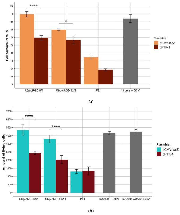
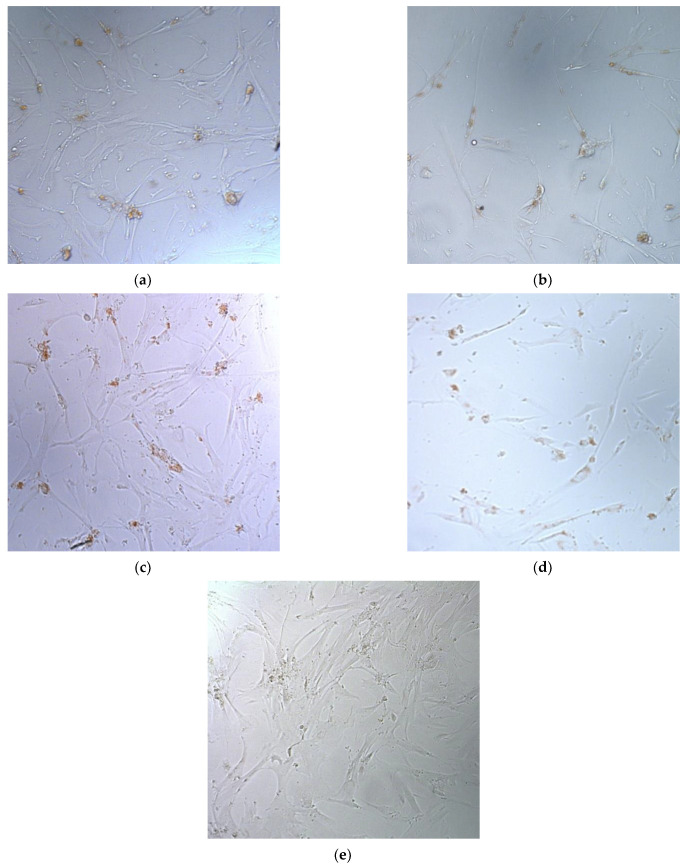
Suicidegene therapy is considered a promising approach for the treatment of uterine leiomyoma (UL), a benign tumor in women characterized by precise localization. In this study, we investigate the efficiency of αvβ3 integrin-targeted arginine-rich peptide carrier R6p-cRGD electrostatically bound to magnetic nanoparticles (MNPs) for targeted DNA delivery into the UL cells. The physico-chemical and cytotoxic properties, transfection efficiency, and specificity of R6p-cRGD/DNA/MNPs polyplexes were evaluated. The addition of MNPs resulted in a decrease in the time needed for successful transfection with simultaneous increase in efficiency. We revealed a therapeutic effect on primary UL cells after delivery of plasmid encoding the herpes simplex virus type 1 (HSV-1) thymidine kinase gene. Treatment with ganciclovir resulted in 20% efficiency of suicide gene therapy in UL cells transfected with the pPTK-1 plasmid. Based on these results, we conclude that the use of cationic peptide carriers with MNPs can be promising for the development of modular non-viral carriers for suicide gene delivery to UL cells.
Keywords: DNA delivery; gene therapy; integrins; magnetic nanoparticles; peptide-based carriers; thymidine kinase; uterine leiomyoma.
Conflict of interest statement
The authors declare no conflict of interest.
Figures
Similar articles
-
Serum-Resistant Ternary DNA Polyplexes for Suicide Gene Therapy of Uterine Leiomyoma.Int J Mol Sci. 2023 Dec 19;25(1):34. doi: 10.3390/ijms25010034. Int J Mol Sci. 2023. PMID: 38203202 Free PMC article.
-
Peptide-Based Nanoparticles for αvβ3 Integrin-Targeted DNA Delivery to Cancer and Uterine Leiomyoma Cells.Molecules. 2022 Nov 30;27(23):8363. doi: 10.3390/molecules27238363. Molecules. 2022. PMID: 36500454 Free PMC article.
-
[Cys-Flanked Cationic Peptides For Cell Delivery of the Herpes Simplex Virus Thymidine Kinase Gene for Suicide Gene Therapy of Uterine Leiomyoma].Mol Biol (Mosk). 2020 May-Jun;54(3):497-511. doi: 10.31857/S0026898420030064. Mol Biol (Mosk). 2020. PMID: 32492014 Russian.
-
Development of iRGD-Modified Peptide Carriers for Suicide Gene Therapy of Uterine Leiomyoma.Pharmaceutics. 2021 Feb 2;13(2):202. doi: 10.3390/pharmaceutics13020202. Pharmaceutics. 2021. PMID: 33540912 Free PMC article.
-
Gene therapy and uterine leiomyoma: a review.Hum Reprod Update. 2006 Jul-Aug;12(4):385-400. doi: 10.1093/humupd/dml015. Epub 2006 Apr 7. Hum Reprod Update. 2006. PMID: 16603566 Review.
Cited by
-
Serum-Resistant Ternary DNA Polyplexes for Suicide Gene Therapy of Uterine Leiomyoma.Int J Mol Sci. 2023 Dec 19;25(1):34. doi: 10.3390/ijms25010034. Int J Mol Sci. 2023. PMID: 38203202 Free PMC article.
-
Genetic and biomarker approaches to uterine fibroids: toward precision medicine.Front Glob Womens Health. 2025 Apr 22;6:1581823. doi: 10.3389/fgwh.2025.1581823. eCollection 2025. Front Glob Womens Health. 2025. PMID: 40330123 Free PMC article. Review.
-
Functionalized Peptide-Based Nanoparticles for Targeted Cancer Nanotherapeutics: A State-of-the-Art Review.ACS Omega. 2022 Oct 5;7(41):36092-36107. doi: 10.1021/acsomega.2c03974. eCollection 2022 Oct 18. ACS Omega. 2022. PMID: 36278104 Free PMC article. Review.
-
Rethinking the application of nanoparticles in women's reproductive health and assisted reproduction.Nanomedicine (Lond). 2024;19(14):1231-1251. doi: 10.2217/nnm-2023-0346. Epub 2024 May 21. Nanomedicine (Lond). 2024. PMID: 38686941 Free PMC article. Review.
-
Peptide-Based Nanoparticles for αvβ3 Integrin-Targeted DNA Delivery to Cancer and Uterine Leiomyoma Cells.Molecules. 2022 Nov 30;27(23):8363. doi: 10.3390/molecules27238363. Molecules. 2022. PMID: 36500454 Free PMC article.
References
-
- Packenham J.P., Du Manoir S., Schrock E., Risinger J.I., Dixon D., Denz D.N., Evans J.A.C., Berchuck A., Barrett J.C., Devereux T.R., et al. Analysis of Genetic Alterations in Uterine Leiomyomas and Leiomyosarcomas by Comparative Genomic Hybridization. Mol. Carcinog. 1997;19:273–279. doi: 10.1002/(SICI)1098-2744(199708)19:4<273::AID-MC9>3.0.CO;2-D. - DOI - PubMed
Grants and funding
LinkOut - more resources
Full Text Sources
