. 2022 Feb 24;9(3):99.
doi: 10.3390/vetsci9030099.

Molecular Mechanisms of Lipopolysaccharide (LPS) Induced Inflammation in an Immortalized Ovine Luteal Endothelial Cell Line (OLENDO)

Affiliations

Molecular Mechanisms of Lipopolysaccharide (LPS) Induced Inflammation in an Immortalized Ovine Luteal Endothelial Cell Line (OLENDO)

Aykut Gram et al. Vet Sci. .

Abstract

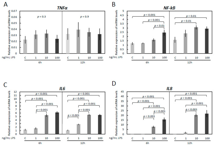
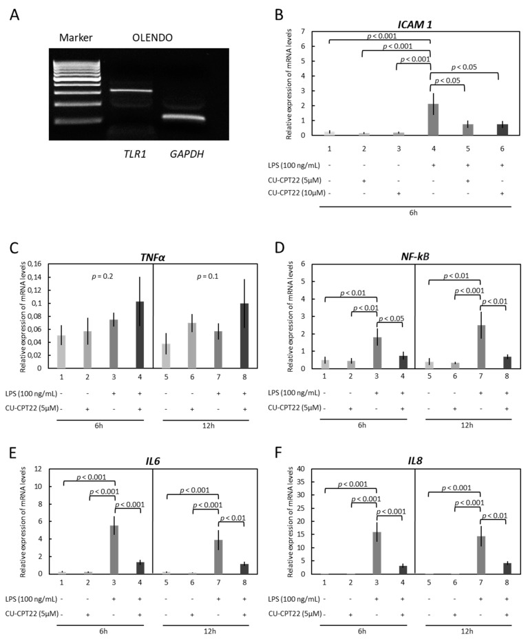
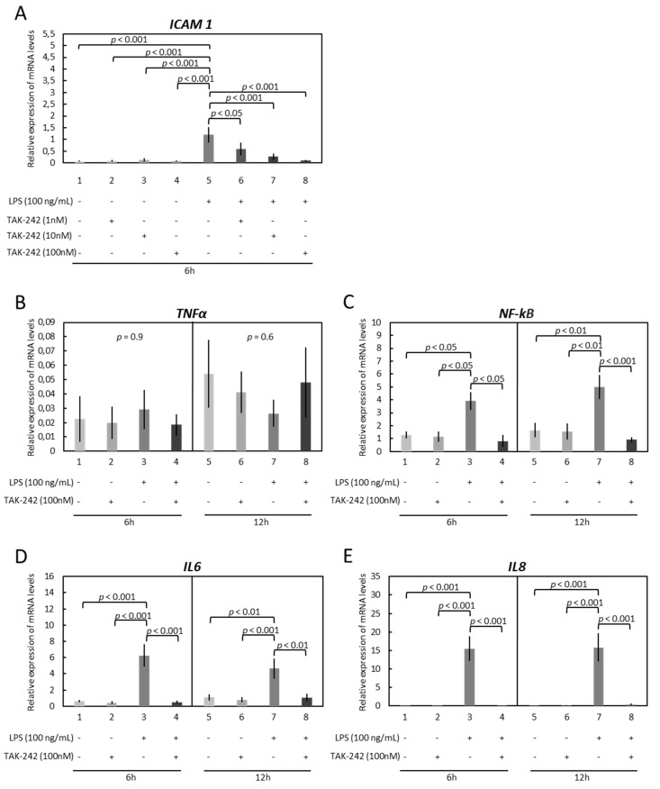
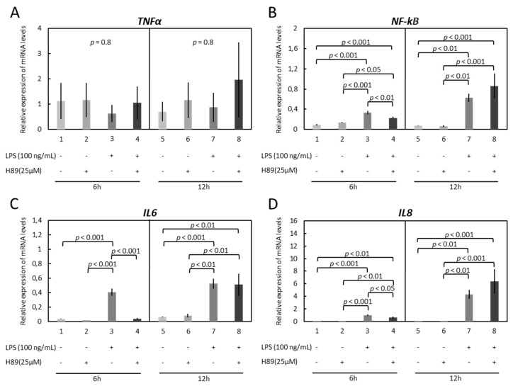
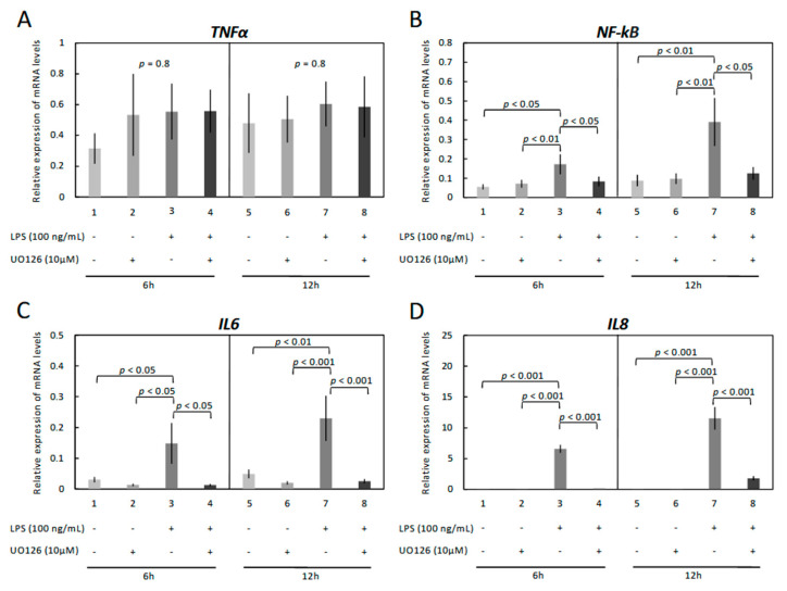
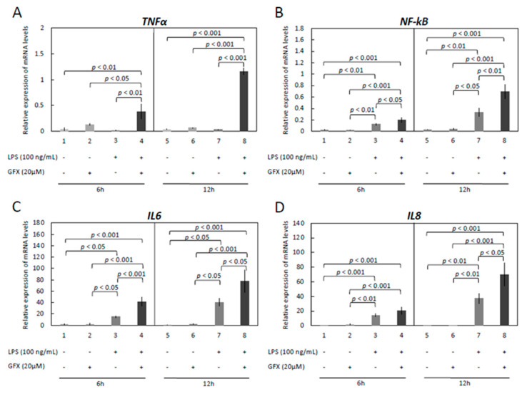
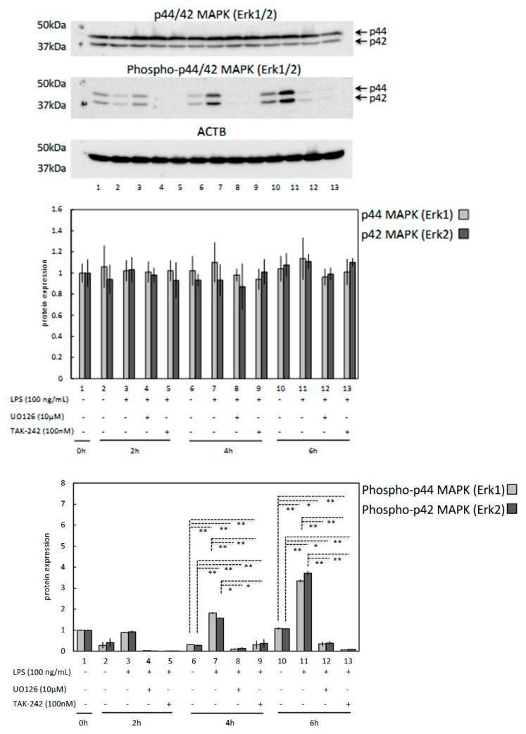
Escherichia coli (E. coli) is the most common Gram-negative bacterium causing infection of the uterus or mammary gland and is one of the major causes of infertility in livestock. In those animals affected by E. coli driven LPS-mediated infections, fertility problems occur in part due to disrupted follicular and luteal functionality. However, the molecular mechanisms by which LPS induces inflammation, and specifically, the role of LPS in the disruption of capillary morphogenesis and endothelial barrier function remain unclear. Here, we hypothesized that LPS may lead to alterations in luteal angiogenesis and vascular function by inducing inflammatory reactions in endothelial cells. Accordingly, OLENDO cells were treated with LPS followed by evaluation of the expression of selected representative proinflammatory cytokines: NF-kB, IL6, IL8, TNFα, and ICAM 1. While TNFα was not affected by treatment with LPS, transcripts of NF-kB, IL6, and IL8 were affected in a dosage-dependent manner. Additionally, the activity of TLR2 and TLR4 was blocked, resulting in suppression of the LPS-induced expression of ICAM 1, NF-kB, IL6, and IL8. Inhibition of the PKA or MAPK/ERK pathways suppressed the LPS-stimulated expression of NF-kB, IL6, and IL8, whereas blocking the PKC pathway had the opposite effect. Furthermore, LPS-induced phosphorylation of Erk1 and Erk2 was inhibited when the TLR4 or MAPK/ERK pathways were blocked. Finally, LPS seems to induce inflammatory processes in OLENDO cells via TLR2 and TLR4, utilizing different signaling pathways.

Keywords: MAPKs; PKA; PKC; inflammation; interleukins; ovary; toll-like receptor.


Conflict of interest statement

The authors declare no conflict of interest.

Figures

Figure 1
Figure 1
Expression of TNFα (A), NF-kB (B), IL6 (C) and IL8 (D) by real-time (TaqMan) PCR after treatment with 0–100 ng/mL LPS for 6 and 12 h. A parametric one-way ANOVA was applied followed by the Tukey–Kramer multiple comparisons post-test. All numerical data are presented as the means ± S.D. p-values < 0.05 were considered significant and are indicated.
Figure 2
Figure 2
The expression of TLR1 and effects of LPS treatment and functional blocking of TLR1/TLR2 on expression of ICAM 1, TNFα, NF-kB, IL6, and IL8 in immortalized OLENDO cells. (A) The expression of TLR1 in OLENDO cells (317 bp) by qualitative PCR (M = marker, GAPDH as internal control). Expression of ICAM 1 (B), TNFα (C), NF-kB (D), IL6 (E), and IL8 (F) as determined by real-time (TaqMan) PCR. A selective blocker of TLR1/TLR2 (CU-CPT22) was used as described in the Materials and Methods. A parametric one-way ANOVA was applied followed by the Tukey–Kramer multiple comparisons post-test. All numerical data are presented as the means ± S.D. p-values < 0.05 were considered significant and are indicated. The original western blotting figures in Figure 2A can be found in Figure S1.
Figure 3
Figure 3
Effects of LPS treatment and functional blocking of TLR4 on expression of ICAM 1, TNFα, NF-kB, IL6, and IL8 in immortalized OLENDO cells. Expression of ICAM 1 (A), TNFα (B), NF-kB (C), IL6 (D), and IL8 (E) as determined by real-time (TaqMan) PCR. A selective blocker of TLR4 (TAK-242) was used as described in the Materials and Methods. A parametric one-way ANOVA was applied followed by the Tukey–Kramer multiple comparisons post-test. All numerical data are presented as the means ± S.D. p-values < 0.05 were considered significant and are indicated.
Figure 4
Figure 4
Effects of LPS treatment and inhibition of PKA (H89) activity on expression of TNFα, NF-kB, IL6 and IL8 in immortalized OLENDO cells. Expression of TNFα (A), NF-kB (B), IL6 (C), and IL8 (D) as determined by real-time (TaqMan) PCR. Inhibition of PKA (H89) activity was performed as described in the Materials and Methods. A parametric one-way ANOVA was applied followed by the Tukey–Kramer multiple comparisons post-test. All numerical data are presented as the means ± S.D. p-values < 0.05 were considered significant and are indicated.
Figure 5
Figure 5
Effects of LPS treatment and inhibition of MAPK (UO126) activity on expression of TNFα, NF-kB, IL6 and IL8 in immortalized OLENDO cells. Expression of TNFα (A), NF-kB (B), IL6 (C), and IL8 (D) as determined by real-time (TaqMan) PCR. Inhibition of MAPK (UO126) activity was performed as described in the Materials and Methods. A parametric one-way ANOVA was applied followed by the Tukey–Kramer multiple comparisons post-test. All numerical data are presented as the means ± S.D. p-values < 0.05 were considered significant and are indicated.
Figure 6
Figure 6
Effects of LPS treatment and inhibition of PKC (GF-109203X) activity on expression of TNFα, NF-kB, IL6 and IL8 in immortalized OLENDO cells. Expression of TNFα (A), NF-kB (B), IL6 (C), and IL8 (D) as determined by real-time (TaqMan) PCR. Inhibition of PKC (GF-109203X; GFX) activity was performed as described in the Materials and Methods. A parametric one-way ANOVA was applied followed by the Tukey–Kramer multiple comparisons post-test. All numerical data are presented as the means ± S.D. p-values < 0.05 were considered significant and are indicated.
Figure 7
Figure 7
Effects of LPS treatment and MAPK (UO126, 10 μM) and TLR4 (TAK-242, 100 nM) inhibitors on protein expression of p44/42 MAPK (Erk1/2) and phosphorylated p44/42 MAPK (Erk1/2) proteins for 0–6 h. ACTB expression (45 kDa) was used as a loading control. Representative immunoblots are shown. Lower panels represent densitometric values (standardized optical density; SOD) for Erk1 (p44), Erk2 (p42) and pErk1 (p44), pErk2 (p42) expression normalized against ACTB. A parametric one-way ANOVA was applied followed by the Tukey–Kramer multiple comparisons post-test. All numerical data are presented as the means ± S.D. p-values < 0.05 were considered significant: * indicates p < 0.01, ** indicates p < 0.001. The original western blotting figures in Figure 7 can be found in Figure S2.

Similar articles

Cited by

References

    1. Sheldon I.M., Rycroft A.N., Dogan B., Craven M., Bromfield J.J., Chandler A., Roberts M.H., Price S.B., Gilbert R.O., Simpson K.W. Specific strains of Escherichia coli are pathogenic for the endometrium of cattle and cause pelvic inflammatory disease in cattle and mice. PLoS ONE. 2010;5:e9192. doi: 10.1371/journal.pone.0009192. - DOI - PMC - PubMed
    1. Hertl J.A., Grohn Y.T., Leach J.D., Bar D., Bennett G.J., Gonzalez R.N., Rauch B.J., Welcome F.L., Tauer L.W., Schukken Y.H. Effects of clinical mastitis caused by gram-positive and gram-negative bacteria and other organisms on the probability of conception in New York State Holstein dairy cows. J. Dairy Sci. 2010;93:1551–1560. doi: 10.3168/jds.2009-2599. - DOI - PubMed
    1. Lavon Y., Leitner G., Klipper E., Moallem U., Meidan R., Wolfenson D. Subclinical, chronic intramammary infection lowers steroid concentrations and gene expression in bovine preovulatory follicles. Domest. Anim. Endocrinol. 2011;40:98–109. doi: 10.1016/j.domaniend.2010.09.004. - DOI - PubMed
    1. Herath S., Williams E.J., Lilly S.T., Gilbert R.O., Dobson H., Bryant C.E., Sheldon I.M. Ovarian follicular cells have innate immune capabilities that modulate their endocrine function. Reproduction. 2007;134:683–693. doi: 10.1530/REP-07-0229. - DOI - PMC - PubMed
    1. Sheldon I.M., Noakes D.E., Rycroft A.N., Pfeiffer D.U., Dobson H. Influence of uterine bacterial contamination after parturition on ovarian dominant follicle selection and follicle growth and function in cattle. Reproduction. 2002;123:837–845. doi: 10.1530/rep.0.1230837. - DOI - PubMed

LinkOut - more resources