. 2022 Feb 24;9(3):100.
doi: 10.3390/vetsci9030100.

Molecular and Pathological Detection of Hepatitis E Virus in Roe Deer (Capreolus capreolus) and Fallow Deer (Dama dama) in Central Italy

Affiliations

Molecular and Pathological Detection of Hepatitis E Virus in Roe Deer (Capreolus capreolus) and Fallow Deer (Dama dama) in Central Italy

Niccolò Fonti et al. Vet Sci. .

Abstract

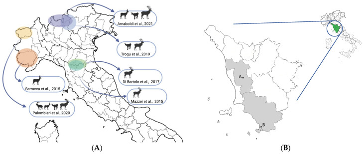
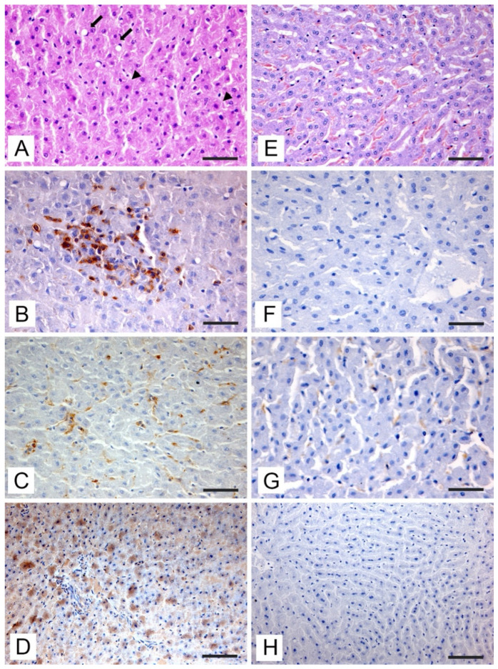
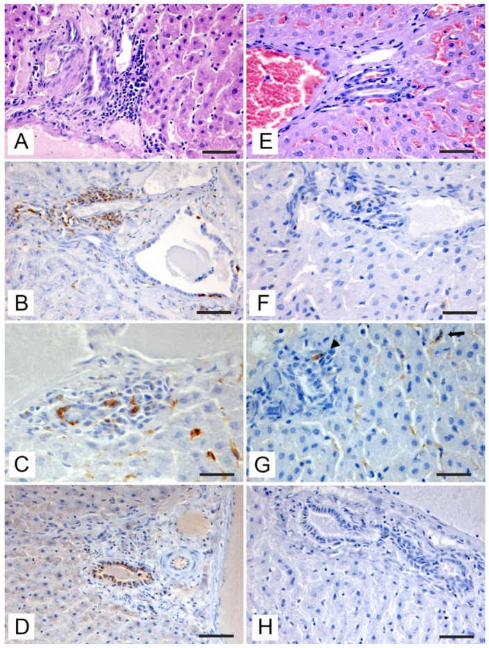
Hepatitis E virus (HEV) is a common causative agent of acute hepatitis in the world, with a serious public health burden in both developing and industrialized countries. Cervids, along with wild boars and lagomorphs, are the main wild hosts of HEV in Europe and constitute a documented source of infection for humans. The aim of this study was to evaluate the presence of HEV in roe deer (Capreolus capreolus) and fallow deer (Dama dama) living in Tuscany, Central Italy. Liver samples from 48 roe deer and 60 fallow deer were collected from carcasses during the hunting seasons. Following the results obtained from molecular and histopathologic studies, 5/48 (10.4%) roe deer and 1/60 (1.7%) fallow deer liver samples were positive for the presence of HEV RNA. All PCR-positive livers were also IHC-positive for viral antigen presence, associated with degenerative and inflammatory lesions with predominantly CD3+ cellular infiltrates. This study represents the first identification in Italy of HEV RNA in roe and fallow deer and the first study in literature describing liver alterations associated with HEV infection in cervids. These results demonstrate that HEV is present in wild cervid populations in Italy and confirm the potential zoonotic role of these species.

Keywords: ELISA; HEV; PCR; deer; immunohistochemistry; inflammatory infiltrates; liver pathology; wildlife; zoonosis.


Conflict of interest statement

The authors declare no conflict of interest.

Figures

Figure 1
Figure 1
(A) Map of Northern and Central Italy with the geographical areas in which previous studies were conducted. No studies are reported in deer populations from Southern Italy. The references and the species included in each study are provided [22,24,25,26,27,28]. Created with BioRender.com (accessed on 10 February 2022); (B) Map of Tuscany with the two sampled areas in this study (Pisa and Grosseto provinces). A = Lajatico (PI), B = Orbetello (GR).
Figure 2
Figure 2
Liver tissue sections (pericentrilobular areas) from PCR-positive roe deer (AD) and PCR-negative subject (EH). (A) Focal area of degeneration with Ito cell hyperplasia (arrow) and single hepatocytes with nucleus loss and cytoplasmic hypereosinophilia (arrowheads) (H,E, bar = 50 μm). (B) CD3+ inflammatory infiltrate associated with degenerating hepatocytes (IHC, bar = 50 μm). (C) Hyperplasia of Kupffer cells labeled with anti-Iba-1 (IHC, bar = 50 μm). (D) Immunohistochemical staining with cytoplasmic pattern of hepatocytes incubated with anti-HEV antibody (IHC, bar = 100 μm). (E) Normal hepatocytes in the pericentrilobular area (H,E, bar = 50 μm). (F) Absence of CD3+ lymphocytic infiltrate in hepatic sinusoids (IHC, bar = 50 μm). (G) Scattered Kupffer cells labeled with anti-Iba-1 antibody (IHC, bar = 50 μm). (H) Absence of immunostaining after incubation with anti-HEV antibody (IHC, bar = 100 μm).
Figure 3
Figure 3
Liver tissue sections from PCR investigated samples. (A) HEV-positive roe deer; periductal lymphocytic infiltration in the portal space (H,E, bar = 50 μm). (B) HEV-positive roe deer; CD3+ periductal inflammatory infiltrate with small clusters of cells in contact with the basal dominium of positive cholangiocytes (IHC, bar = 100 μm). (C) HEV-positive roe deer; cluster of macrophages infiltrating the portal space stained with anti-Iba-1 antibody (IHC, bar = 50 μm). (D) HEV-positive roe deer; immunohistochemical staining with granular cytoplasmic pattern in cholangiocytes incubated with anti-HEV antibody (IHC, bar = 100 μm). (E) Roe deer HEV-negative; absence of periductal inflammatory infiltrate (H,E, bar = 50 μm). (F) HEV-negative roe deer; little amount of CD3+ periductal lymphocytes (IHC, bar = 50 μm). (G) HEV-negative roe deer; resident Kupffer cells labeled with anti-Iba-1 antibody (arrow) and a single macrophage cell, also labeled (arrowhead) (IHC, bar = 50 μm). (H) HEV-negative roe deer; cholangiocytes with no staining after incubation with anti-HEV antibody (IHC, bar = 100 μm).

Similar articles

Cited by

References

    1. Nimgaonkar I., Ding Q., Schwartz R.E., Ploss A. Hepatitis e virus: Advances and challenges. Nat. Rev. Gastroenterol. Hepatol. 2018;15:96–110. doi: 10.1038/nrgastro.2017.150. - DOI - PMC - PubMed
    1. Purdy M.A., Harrison T.J., Jameel S., Meng X.J., Okamoto H., Van Der Poel W.H.M., Smith D.B. ICTV virus taxonomy profile: Hepeviridae. J. Gen. Vir. 2017;98:2645–2646. doi: 10.1099/jgv.0.000940. - DOI - PMC - PubMed
    1. Lhomme S., Marion O., Abravanel F., Izopet J., Kamar N. Clinical Manifestations, Pathogenesis and Treatment of Hepatitis E Virus Infections. J. Clin. Med. 2020;9:331. doi: 10.3390/jcm9020331. - DOI - PMC - PubMed
    1. King N.J., Hewitt J., Perchec-Merien A.M. Hiding in Plain Sight? It’s Time to Investigate Other Possible Transmission Routes for Hepatitis E Virus (HEV) in Developed Countries. Food Environ. Virol. 2018;10:225–252. doi: 10.1007/s12560-018-9342-8. - DOI - PubMed
    1. Nicot F., Dimeglio C., Migueres M., Jeanne N., Latour J., Abravanel F., Ranger N., Harter A., Dubois M., Lameiras S., et al. Classification of the Zoonotic Hepatitis E Virus Genotype 3 Into Distinct Subgenotypes. Front. Microbiol. 2021;11:634430. doi: 10.3389/fmicb.2020.634430. - DOI - PMC - PubMed

LinkOut - more resources