Case Reports
. 2022 Mar 15;9(3):136.
doi: 10.3390/vetsci9030136.

Use of Electrodiagnostics in the Diagnosis and Follow-Up of Brachial Plexus Syndrome in a Calf

Affiliations
Case Reports

Use of Electrodiagnostics in the Diagnosis and Follow-Up of Brachial Plexus Syndrome in a Calf

Marilena Bolcato et al. Vet Sci. .

Abstract

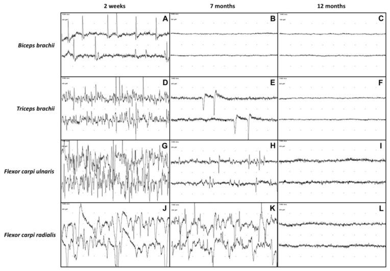
Electrodiagnostic testing by using electromyography (EMG) and nerve conduction studies (NCS) is essential in the evaluation of patients with traumatic brachial plexus injury as it facilitates the localization of the lesion and the prognosis. In this case report, we present a long-term electrodiagnostic follow-up of a 5-day-old female Holstein calf with brachial plexus syndrome. Electrodiagnostic studies were carried out at 2 weeks, 5 weeks, 7 months and 12 months after admission. Initially, EMG confirmed the damage to the brachial plexus, potentially indicating a condition of neurotmesis or axonotmesis. However, motor NCS and the repeated electrodiagnostic follow-up, along with the evolution of the clinical signs, allowed a more favorable diagnosis of axonotmesis to be made. In fact, EMG showed a slow but gradual reduction and finally the disappearance of spontaneous pathological activity, while motor NCS revealed an increase in the amplitude and areas of the compound muscle action potentials. The animal was deemed fully recovered 12 months after admission. To the authors' knowledge, this is the first report on the use of motor NCS in bovine medicine and it demonstrates that electrodiagnostics represent a useful and practical tool for the evaluation and prognosis of brachial plexus injury cases in cattle.

Keywords: bovine; brachial plexus; electromyography; motor nerve conduction study; neurology; traumatic injury.


Conflict of interest statement

The authors declare no conflict of interest.

Figures

Figure 1
Figure 1
Presentation of the calf at admission. Observe the posture of the left thoracic limb: flexed and dropped elbow, flexed carpus, overextended fetlock joint.
Figure 2
Figure 2
Photographs demonstrating the position of electrodes during motor NCS on an anatomical model of bovine right thoracic limb. (A) Lateral view. The stimulating needles for the radial nerve were placed at the angle between the head of the humerus and the ventral portion of the scapula (PS) and at the third distal of the humerus, caudally to the brachialis muscle (DS). The position of ground needle (=) and recording needle (R) is also shown. (B) Medial view. The stimulating needles for the ulnar nerve (PSu, DSu) were placed caudally to the medial humerus epicondyle; the stimulating needles for the median nerve (PSm, DSm) were placed cranially to the medial humerus epicondyle. The position of ground needle (=) and recording needles (R) is also shown. (Courtesy of Domestic Animal Anatomy Collection, Sistema Museale d’Ateneo, Alma Mater Studiorum, University of Bologna).
Figure 3
Figure 3
EMG recording of the left thoracic limb’s muscles (biceps brachii, triceps brachii, flexor carpi ulnaris, flexor carpi radialis) at 2 weeks, 7 months and 12 months after admission. Sweep speed: 10 ms/div. Biceps brachii muscle at 2 weeks (A), 7 months (B) and 12 months (C) after admission. Triceps brachii muscle at 2 weeks (D), 7 months (E) and 12 months (F) after admission. Third row: Flexor carpi ulnaris muscle at 2 weeks (G), 7 months (H) and 12 months (I) after admission. Flexor carpi radialis muscle at 2 weeks (J), 7 months (K) and 12 months (L) after admission. An evident reduction of spontaneous pathologic activity can be observed at 7 months after admission up to normalization of EMG tracings in all the recorded muscles at 12 months after admission.
Figure 4
Figure 4
Motor NCS (CMAPs) of the left thoracic limb’s nerves (radial nerve, ulnar nerve, median nerve) at 5 weeks, 7 months and 12 months after admission (distal stimulation). Sweep speed: 5 ms/div. Sensitivity: 2 mV/div. A progressive increase of the amplitudes and areas of the CMAPs over time can be observed in all the tested nerves. (A) CMAP amplitude 1.8 mV, area 6.3 uVs; (B) CMAP amplitude 7.1 mV, area 23.1 uVs; (C) CMAP amplitude 11.2 mV, area 45.7 uVs; (D) CMAP amplitude 1.5 mV, area 3 uVs; (E) CMAP amplitude 3.9 mV, area 7.5 uVs; (F) CMAP amplitude 25.1 mV, area 54.3 uVs; (G) CMAP amplitude 0.5 mV, area 0.9 uVs; (H) CMAP amplitude 2.5 mV, area 5.3 uVs; (I) CMAP amplitude 3.1 mV, area 8.9 uVs.
Figure 5
Figure 5
Motor NCS (CMAPs) of the right thoracic limb’s nerves (healthy control) at 5 weeks after admission. Sweep speed: 5 ms/div. CMAP amplitudes, areas, and nerve conduction velocity (NCV) for each tested nerve are reported: (A) Radial nerve (proximal CMAP amplitude 23.7 mV, area 73.3 uVs; distal CMAP amplitude 16.3 mV, area 62.4 uVs; NCV 53.6 m/s). Sensitivity: 5 mV/div; (B) Ulnar nerve (proximal CMAP amplitude 5.5 mV, area 9.3 uVs; distal CMAP amplitude 3.2 mV, area 7.8 uVs; NCV 44.4 m/s). Sensitivity: 2 mV/div; (C) Median nerve (proximal CMAP amplitude 2.8 mV, area 6.6 uVs; distal CMAP amplitude 3.9 mV, area 8 uVs; NCV 125 m/s). Sensitivity: 2 mV/div.

Similar articles

References

    1. Lorenz M.D., Coates J.R., Kent M. Handbook of Veterinary Neurology. 5th ed. Elsevier-Saunders; St. Louis, MO, USA: 2011. pp. 94–108.
    1. Barone R., Simoens P. Anatomia Comparata dei Mammiferi Domestici—Vol. 7° Neurologia. 1st ed. Edagricole New Business Media; Milano, Italy: 2012. pp. 133–275.
    1. Greenough P.R., Weaver A.D. Lameness in Cattle. 3rd ed. Saunders; Philadelphia, PA, USA: 1997. pp. 203–218.
    1. Ciszewski D.K., Ames N.K. Diseases of the Peripheral Nerves. Vet. Clin. N. Am. Food Anim. Pract. 1987;3:193–212. doi: 10.1016/S0749-0720(15)31190-7. - DOI - PubMed
    1. De Lahunta A., Glass E. Veterinary Neuroanatomy and Clinical Neurology. 3rd ed. Saunders-Elsevier; St. Louis, MO, USA: 2009. pp. 77–133.

Publication types

LinkOut - more resources