. 2022 Mar 24;17(3):e0265890.
doi: 10.1371/journal.pone.0265890. eCollection 2022.

Adrenomedullin and its receptors are expressed in mouse pancreatic β-cells and suppresses insulin synthesis and secretion

Affiliations

Adrenomedullin and its receptors are expressed in mouse pancreatic β-cells and suppresses insulin synthesis and secretion

Yuanlin Dong et al. PLoS One. .

Abstract

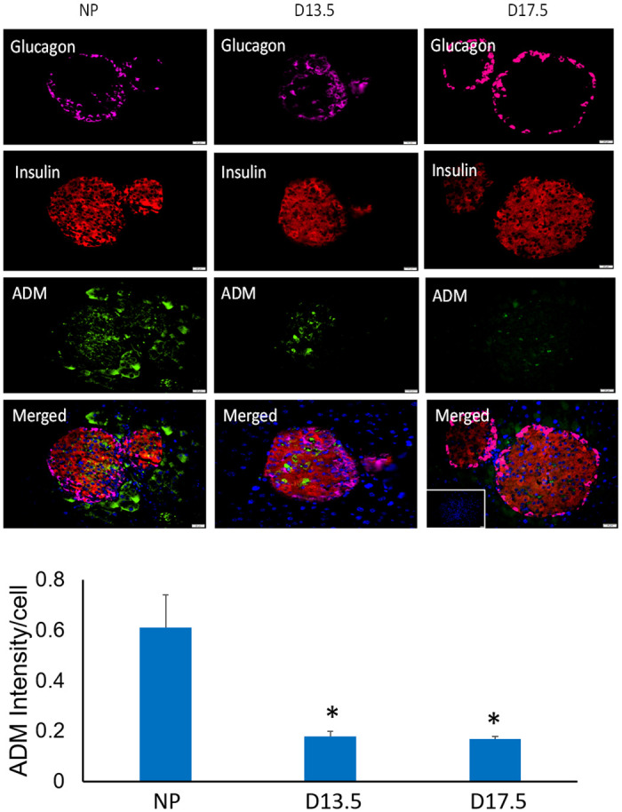
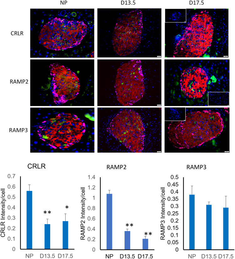
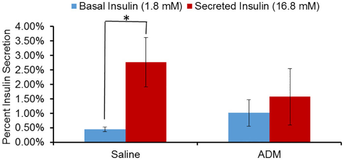
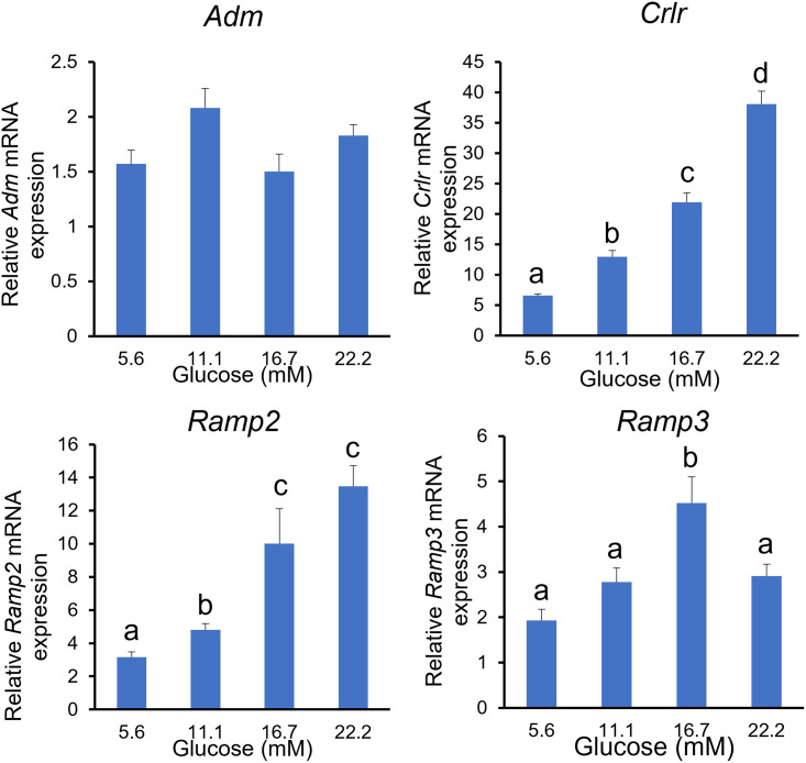
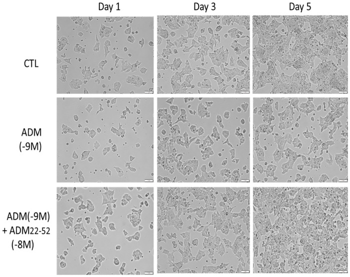
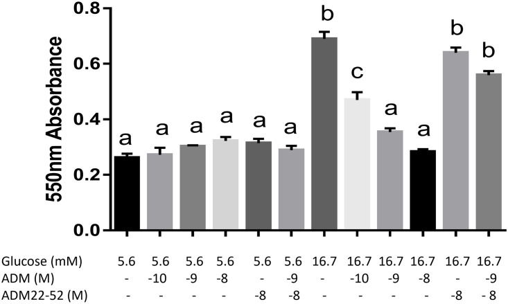
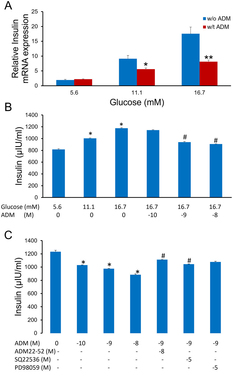
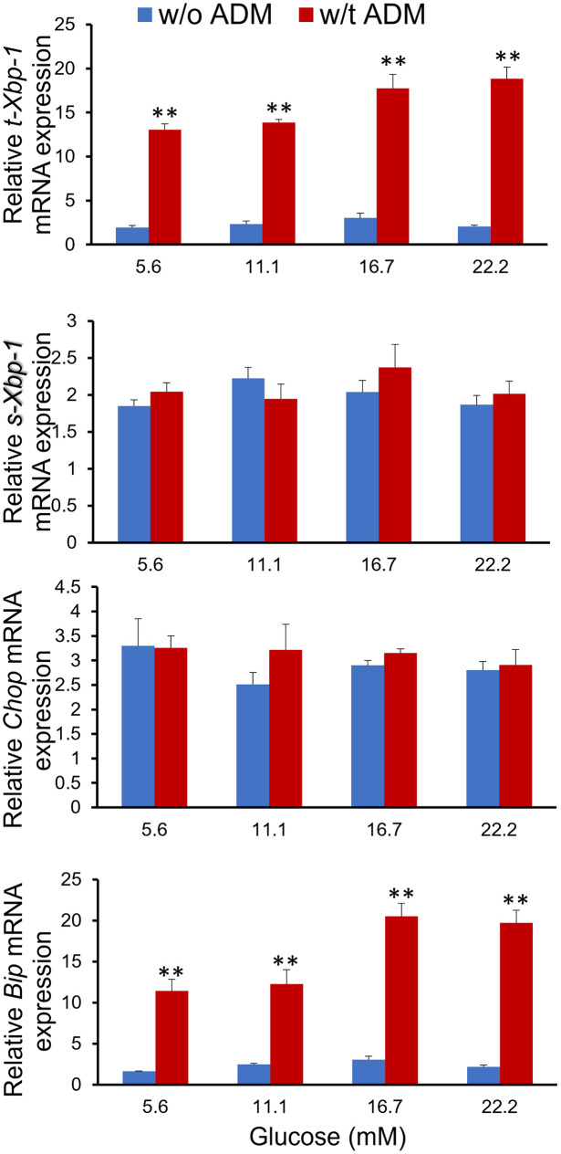
Gestational diabetes mellitus (GDM) is associated with defective pancreatic β-cell adaptation in pregnancy, but the underlying mechanism remains obscure. Our previous studies demonstrated that GDM women display increased plasma adrenomedullin (ADM) levels, and non-obese GDM mice show decreased serum concentrations of insulin and the number of β-cells in pancreas islets. The aims of this study is to examine if ADM and its receptors are expressed in female mouse pancreas, and if so, whether insulin secretion is regulated by ADM in mouse β-cell line, NIT-1 cells and isolated mouse pancreatic islets. Present study shows that ADM and its receptor components CRLR, RAMPs are present in mouse pancreatic islets and co-localized with insulin. The expressions of ADM, CRLR and RAMP2 in islets from pregnant mice are reduced compared to that of non-pregnant mice. NIT-1-β cells express ADM and its receptor mRNA, and glucose dose-dependently stimulates expressions. Furthermore, ADM inhibits NIT-1-β cell growth, and this inhibition is reversed by ADM antagonist, ADM22-52. The glucose-induced insulin secretion was suppressed by ADM in NIT-1-β cells and isolated pancreatic islets from pregnant mice. These inhibitory effects are accompanied by upregulation of endoplasmic reticulum (ER) stress biomarker genes in NIT-1-β cells. This study unveils that reduced ADM and its receptors may play a role in β-cell adaptation during pregnancy, while increased plasma ADM in GDM may contribute to the β-cells dysfunction, and blockade of ADM may reverse β-cell insulin production.


Conflict of interest statement

The authors have declared that no competing interests exist.

Figures

Fig 1
Fig 1. Cellular localization of ADM in mouse pancreas.
Representative illustrative images showing triple immunofluorescence of glucagon (magenta), insulin (red), and ADM (green) in pancreas from nonpregnant (NP) and pregnant mouse on day 13.5 (D13.5) and day 17.5 (D17.5) of pregnancy. The merged column represents a composite of the above three images with cell nuclei (identified by DAPI, blue). Magnification, x 400. The labelings for ADM are generally restricted to the islets and colocalized with insulin. The data for ADM intensity per cell in the islets were displayed as mean+/-SEM (n = 4). * indicate significant difference when compared with NP (P<0.05). Inset is secondary only negative control.
Fig 2
Fig 2. Cellular localization of ADM receptor components CRLR, RAMP2, and RAMP3 in mouse pancreas.
Representative illustrative images represent a composite of the glucagon (magenta), insulin (red), and CRLR, RAMP2, RAMP3 (green) with cell nuclei (identified by DAPI, blue) in pancreas from nonpregnant mice (NP) and mice on day 13.5 (D13.5) and day 17.5 (D17.5) of pregnancy. Magnification, x 400. The labelings for CRLR, RAMP2 and RAMP3 are generally restricted to the islets and colocalized with insulin. The data for CRLR, RAMPs intensity per cell in the islets were displayed as mean+/-SEM (n = 4). * indicate significant difference compared with NP (P<0.05), and ** indicate P<0.01. Inset is secondary only negative control.
Fig 3
Fig 3. ADM inhibits glucose stimulated insulin secretion in pregnant mouse islets.
Pancreatic islets were isolated from day 13.5 pregnant dams. GSIS assays were performed in the presence or absence of ADM. The data is displayed as percent secreted insulin +/- SEM (n = 4). * indicates significant difference between basal (in presence of 1.8 mM glucose) and secreted (in presence of 16.8 mM glucose) insulin.
Fig 4
Fig 4. Glucose stimulates mRNA for ADM receptor components in mouse NIT-1 β cells.
NIT-1 cells were incubated in Ham’s F12K medium with increasing doses of glucose (5.6–22.2 mM) for 24 hours, and the mRNA expression for ADM, CRLR, RAMP2, and RAMP3 were determined by using Real-time PCR with specific primers. The data were normalized to β-actin and GAPDH, and displayed as mean+/-SEM (n = 6). Different letters on each bar indicate significant difference between groups (P<0.05). Data showed that mRNA for CRLR, RAMP2, and RAMP3, but not ADM, are stimulated by glucose in a dose-dependent manner.
Fig 5
Fig 5. ADM inhibits NIT-1 β cell proliferation.
Representative illustrative images showing the NIT-1 β cells grown in Ham’s F12K medium with 16.7 mM glucose and treated with ADM (10−9 M) in the presence or absence of ADM22-52 (10−8 M). The micrographs were taken on day 1, day 3, and day 5 after treatments. Compared with controls, profoundly decreased cell density was observed after 5 days ADM treatments, and this reduction was reversed by pretreatment of the cells with ADM22-52. Magnification, x 200.
Fig 6
Fig 6. ADM inhibits glucose-stimulated NIT-1 β cell growth in MTT reduction test.
NIT-1 β cells were cultured as described in the Materials and Methods. Reduction of MTT was colorimetrically determined after 72 hours treatments with ADM in the presence or absence of ADM22-52. ADM did not significantly affect the growth of NIT-1 cells in medium containing 5.6 mM glucose, but did significantly inhibit cell growth in medium containing 16.7 mM glucose, and this inhibition was reversed by ADM antagonist. The data displayed as mean+/-SEM (n = 4). Different letters on each bar indicate significant difference between groups (P<0.05).
Fig 7
Fig 7. ADM inhibits glucose-stimulated insulin mRNA expression, secretion and synthesis in NIT-1 β-cells.
(A) NIT-1 β-cells were incubated in Ham’s F12K medium containing increasing doses of glucose (5.6–16.7mM) with or without ADM (10−9 M) for 24 hours (n = 6). Real-time PCR was performed to determine the mRNA expression of insulin by NIT-1 cells. mRNA was normalized to housekeeping genes of β-actin and GAPDH. (B) NIT-1 β-cells were grown in medium containing increasing doses of glucose (5.6–16.7mM) with or without ADM for 30-min, after which insulin concentration in the medium was measured using ELISA kit. (C) NIT-1 β-cells were grown in medium containing 16.7mM glucose and ADM in the presence or absence of ADM22-52, SQ22536, and PD98059 for 24 hours, after which insulin concentration in the medium was measured using ELISA kit. Data displayed as mean+/-SEM (n = 3). * and # indicate significant difference vs. paired controls (P<0.05).
Fig 8
Fig 8. ADM induces ER stress in NIT-1 β cells.
NIT-1 cells were cultured in Ham’s F12K medium containing increasing doses of glucose with or without ADM (10−9 M) for 24 hours. mRNA expressions for ER stress markers t-Xbp-1, s-Xbp-1, Chop, and BIP were determined by Real-time PCR, and normalized to housekeeping genes of β-actin and GAPDH. The data displayed as mean+/-SEM (n = 6). ** indicate significant difference between ADM treated and control groups (P<0.01).

Similar articles

Cited by

References

    1. Bellmann O, Hartmann E. Influence of pregnancy on the kinetics of insulin. Am J Obstet Gynecol. 1975;122(7):829–33. Epub 1975/08/01. doi: 10.1016/0002-9378(75)90723-1 . - DOI - PubMed
    1. Parsons JA, Brelje TC, Sorenson RL. Adaptation of islets of Langerhans to pregnancy: increased islet cell proliferation and insulin secretion correlates with the onset of placental lactogen secretion. Endocrinology. 1992;130(3):1459–66. doi: 10.1210/endo.130.3.1537300 . - DOI - PubMed
    1. Ferrara A. Increasing prevalence of gestational diabetes mellitus: a public health perspective. Diabetes Care. 2007;30 Suppl 2:S141–6. Epub 2007/07/13. doi: 10.2337/dc07-s206 . - DOI - PubMed
    1. Dabelea D, Hanson RL, Lindsay RS, Pettitt DJ, Imperatore G, Gabir MM, et al.. Intrauterine exposure to diabetes conveys risks for type 2 diabetes and obesity: a study of discordant sibships. Diabetes. 2000;49(12):2208–11. Epub 2000/12/16. doi: 10.2337/diabetes.49.12.2208 . - DOI - PubMed
    1. Chavey A, Ah Kioon MD, Bailbe D, Movassat J, Portha B. Maternal diabetes, programming of beta-cell disorders and intergenerational risk of type 2 diabetes. Diabetes Metab. 2014;40(5):323–30. Epub 2014/06/21. doi: 10.1016/j.diabet.2014.02.003 . - DOI - PubMed

Publication types