. 2022 Mar 24;18(3):e1010365.
doi: 10.1371/journal.ppat.1010365. eCollection 2022 Mar.

The Cdkn2a gene product p19 alternative reading frame (p19ARF) is a critical regulator of IFNβ-mediated Lyme arthritis

Affiliations

The Cdkn2a gene product p19 alternative reading frame (p19ARF) is a critical regulator of IFNβ-mediated Lyme arthritis

Jinze Li et al. PLoS Pathog. .

Abstract

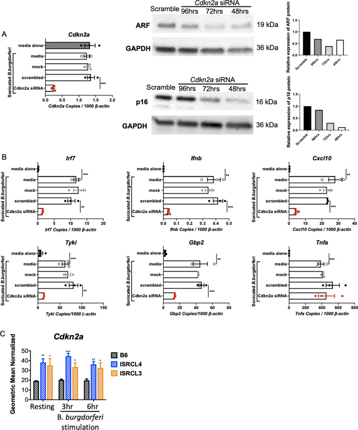
Type I interferon (IFN) has been identified in patients with Lyme disease, and its abundant expression in joint tissues of C3H mice precedes development of Lyme arthritis. Forward genetics using C3H mice with severe Lyme arthritis and C57BL/6 (B6) mice with mild Lyme arthritis identified the Borrelia burgdorferi arthritis-associated locus 1 (Bbaa1) on chromosome 4 (Chr4) as a regulator of B. burgdorferi-induced IFNβ expression and Lyme arthritis severity. B6 mice introgressed with the C3H allele for Bbaa1 (B6.C3-Bbaa1 mice) displayed increased severity of arthritis, which is initiated by myeloid lineage cells in joints. Using advanced congenic lines, the physical size of the Bbaa1 interval has been reduced to 2 Mbp, allowing for identification of potential genetic regulators. Small interfering RNA (siRNA)-mediated silencing identified Cdkn2a as the gene responsible for Bbaa1 allele-regulated induction of IFNβ and IFN-stimulated genes (ISGs) in bone marrow-derived macrophages (BMDMs). The Cdkn2a-encoded p19 alternative reading frame (p19ARF) protein regulates IFNβ induction in BMDMs as shown by siRNA silencing and overexpression of ARF. In vivo studies demonstrated that p19ARF contributes to joint-specific induction of IFNβ and arthritis severity in B. burgdorferi-infected mice. p19ARF regulates B. burgdorferi-induced IFNβ in BMDMs by stabilizing the tumor suppressor p53 and sequestering the transcriptional repressor BCL6. Our findings link p19ARF regulation of p53 and BCL6 to the severity of IFNβ-induced Lyme arthritis in vivo and indicate potential novel roles for p19ARF, p53, and BCL6 in Lyme disease and other IFN hyperproduction syndromes.


Conflict of interest statement

The authors have declared that no competing interests exist.

Figures

Fig 1
Fig 1. Silencing of Cdkn2a suppressed expression of Ifnb and ISGs.
A) Cdkn2a was silenced in B6.C3-Bbaa1 BMDMs upon transfection of 100 nM Cdkn2a Smartpool siRNA using the Neon electroporation system. Scrambled siRNA was transfected as the control. Transfected macrophages were treated with 5 μg/ml sonicated B. burgdorferi for 6 h prior to collection for RNA and protein extraction. The induction of IFN responses by B. burgdorferi treatment is confirmed by comparing B. burgdorferi treated media (no-silencing) and BSK media alone (no stimulus) groups. Cdkn2a knockdown efficiency was assessed by qPCR with normalization to β-actin and Western blot. The quantification of Cdkn2a-encoded ARF and p16 protein levels is shown in the bar graph on the right side of the western blots. Significance was determined by Student t-test. Data are shown as mean ± SEM (n = 3 per group). B) The impact of Cdkn2a silencing on expression of Ifnb and the IFN downstream genes Cxcl10, Tyki, and Gbp2 was determined by qPCR normalized to β-actin. All transfection experiments were repeated at least twice. Significance was determined by Student t-test. Data are shown as mean ± SEM (n = 3 per group). C) RNA-seq data demonstrated that Cdkn2a was expressed constitutively at a higher level in BMDMs from ISRCL3 and ISRCL4 mice than in BMDMs from B6 mice following stimulation with live B. burgdorferi for 3 and 6 h. Significance was assessed by 1-way ANOVA followed by Dunnett’s multiple comparison test versus B6. Error bars indicate SEM (n = 3 or 4 per group). *p < 0.05, **p < 0.01, ***p < 0.001, ****p < 0.0001.
Fig 2
Fig 2. ARF regulates the IFNβ pathway.
A) Cdkn2a encodes two proteins, p16 and ARF, and possesses 2 SNPs in the transcribed sequences. B) Arf expression was silenced with Arf siRNA (100 nM) in B6.C3-Bbaa1 BMDMs. ARF and p16 expression levels were evaluated by qPCR with normalization to β-actin and Western blot following treatment with 5 μg/ml sonicated B. burgdorferi for 6 h. IFN responses induced by B. burgdorferi treatment are indicated as B. burgdorferi-treated media, while those not receiving B. burgdorferi are indicated by media alone. Significance was determined by Student t-test. Error bars indicate the SEM (n = 3 per group). C) The impact of Arf silencing on expression of B. burgdorferi-stimulated IFN responses in B6.C3-Bbaa1 BMDMs were determined by qPCR normalized to β-actin. Significance of differences was measured by Student t-test. Data are shown as mean ± SEM (n = 3 per group). **p < 0.01, ***p < 0.001, ****p < 0.0001.
Fig 3
Fig 3. IFNβ modulating activity of Cdkn2a is not due to p16 protein.
A) Transfection of p16 siRNA (100 nM) into B6.C3-Bbaa1 BMDMs silenced p16. ARF and p16 expression levels were evaluated by qPCR with normalization to β-actin and Western blot following treatment with 5 μg/ml sonicated B. burgdorferi for 6 h. Significance was determined by Student t-test. Data are shown as mean ± SEM (n = 3 per group). B) The impact of selective silencing of p16 on expression of B. burgdorferi-stimulated Ifnb and the IFN downstream genes Cxcl10, Tyki, and Gbp2 in B6.C3-Bbaa1 BMDMs was determined by qPCR normalized to β-actin. Error bars indicate SEM (n = 3 per group). **p < 0.01, ***p < 0.001, ****p < 0.0001.
Fig 4
Fig 4. ARF impacts the magnitude of IFNβ expression in response to living B. burgdorferi and pathogenic E.coli.
A) The impact of Cdkn2a, Arf and p16 silencing on IFN expression in response to 7.4×10⁶ live B. burgdorferi (MOI of 10) for 6 h is shown. Data are shown as mean ± SEM (n = 3 per group). B) The impact of silencing of Cdkn2a, Arf and p16 on live E. coli CFT073 stimulated IFN responses was determined by qPCR normalized to β-actin following stimulation for 6 h, at an MOI of 10. Error bars indicate SEM (n = 3 per group). Significance of the differences was measured by Student t-test. *p < 0.05, **p < 0.01, ***p < 0.001, ****p < 0.0001.
Fig 5
Fig 5. Overexpression of ARF in B6 macrophages enhances Ifnb expression.
A) ARF was overexpressed in B6 BMDMs by transfection of the MSCV-p19ARF plasmid. The MSCV vector was transfected as the control. The transfected cells were allowed to rest for 72 h before the addition of sonicated B. burgdorferi for 6 h. The induction of IFN responses by B. burgdorferi treatment is confirmed by comparing the data from B. burgdorferi treated media with media alone. Total RNA and protein were isolated for analysis. B) The impact of ARF overexpression on activation of Ifnb and ISGs in response to sonicated B. burgdorferi stimulation in B6 BMDMs was determined by qPCR normalized to β-actin. Significance was calculated by Student t-test. Error bars indicate SEM (n = 3 per group). *p < 0.05, **p < 0.01.
Fig 6
Fig 6. The C3H Bbaa1 allele of ARF enhances arthritis severity in B. burgdorferi-infected mice.
A) BMDMs isolated from B6 and B6 Arf-/- mice were treated with sonicated B. burgdorferi for 6 h to induce the IFN response. The impact of ablation only on ARF expression, but not p16, was confirmed by qPCR and Western blot, and performed twice. Significance was determined by Student t-test. Error bars indicate SEM (n = 3 per group). B) Sonicated B. burgdorferi-stimulated Ifnb, Gbp2, Tyki, and Cxcl9 expression levels in B6 Arf-/- and B6 BMDMs were determined by qPCR normalized to β-actin. The B. burgdorferi-stimulated IFN response is compared with the BSK media alone group. Significance of differences was measured by Student t-test. Data are shown as mean ± SEM (n = 3 per group). C) B6 Arf-/-, B6.C3-Bbaa1, and B6 mice were infected with live B. burgdorferi at 6 or 7 weeks of age. Arthritis severity was measured by ankle measurement (ankle swelling) and by histopathologic assessment (total score) at 4 weeks following infection. There were 5 mice of mixed sex in each infected mouse group. Data were pooled from 2 separate experiments. Statistical analysis was assessed by Student t-test. Error bars indicate SEM. D) Reconstitution of B6 Arf-/- mice with splenocytes from B6.C3-Bbaa1 congenic mice or B6 mice revealed the contribution of the C3H Bbaa1 allele of ARF to the development of severe Lyme arthritis. Mice were infected 3 weeks after reconstitution, and arthritis severity was measured at 4 weeks post-infection by ankle measurement (ankle swelling) and by histopathologic assessment (total score). The direction of transplantation from donor to recipient is shown on the figure. Results from two separate experiments with ≥ 5 mice in each infected mouse group were pooled. Statistical analysis was assessed by Student t-test. Error bars indicate SEM (n = 5 or 8 or 10 per group). E) B. burgdorferi in joint tissue was quantified by 16S rRNA PCR (normalized to β-actin) and F) by ELISA quantification of serum anti-B. burgdorferi IgG. Significance was calculated by Student t-test. (n = 5 or 8 or 10 per group). G) The efficiency of ARF reconstitution in recipient B6 Arf-/- mice was determined by qPCR of RNA isolated from whole blood. qPCR analysis was normalized to β-actin. Significance was calculated by Student t-test. Error bars indicate SEM (n = 5 or 8 or 10 per group). *p < 0.05, **p < 0.01, ***p < 0.001, ****p < 0.0001.
Fig 7
Fig 7. The involvement of p53 in IFNβ expression.
A) ARF was overexpressed in B6 BMDMs by transfection of the MSCV-p19ARF plasmid and expression assessed by Western Blot. The transfected cells were allowed to rest for 72 h before the addition of sonicated B. burgdorferi for 6 h. B) p53 was silenced in B6.C3-Bbaa1 BMDMs by transfection with 100 nM p53 siRNA. Transfected cells were stimulated with sonicated B. burgdorferi prior to collection for RNA and protein extraction. IFN responses was induced by B. burgdorferi treatment as indicated by B. burgdorferi treated media, while samples receiving no stimulus are indicated as media alone. Cells were collected and protein was isolated at 48 h, 72 h, and 96 h to assess for silencing efficiency by Western Blot. p53 transcripts were analyzed by qPCR normalized to β-actin. Significance was determined by Student t-test. Data are shown as mean ± SEM (n = 3 per group). The impact of p53 silencing on expression of sonicated B. burgdorferi-stimulated IFN responses in B6.C3-Bbaa1 BMDMs was determined by qPCR normalized to β-actin. Significance of differences was calculated by Student t-test. Data are shown as mean ± SEM (n = 3 per group). C) BMDMs were treated with PFTα for 1 h prior to stimulation with sonicated B. burgdorferi for 6 h. qPCR was used to assess the impact of the inhibitor on expression of Ifnb and downstream genes, and data were normalized to β-actin. Significance was determined by Student t-test. Data are shown as mean ± SEM (n = 3 per group). D) Expression of a p53 downstream gene, p21, was used to verify the efficiency of blocking p53 with siRNA and the PFTα inhibitor. Expression was measured by qPCR normalized to β-actin. Significance of difference was calculated by Student t-test (n = 3 per group). *p < 0.05, **p < 0.01, ***p < 0.001.
Fig 8
Fig 8. BCL6 plays a role in regulating IFNβ activity.
A) Bcl6 was silenced in B6.C3-Bbaa1 BMDMs by transfection of Bcl6 siRNA. Transfected cells were allowed to rest for 48 h and were then incubated with sonicated B. burgdorferi for 6 h prior to collection for RNA isolation. Transfected cells were incubated for 48 h, 72 h, and 96 h before treatment with sonicated B. burgdorferi for 6 h prior to collection for protein isolation. The induction of IFN responses is indicated by comparing B. burgdorferi treated samples (media) with unstimulated media alone. Bcl6 expression was measured by qPCR normalized to β-actin. Significance was determined by Student t-test. Data are shown as mean ± SEM (n = 3 per group). B) The impact of Bcl6 silencing on expression of B. burgdorferi-stimulated IFN responses in B6.C3-Bbaa1 BMDMs was determined by qPCR normalized to β-actin. Significance of differences was determined by Student t-test. Data are shown as mean ± SEM (n = 3 per group). C) B6.C3-Bbaa1 BMDMs were treated with 13 μM FX1 for 0.5 h before the addition of sonicated B. burgdorferi for 6 h. Blocking of BCL6 induced expression of Irf7, Ifnb, and ISGs, which was determined by qPCR normalized to β-actin. Significance of differences was calculated by Student t-test. Error bars indicate SEM (n = 3 per group). D) Overexpression of BCL6 by transfection of B6.C3-Bbaa1 BMDMs with 1 μg MSCV-BCL6 plasmid was performed to confirm transcriptional repression by BCL6. Transfected cells were allowed to rest for 48 h and were then incubated with sonicated B. burgdorferi for 6 h prior to isolation of RNA and protein. The impact of BCL6 overexpression on activation of Ifnb and ISGs upon stimulation with B. burgdorferi in B6.C3-Bbaa1 BMDMs was determined by qPCR normalized to β-actin. Significance was calculated by Student t-test. Error bars indicate SEM (n = 3 per group). *p < 0.05, **p < 0.01, ****p < 0.0001.
Fig 9
Fig 9. Suppression of BCL6 enhances induction of IFNβ and development of severe arthritis in joint tissues of B6 mice and B6 Rag1-/- mice.
A) B6 mice (6–7 weeks of age) that were infected with live B. burgdorferi on day 1 received daily i.p. injections of FX1 (50 mg/kg) starting the day before infection and continuing for 12 days. B) Arthritis was assessed at 28 days post-infection by measuring ankle swelling and by histopathologic determination (total score). Data were pooled from 2 separate experiments. Statistical analysis was performed using the Student t-test. Error bars indicate SEM (n = 9 or 10 per group). C) B. burgdorferi was quantified in joint tissues by 16S rRNA PCR normalized to β-actin and by ELISA quantification of serum anti-B. burgdorferi IgG. Significance was calculated by Student t-test (n = 9 or 10 per group). D) The impact of blocking BCL6 on Ifnb expression was assessed at 1week post-infection in mice treated daily with 50 mg/kg FX1 beginning the day before infection and continuing until the mice were sacrificed at day 7 post-infection. E) RNA was isolated from joint tissues and Ifnb expression was measured by qPCR normalized to β-actin. Data were pooled from 2 separate experiments. Significance was calculated by Student t-test (n = 9 or 10 per group). F) Host defense was assessed by qPCR of B. burgdorferi 16S rRNA in joint tissues normalized to β-actin and by quantification of serum anti-B. burgdorferi IgM. Significance of differences was determined by Student t-test (n = 9 or 10 per group). G) B. burgdorferi-infected B6 Rag1-/- mice (6–7 weeks of age) received daily i.p. injections of FX1 (50 mg/kg) starting the day before the infection and continuing for 12 days as described in Fig 7A. Arthritis was measured at 28 days post-infection and assessed by ankle swelling and histopathologic determination (total score). This experiment was repeated 3 times. Statistical analysis was performed using the Student t-test. Error bars indicate SEM (n = 5 per group). H) B. burgdorferi was quantified in joint tissue by 16S rRNA PCR normalized to β-actin. Significance of difference was calculated by Student t-test (n = 5 per group). *p < 0.05, **p < 0.01, ***p < 0.001, ****p < 0.0001. (I-J) Responses of rear ankle joints (i.e., tibiotarsal and other connected joints) to injury in IFNβ mediated Lyme arthritis. I1) Vehicle treated infected B6 mice. Note minimal to mild thickening of the sheaths (arrows) of the tendons (t) and minimal to mild thickening of the periosteum (arrowheads) of the bone (b). Minimal numbers of neutrophils and/or mononuclear inflammatory cells are present in the tendon sheath spaces (not visible in the image). H&E stain. I2) FX1 treated infected B6 mice. Note moderate thickening of the tendon sheaths (arrows) and hypertrophy/hyperplasia of synoviocytes covering the tendons (t). Mild to moderate thickening of the periosteum (arrowheads) of the bone (b) is also present. Moderate numbers of neutrophils and mononuclear inflammatory cells are present in the tendon sheath spaces (not visible in the image). A nerve (n) is present in the specimen. H&E stain. J1) Vehicle treated infected B6 Rag-/- mice. Note minimal to mild thickening (hypertrophy/hyperplasia) of the tendon sheath (ts) and minimal numbers of neutrophils and/or mononuclear inflammatory cells in the tendon sheath space (tss). H&E stain. J2) FX1 treated infected B6 Rag-/- mice. Note moderate thickening of the tendon sheath (ts) and hypertrophy/hyperplasia of synoviocytes (arrow) covering the tendon (t). Moderate numbers of neutrophils and minimal numbers of mononuclear inflammatory cells are present in the tendon sheath space (tss). H&E stain.
Fig 10
Fig 10. Proposed model of ARF-IFNβ pathway.
A) The constitutively heightened expression of ARF in B6.C3-Bbaa1 congenic mice results in a greater magnitude of IFNβ production and more severe Lyme arthritis following infection with B. burgdorferi. B) The heightened expression of the C3H allele of ARF blocks the activity of ubiquitin ligase MDM2, resulting in stabilized p53. p53 acts to enhance the expression of IRF7 which induces IFNβ production, but not downstream ISGs. C) The greater expression of the C3H allele of ARF also binds and suppresses BCL6, resulting in increased IRF7 expression and greater production of IFNβ. Expression of downstream ISGs is mediated through activation of signal transducer and activator transcription 1 (STAT1)-STAT2-IRF9 signaling complex. Parallel ARF interactions with p53 and BCL6 modulate IFNβ hyper production in resident myeloid cells. IFNβ leads to the upregulation of the muscle regulatory protein myostatin in CD45- cells, a previously identified mediator of Lyme arthritis development in B6.C3-Bbaa1 mice. (Created with BioRender.com).

Similar articles

Cited by

References

    1. Steere AC, Schoen RT, Taylor E. The clinical evolution of Lyme arthritis. Ann Intern Med. 1987;107(5):725–31. doi: 10.7326/0003-4819-107-5-725 - DOI - PubMed
    1. Kugeler KJ, Schwartz AM, Delorey MJ, Mead PS, Hinckley AF. Estimating the Frequency of Lyme Disease Diagnoses, United States, 2010–2018. Emerg Infect Dis. 2021;27(2):616–9. doi: 10.3201/eid2702.202731 - DOI - PMC - PubMed
    1. Steere AC, Glickstein L. Elucidation of Lyme arthritis. Nat Rev Immunol. 2004;4(2):143–52. doi: 10.1038/nri1267 - DOI - PubMed
    1. Schwartz AM, Hinckley AF, Mead PS, Hook SA, Kugeler KJ. Surveillance for Lyme Disease—United States, 2008–2015. MMWR Surveill Summ. 2017;66(22):1–12. doi: 10.15585/mmwr.ss6622a1 - DOI - PMC - PubMed
    1. Hu LT. Lyme Disease. Ann Intern Med. 2016;164(9):Itc65–itc80. doi: 10.7326/AITC201605030 - DOI - PubMed

Publication types

Substances