Review
. 2022 Oct 3;24(10):1647-1657.
doi: 10.1093/neuonc/noac055.

T lymphocytes as dynamic regulators of glioma pathobiology

Affiliations
Review

T lymphocytes as dynamic regulators of glioma pathobiology

Elizabeth C Cordell et al. Neuro Oncol. .

Abstract

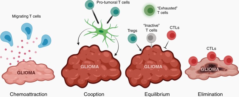
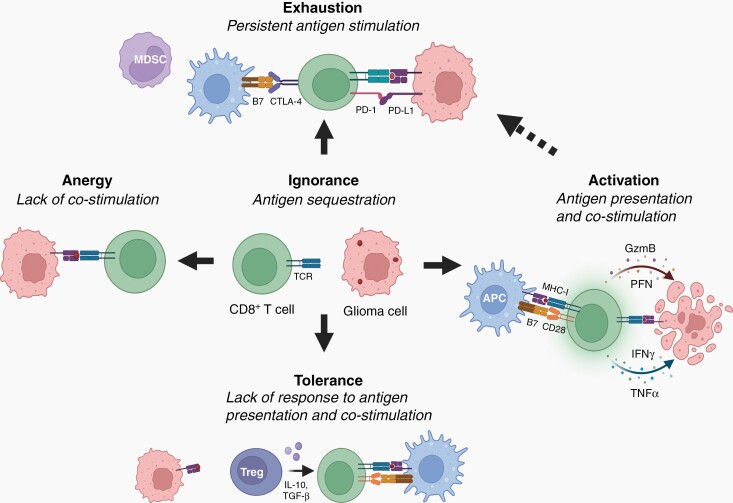
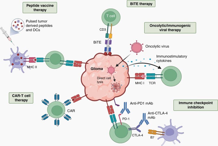
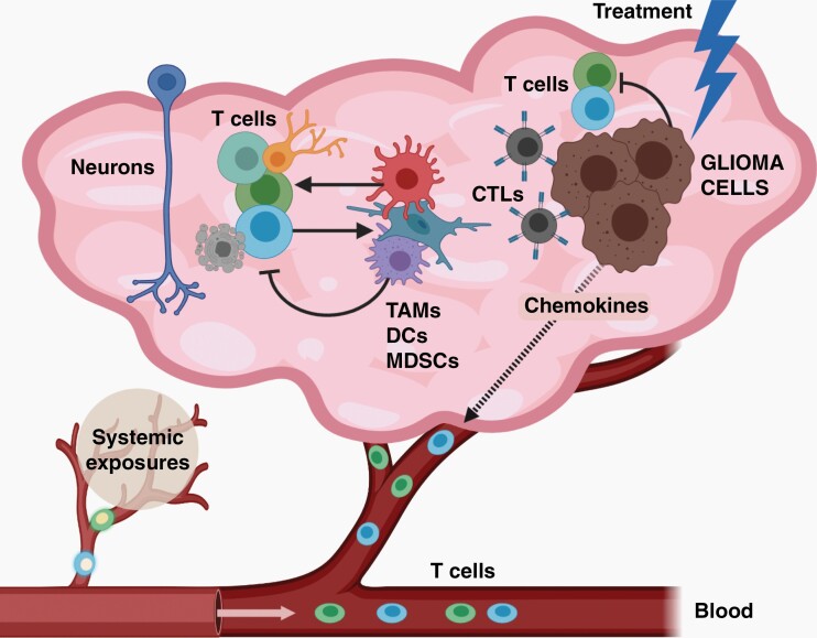
The brain tumor microenvironment contains numerous distinct types of nonneoplastic cells, which each serve a diverse set of roles relevant to the formation, maintenance, and progression of these central nervous system cancers. While varying in frequencies, monocytes (macrophages, microglia, and myeloid-derived suppressor cells), dendritic cells, natural killer cells, and T lymphocytes represent the most common nonneoplastic cellular constituents in low- and high-grade gliomas (astrocytomas). Although T cells are conventionally thought to target and eliminate neoplastic cells, T cells also exist in other states, characterized by tolerance, ignorance, anergy, and exhaustion. In addition, T cells can function as drivers of brain cancer growth, especially in low-grade gliomas. Since T cells originate in the blood and bone marrow sinuses, their capacity to function as both positive and negative regulators of glioma growth has ignited renewed interest in their deployment as immunotherapeutic agents. In this review, we discuss the roles of T cells in low- and high-grade glioma formation and progression, as well as the potential uses of modified T lymphocytes for brain cancer therapeutics.

Keywords: T cells; astrocytoma; glioblastoma; gliomagenesis; microglia; pediatric low-grade glioma; tumor microenvironment; tumor-associated monocytes.


Figures

Fig. 1
Fig. 1
T cells shape glioma evolution. Glioma cells recruit T cells into the tumor microenvironment (TME) through the elaboration of chemokines. In addition, T cells produce paracrine factors that can act on other cells in the TME (eg, microglia) to increase tumor growth (“cooption”). Pro-tumoral regulatory T cells (Tregs) can also function to increase glioma growth (“equilibrium”), while cytotoxic T lymphocytes (CTLs) lyse tumor cells by secreting perforin (PFN) and granzyme B (GZMB) or by direct cell-to-cell interaction (“elimination”). Other CTLs can adopt an “exhausted” phenotype or become inactivated by glioma cells through the expression of immune checkpoint proteins. In response to extrinsic or intrinsic pressures, the balance of pro- and anti-tumoral T cells can shift to favor CTLs, resulting in glioma elimination. Created with BioRender.
Fig. 2
Fig. 2
Glioma-mediated T-cell dysfunction during gliomagenesis. During early gliomagenesis, CD8+ T cells may remain “ignorant” due to inaccessibility of tumor-associated antigens (lack of Major Histocompatibility I (MHC-I) molecule interactions with T cell receptors (TCRs)). As gliomas progress and sufficient glioma antigen is produced, glioma-specific T cells can exist in different dysfunctional states, depending upon the specific signals from the glioma cells and the immune microenvironment. T cells can also be activated as a consequence of a lack of costimulation (B7-CD28 interactions), inducing a state of T cell “anergy”. Additionally, glioma cells can express immunosuppressive molecules (eg, Programmed Death Ligand-1 (PD-L1)) and recruit immunosuppressive cells (eg, myeloid-derived suppressor cells (MDSCs)) into the tumor microenvironment. Persistent tumor antigen and inhibitory signals can drive T cell “exhaustion”, a state of nontumor reactivity. Finally, in the presence of other immunosuppressive cells (eg, regulatory T cells (Tregs)), T cells can exhibit “tolerance”, in which they do not respond to antigen stimulation due to the presence of immunosuppressive cytokines (eg, Interleukin-10 (IL-10) and transforming growth factor-β (TGF-β)). The goal of immunotherapies is to induce T cell activation, instead of progression to these dysfunctional states. Activated T cells, after interacting with antigen-presenting cells (APCs) function to lyse tumor cells through the release of perforin (PFN), granzyme-B (GZMB), interferon-γ (IFNγ), and tumor necrosis factor-α (TNF-α). Created with BioRender.
Fig. 3
Fig. 3
T cells as immunotherapeutic agents. T cells can be deployed as therapeutic modalities, serving as cancer vaccine inducers, cytotoxic effectors, or as targets for immune checkpoint de-repression. Bispecific T cell engager (BiTE) therapy uses a bispecific antibody that binds to CD3 and a tumor specific antigen, activating T cells to lyse the tumor. Oncolytic viruses cause direct tumor cell lysis, but can also be designed to stimulate the immune system by producing immunostimulatory cytokines or through tumor cell-mediated viral antigen presentation. Immune checkpoint inhibitors target Programmed Death Receptor-1 (PD-1) or Cytotoxic T-lymphocyte antigen 4 (CTLA-4) receptors for PDL-1 and B7 respectively, on T cells to promote cell cycle arrest and T cell exhaustion. Many tumors express PD-L1, which suppresses cytotoxic T cell function. Chimeric Antigen Receptor (CAR) T cells are autologous or donor T cells primed with an antigen receptor recognizing one or multiple glioma-expressed antigens or neoantigens. Cancer vaccines bolster cytotoxic T cell responses by overloading the immune system with neoantigens (peptides), either alone or presented on dendritic cells (DCs). With an increase in neoantigen content, antigen-presenting cells (APCs) can express these neoantigens on their cell surface, thus enabling T cells to generate more robust anti-tumoral responses. Created with BioRender.
Fig. 4.
Fig. 4.
The complex and ever-evolving immune microenvironment in glioma. In gliomas, numerous distinct populations of T cells exist, which could either increase or inhibit tumor cell expansion. In addition, both T cell infiltration and function can be controlled by both neoplastic (glioma cells) and nonneoplastic (neurons, TAMs, DCs, MDSCs) cells through paracrine factor signaling. Importantly, T cell function is also influenced by systemic exposures (eg, atopic conditions, like asthma), but also by tumor-directed therapies. Created with BioRender.

Comment in

References

    1. Friebel E, Kapolou K, Unger S, et al. Single-cell mapping of human brain cancer reveals tumor-specific instruction of tissue-invading leukocytes. Cell. 2020; 181(7):1626–1642.e20. - PubMed
    1. González-Tablas Pimenta M, Otero A, Arandia Guzman DA, et al. Tumor cell and immune cell profiles in primary human glioblastoma: Impact on patient outcome. Brain Pathol. 2021; 31(2):365–380. - PMC - PubMed
    1. Chen J, Sinha N, Cobb O, et al. Immune cell analysis of pilocytic astrocytomas reveals sexually dimorphic brain region-specific differences in T-cell content. Neurooncol Adv. 2021; 3(1):vdab068. - PMC - PubMed
    1. Gutmann DH, Kettenmann H. Microglia/brain macrophages as central drivers of brain tumor pathobiology. Neuron. 2019; 104(3):442–449. - PMC - PubMed
    1. Robinson MH, Vasquez J, Kaushal A, et al. Subtype and grade-dependent spatial heterogeneity of T-cell infiltration in pediatric glioma. J ImmunoTher Cancer. 2020; 8(2):e001066. - PMC - PubMed

Publication types