. 2022 Apr 14;185(8):1308-1324.e23.
doi: 10.1016/j.cell.2022.02.022. Epub 2022 Mar 23.

Liquid-to-solid phase transition of oskar ribonucleoprotein granules is essential for their function in Drosophila embryonic development

Affiliations

Liquid-to-solid phase transition of oskar ribonucleoprotein granules is essential for their function in Drosophila embryonic development

Mainak Bose et al. Cell. .

Abstract

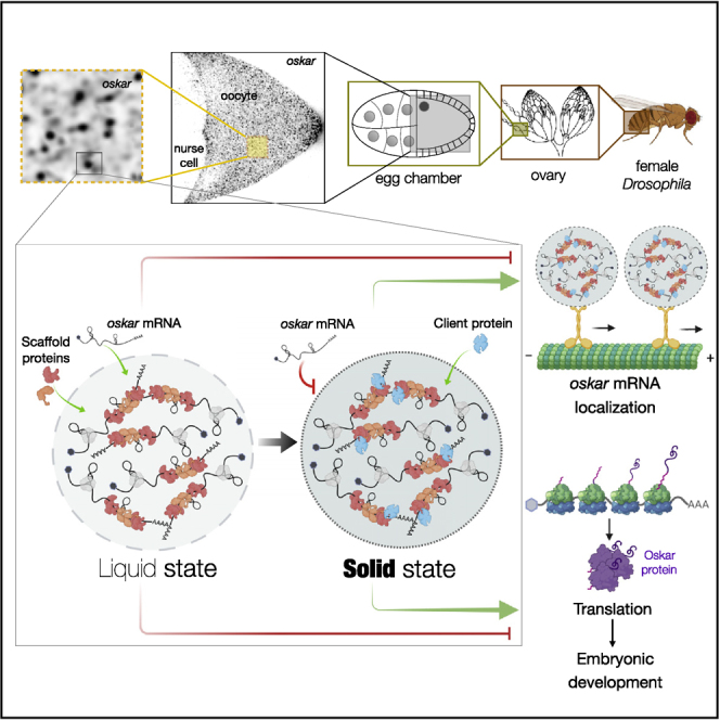
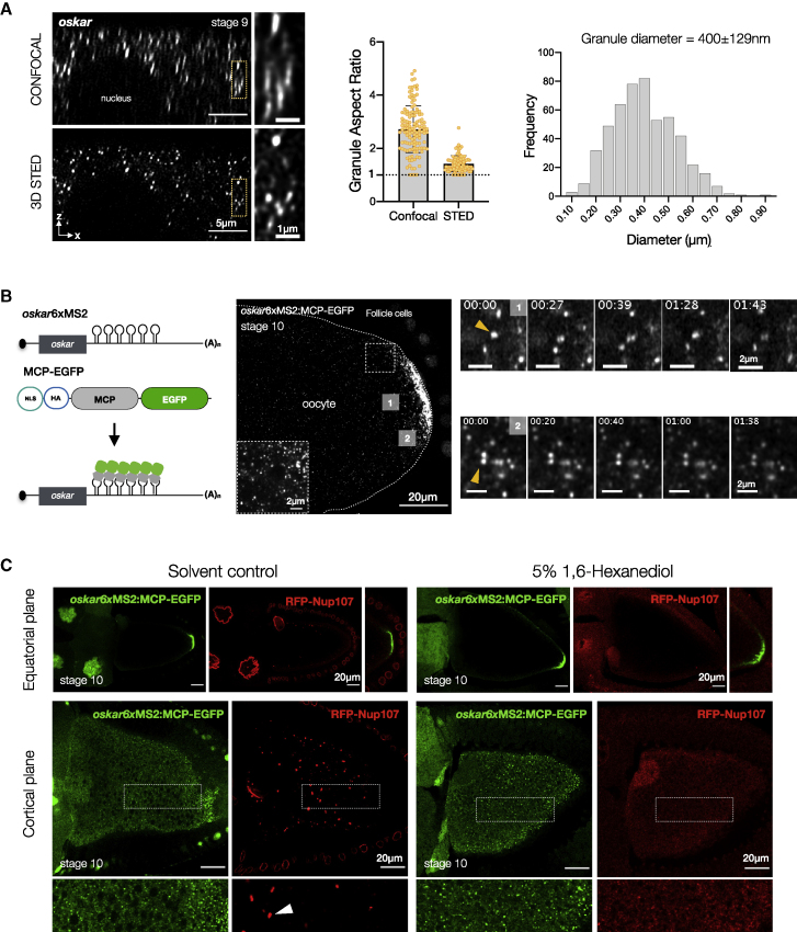
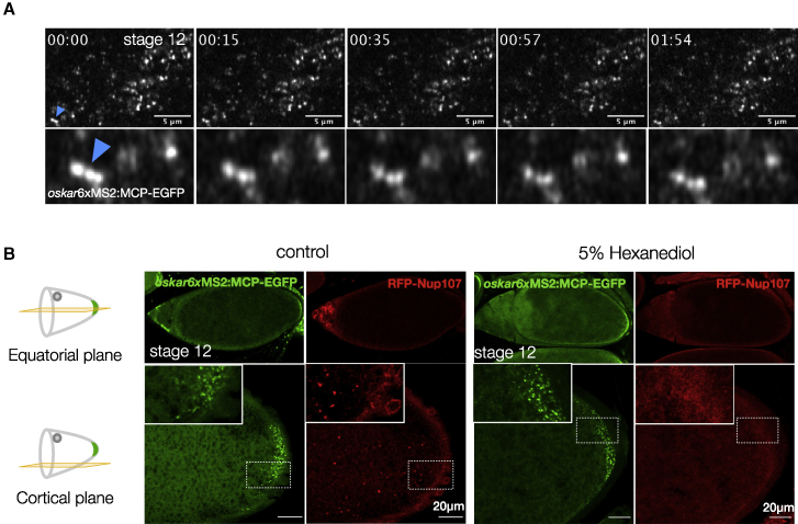
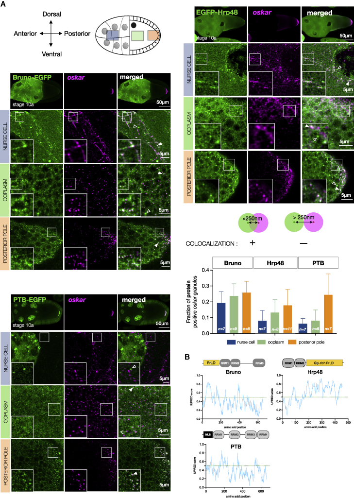
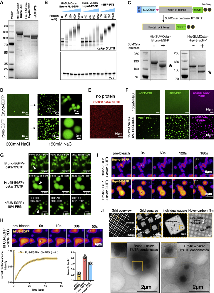
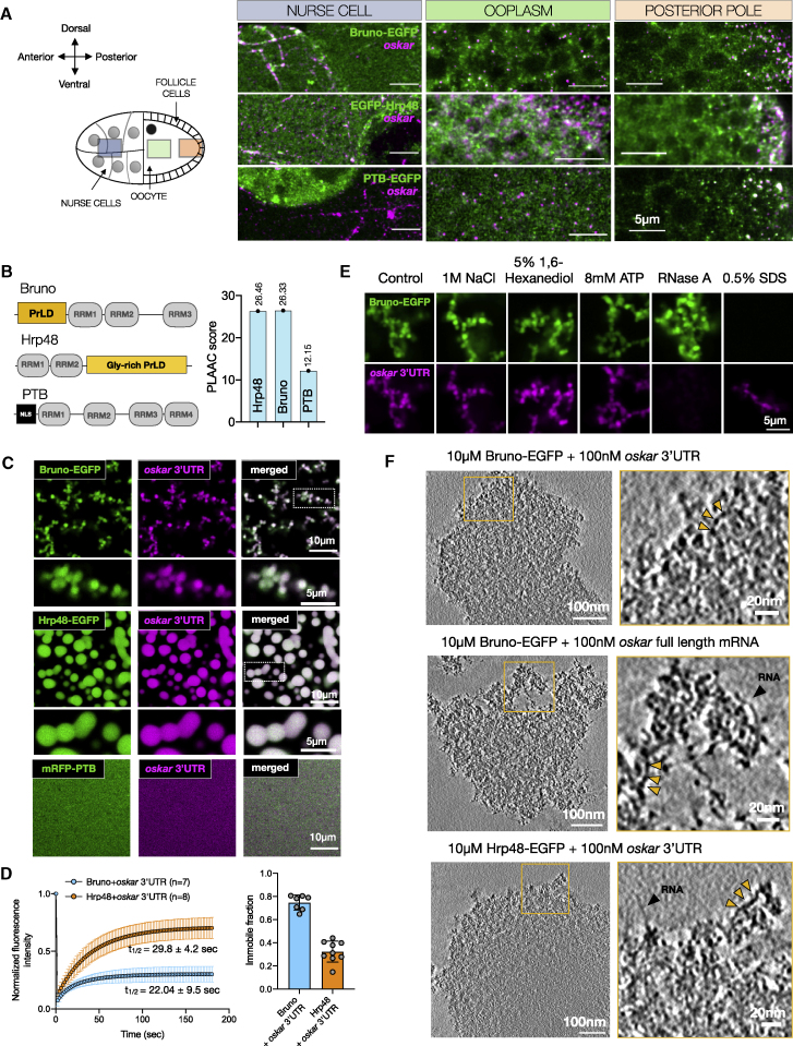
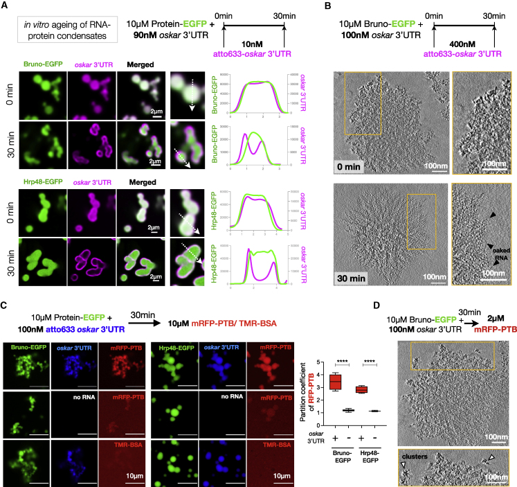
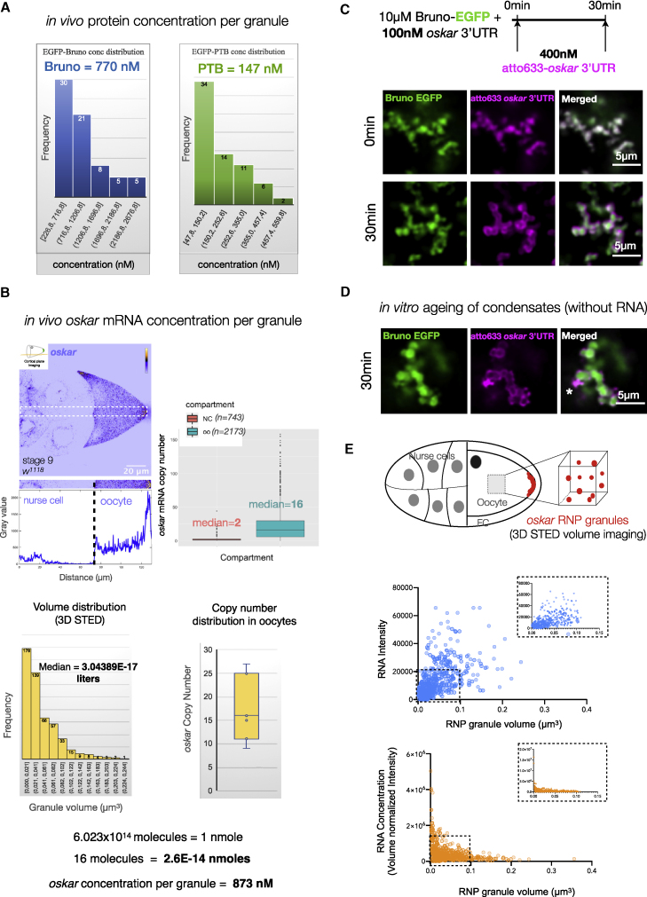
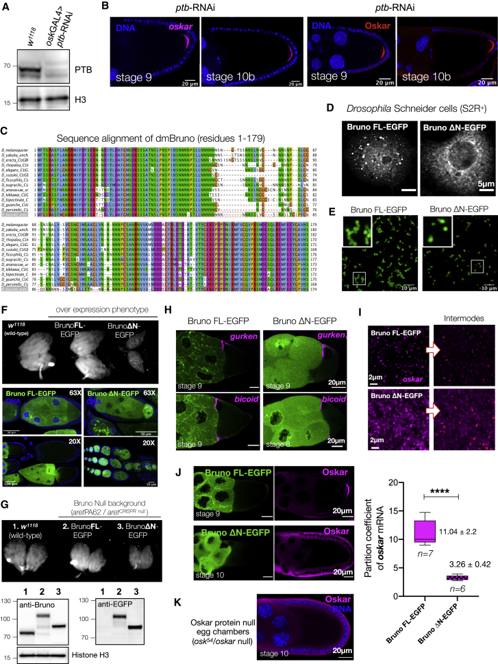
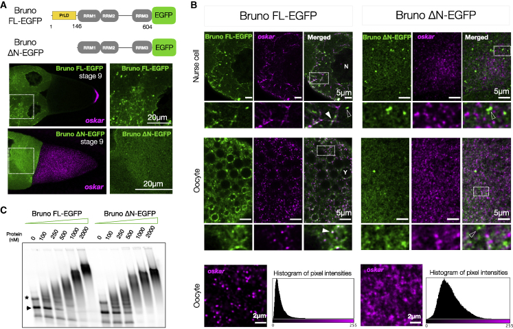
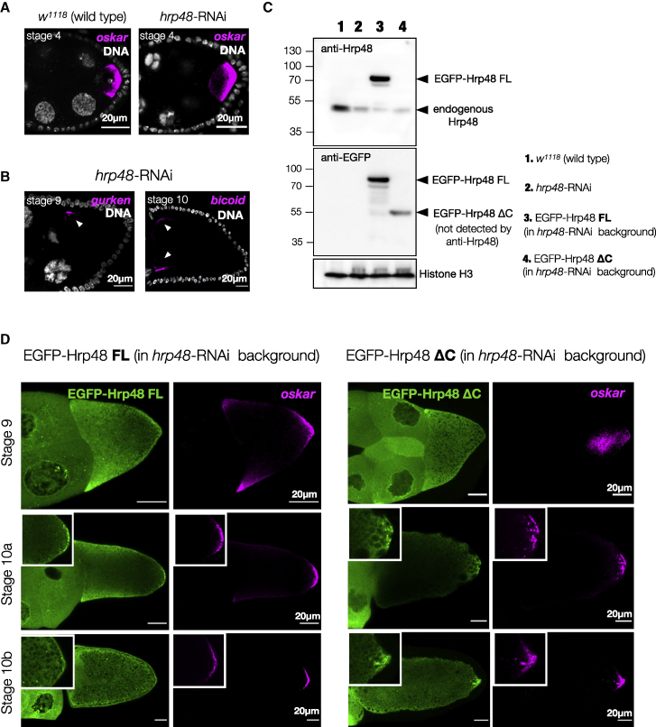
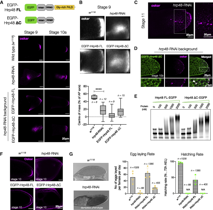
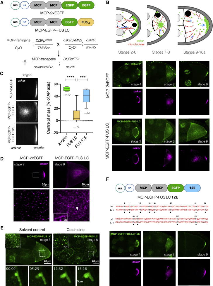
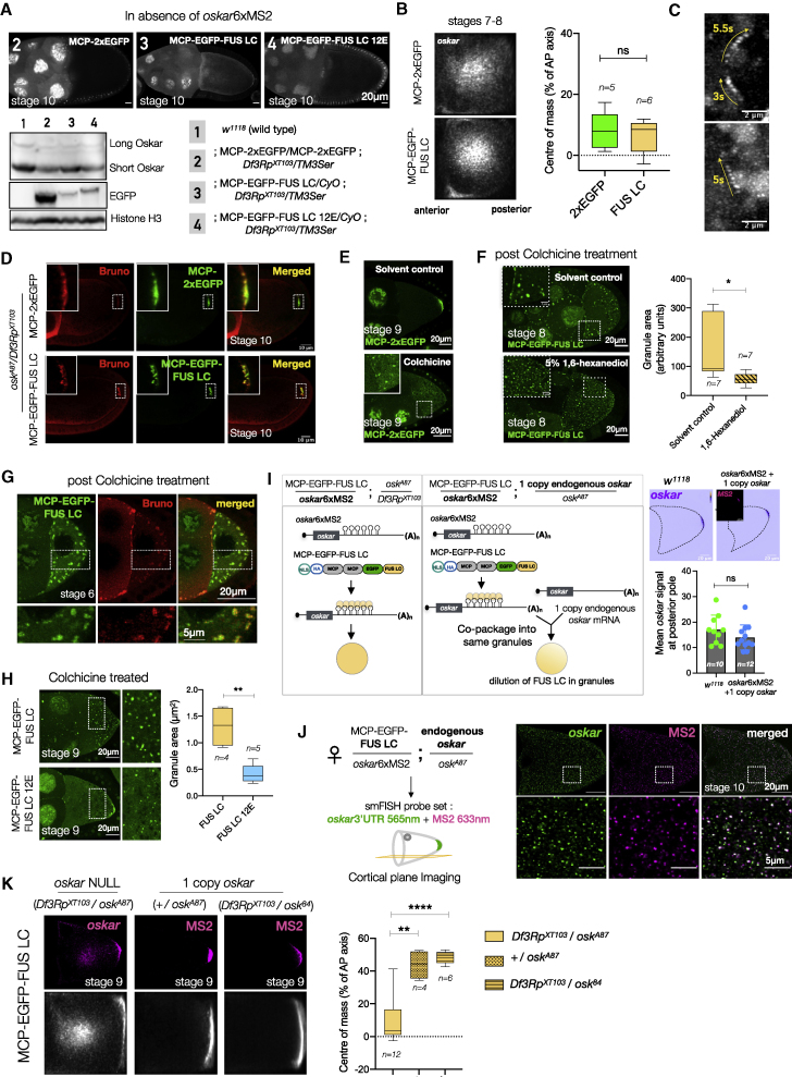
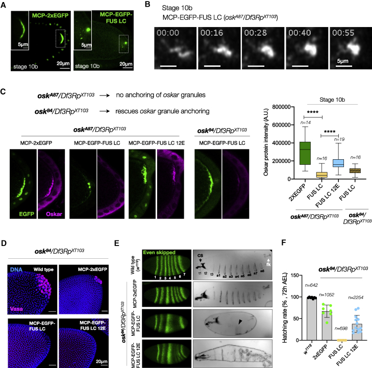
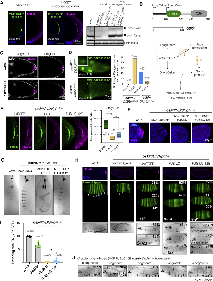
Asymmetric localization of oskar ribonucleoprotein (RNP) granules to the oocyte posterior is crucial for abdominal patterning and germline formation in the Drosophila embryo. We show that oskar RNP granules in the oocyte are condensates with solid-like physical properties. Using purified oskar RNA and scaffold proteins Bruno and Hrp48, we confirm in vitro that oskar granules undergo a liquid-to-solid phase transition. Whereas the liquid phase allows RNA incorporation, the solid phase precludes incorporation of additional RNA while allowing RNA-dependent partitioning of client proteins. Genetic modification of scaffold granule proteins or tethering the intrinsically disordered region of human fused in sarcoma (FUS) to oskar mRNA allowed modulation of granule material properties in vivo. The resulting liquid-like properties impaired oskar localization and translation with severe consequences on embryonic development. Our study reflects how physiological phase transitions shape RNA-protein condensates to regulate the localization and expression of a maternal RNA that instructs germline formation.

Keywords: RNA localization; RNP granules; biomolecular condensates; embryonic development; material properties; oskar mRNA; phase separation; ribonucleoprotein granules; translation control.


Conflict of interest statement

Declaration of interests The authors declare no competing interests.

Figures

None
Graphical abstract
Figure 1
Figure 1
oskar RNP granules are spherical, solid-like assemblies (A) Confocal and STED stacks in XZ plane, with oskar (grayscale) detected by single-molecule fluorescence in situ hybridization (smFISH) in wild-type (w1118) egg chambers. Center: comparison of granule aspect ratio. Error bars represent SD. Right: distribution of granule diameter as measured by STED. (B) Schematic of oskar mRNA in vivo labeling. A representative stage 10 egg chambers with oskar granules in grayscale. Live imaging of granules (min:sec) was performed on a cortical region approximately 15–20 μm from the posterior pole (1) and on granules at the posterior pole (2). A total of ∼70 pairs and clusters of granules were observed from time-lapse movies of six oocytes. (C) Representative images of egg chambers expressing RFP-Nup107 and oskar6xMS2-MCP-EGFP after treatment with solvent control or 5% 1,6-hexanediol for 15 min. White arrowheads, annulate lamellae. See also Figure S1; Video S1.
Figure S1
Figure S1
oskar granules maintain their solid-like behavior in late oogenesis, related to Figure 1 (A) Live imaging of MCP-EGFP tethered oskar granules performed on a cortical region at the posterior pole in a stage 12 egg chamber. The time point (min:sec) is indicated at the top of each frame. Arrowhead indicates a cluster of granules that is zoomed below. Also refer to Video S1. (B) Representative images of stage 12 egg chambers expressing RFP-Nup107 and oskar6xMS2-MCP-EGFP after treatment with solvent control or 5% 1,6-hexanediol for 15 min.
Figure S2
Figure S2
Association of oskar RNA with bona fide granule proteins in the egg chamber, related to Figure 2 (A) oskar mRNA association with three bona fide granule proteins. smFISH for oskar mRNA (magenta) and the respective proteins (green). A maximum intensity projection of a z stack of 1 μm is shown for nurse cell, ooplasm, and posterior pole (cortical plane). For Bruno, BrunoFL-EGFP was conditionally expressed in the germline in an endogenous Bruno-deficient genetic background (aretPA62/aretCRISPRnull). For Hrp48, EGFP-Hrp48 FL was expressed in the germline in an hrp48-RNAi background (refer to Figure S6 for knockdown efficiency). For PTB, a homozygous EGFP-trap line was used in which both alleles of PTB bear the GFP insertion. Insets are marked with a white boxes. White arrowheads, colocalized spots; empty arrowheads, absence of colocalization. Quantification of colocalization frequency in the three cellular compartments using an object-based approach (see STAR Methods). smFISH probe set used for oskar RNA (5′ to 3′): gatccatcagcgtaaatcg, ccaacttaatactccagactcg, ccagaacagatagggttcc, tcgttgattagacaggagtg, acaatagttgcccagcgg, tttgttagaatcggcaccaa, gcatattgtgcatctccttga, ctcgatctgaaccaaaggc, ataatgtccaccgatccga, gacgatgatctgagtaccc, agtccggatacacaaagtcc, cattcgggcgagatatagca, catcgcccataagcggaaag, agataggcatcgtaatccgag, tcgtcagcagagaatcgttg, gtcatttcgtggcgtctct, gctttgggttctgcagct, gagccaaattgattggttcctc, gctgtagatgttgatggg, gcatttacgctggcttgc, aattatcctggtagcaccag, gtttgaagggattcttccag, aggtgctcgtggtatgttc, tagtcgctggtgcgctct, agcaccatatccaggagg, cgttcttcaggctcgctt, aagatccgcttaccggac, ctgcactcagcggtcaca, ggaatggtcagcaggaaa, cgtcacgttgtcgtgcag, aaatggattgcccgtcag, cttgatgctcgatatcgtga, tgggcgtggctcagcaata, cgcgcacctcactatcta, atattcctcgcgcacgga, atagttgctctcgatgatgg, tgttctcgctggtgttgc, gttgtaggtgatttccttgg, tctgagtggacgagaagag, gctacgacttgcaactgc, gagttcatgggccaccaa, cttccacaactccggcaa. (B) Domain architecture of the three oskar granule proteins with disorder prediction using IUPred (Mészáros et al., 2018).
Figure S3
Figure S3
In vitro condensate assembly with oskar 3′UTR and granule RBPs, related to Figure 2 (A) Coomassie-stained SDS-PAGE gel of the purified three granule proteins from insect cells. (B) EMSA demonstrating the intrinsic affinities of the purified granule proteins for oskar 3′UTR RNA. 50 nM atto633-labeled oskar 3′UTR or y14 (control RNA) was used with the indicated concentrations of the proteins, and the reaction was resolved on an agarose gel. (C and D) Schematic representation of the in vitro condensate assembly assay for Bruno and Hrp48. 10 μM of tagged protein incubated with 1 U of SumoStar protease for 30 min at room temperature followed by SDS-PAGE or imaging. SDS-PAGE shows the efficiency of tag cleavage in assay buffer with 300 mM NaCl; ∗ indicates the cleaved protein band (C). Tag cleavage does not induce condensate formation in 300 mM NaCl buffer. Exchange to 150 mM NaCl buffer triggers LLPS of Bruno and Hrp48 (D). (E) 100 nM oskar 3′UTR-atto633 (red) does not self-assemble into condensates in absence of protein under the same conditions. Note that the laser power used for imaging was five times higher than for other conditions. (F) Molecular crowding promotes condensate formation of oskar 3′UTR with PTB. 10 μM RFP-PTB (green) is soluble in 150 mM NaCl buffer in absence or presence of oskar 3′UTR (magenta). Addition of 5% (w/v) PEG-4000 induces formation of spherical condensates of PTB alone and with oskar 3′UTR. (G) Condensates formed with 10 μM Bruno-EGFP or Hrp48-EGFP (green) and 100 nM oskar 3′UTR do not fuse and relax like liquid droplets, unlike hFUS-EGFP condensates assembled with 8-μM hFUS-EGFP (without RNA) in presence of 10% PEG-4000 (Patel et al., 2015). (H) FRAP of hFUS-EGFP condensates assembled with 8 μM hFUS-EGFP (without RNA) and 10% PEG-4000 (Patel et al., 2015). The bleached region of interest (ROI) is marked with a dotted circle. Bottom right: quantification of immobile fractions of hFUS droplets, and Bruno-EGFP (10 μM) or Hrp48-EGFP (10 μM) condensates assembled with 100 nM oskar 3′UTR in FRAP assays where fluorescence recovery was recorded up to 1 min after bleaching. (I) FRAP movie snapshots of 10 μM of Bruno or Hrp48 (heatmap) condensates assembled with 100 nM oskar 3′UTR (unlabeled) at indicated time points. The bleached ROI is marked with a dotted circle. Quantification is provided in Figure 2D. (J) Top panel: cryo-EM image of Bruno-oskar 3′UTR condensates deposited on a holey carbon EM grid. Leftmost panel shows a grid map with varying ice thickness, and yellow dotted box represents grid squares enlarged on the right. Within an individual square, condensates are indicated by black arrows. Small condensates deposited in holes amenable to tilt series acquisition are marked with white arrows, whereas black arrow marks a condensate on the support film that is too thick to be imaged. Bottom panel: cryo-EM overview images of Bruno-oskar 3′UTR (left) and Hrp48-oskar 3′UTR (right) condensates on the EM grid holey-support film. Left panel shows a cluster of spherical condensates too thick for acquiring tilt series; inset shows an enlarged view of spherical condensates. Right panel shows two spherical condensates; inset shows an enlarged view of one that is suitable for tilt series acquisition.
Figure 2
Figure 2
In vitro reconstituted minimal oskar RNP condensates recapitulate properties of in vivo RNP granules (A) oskar mRNA (magenta) association with three bona fide granule RBPs (green) in the nurse cell cytoplasm, ooplasm, and posterior pole of stage 10 egg chambers. (B) Domain architecture and PLAAC score of the three RBPs. RRM, RNA recognition motif; NLS, nuclear localization signal. (C) Condensates formed with 100 nM oskar 3′UTR-atto633 (magenta) and 10 μM of the indicated RBPs (green) imaged with confocal microscopy. (D) Quantification of fluorescent recovery after photobleaching (FRAP) kinetics and immobile fractions of condensates assembled with 100 nM oskar 3′UTR and 10 μM Bruno-EGFP or Hrp48-EGFP. Error bars, SD; N, number of movies. (E) Bruno-EGFP (green)-oskar 3′UTR (magenta) condensates subjected to the indicated treatments after 30 min of aging. Images acquired under identical microscope settings. (F) 4 nm-thick tomographic slices of condensates formed under the indicated conditions and plunge frozen after 30 min of aging. Yellow arrowheads, “beads on a string” structures; black arrowheads, putative naked RNA molecules. See also Figures S2 and S3; Video S2.
Figure 3
Figure 3
In vitro reconstituted oskar RNP condensates are selectively permeable (A) Scheme of the in vitro condensate ageing assay. Single confocal slices and representative line profiles (dotted arrows) shown. (B) 4 nm-thick tomographic slices of condensates at 0 and 30 min after addition of 400 nM atto633-oskar 3′UTR. Black arrowheads, naked RNA strands. (C) Condensates assembled with oskar 3′UTR-atto633 (blue) and EGFP-tagged Bruno or Hrp48 (green). mRFP-PTB (red) or TMR (tetramethyl rhodamine)-BSA (red) was added after 30 min. Single confocal slices shown; partition coefficient of mRFP-PTB calculated from 10 fields of view. (D) Tomographic slices (4 nm thick) of 2 μM mRFP-PTB added to 30 min aged Bruno-oskar 3′UTR condensates. White arrowheads, protein clusters. See also Figure S4; Video S3.
Figure S4
Figure S4
The liquid phase is essential for incorporation of oskar mRNA in vitro, related to Figure 3 (A) Quantification of in vivo protein concentrations per granule. GFP-trap lines of Bruno and PTB were used and absolute concentrations calculated based on a calibration curve of recombinant EGFP imaged under identical conditions in the same imaging session (Xing et al., 2020). Numbers in the histogram refer to the mean number of granules grouped under the indicated range of concentration. (B) For in vivo oskar RNA concentration per granule, w1118 egg chambers were stained for oskar by smFISH, and oskar copy number per granule in the oocyte compartment was calculated. A representative oskar smFISH image of a cortical plane acquisition done in “photon-counting mode” to avoid saturation of the signal in the oocyte. The intensity profile of the boxed area indeed shows the increase in oskar signal intensity along the AP axis. Granule volume obtained from 3D STED experiments was plotted, and absolute molar concentration of oskar RNA per granule was then derived based on average granule volume. Numbers in the histogram refer to the number of granules grouped under the indicated range of volume. (C) Representative light microscopy single plane confocal images of experimental conditions used for cryoelectron tomography in Figure 3B. Images were acquired and processed independently. (D) Condensates with Bruno alone preclude incorporation of oskar 3′UTR. Condensates were assembled with Bruno alone in 150 mM NaCl assay buffer. 10 nM atto633 labeled oskar 3′UTR RNA was added at 30 min of condensate aging. Note that new condensates formed after RNA addition show colocalization of the RNA and protein (marked by ). (E) Plot of mRNA intensity versus granule volume of oskar RNP granules measured by 3D STED on w1118 egg chambers probed for oskar mRNA by smFISH. Intensity of oskar mRNA signal (top plot) was normalized by granule volume to derive RNA concentration per granule, which does not increase with increase in granule volume.
Figure S5
Figure S5
Role of Bruno and its PrLD in oskar function, related to Figure 4 (A) Western blot depicting knockdown of PTB upon RNAi driven by oskarGAL4 driver in the germline. (B) Posterior localization of oskar (detected by smFISH) and translation of Oskar protein are unaffected upon PTB knockdown. (C) Sequence alignment of amino acids 1–179 of Drosophila melanogaster Bruno and orthologs in other Drosophilids. (D) Expression of Bruno FL-EGFP and ΔN-EGFP in Schneider cells (S2R+). Note that Schneider cells do not express oskar mRNA. (E) In vitro reconstitution of 10 μM Bruno FL-EGFP and ΔN-EGFP in 150 mM NaCl buffer. (F) Overexpression of EGFP-tagged FL and ΔN Bruno in the germline by oskarGAL4 driver. Ovary morphology of the different genotypes shows the atrophic ovaries caused by overexpression of ΔN-EGFP. Protein is in green, and nuclei are stained with DAPI. (G) Morphology of ovaries of the indicated genotype is shown along with wild type. Western blot showing levels of expression of Bruno transgenes (in Bruno-deficient genetic background) with respect to wild type. After probing Bruno, the blot was stripped and re-probed with anti-EGFP antibody; histone H3 serves as loading control. Note that ΔN-EGFP expression levels cannot be directly compared with wild type or FL-EGFP, as in the case of ΔN-EGFP oogenesis is arrested, and the ovaries primarily contain younger-stage egg chambers. (H) Localization of gurken and bicoid is not affected upon expression of EGFP-tagged Bruno FL or ΔN in a Bruno-deficient background (aretPA62/aretCRISPRnull). smFISH detected gurken (magenta) localizing correctly at the dorso-anterior corner and bicoid (magenta) at the anterior margin of the oocyte in mid-oogenesis. smFISH probe set used for gurken RNA (5′ to 3′): ggagctgctatatggcctg, ctacacacttgcatctccttg, tcggctcgaacaacaatctg, agcgtatgctctcggagaag, ctccaggcgattgagcaac, atcagtgattggtgtgctgc, tttcgggtgttgtcactgtc, tgaatctctgtctccttgtcg, tgttgttcaccatcggatgc, ggcaggaatggaagactgtg, agtcaccattccagctcttg, cgggaaaggagaagacgatg, gcgcaacgtaaagaaatatgg, tcgagtcgagtcccaatcc, gaacgcacacacacgaaac, gaccgattgtccaccactag, tctcctggatctgctgctg, caggtgtcggtactggatc, accgctctccatcgtagtc, agaacgtagagcgacgacag, ctgcttccggcgataatcc, tgcttatgcaggtgtagttg, tgccatccaacaaagaggag, aagcgaaacaaacgaaactaag, and for bicoid RNA (5′ to 3′): tggcaaaggagtgtggaaac, ctgaagctgcggatgttgg, tcgaagggatttcggaattg, ccatatcttcacctgggctg, gtccttgtgctgatccgat, ctccacccaagctaagagtc, gcgttgaatgactcgctgtag, tgtggcctccattgtagttg, ggtgattatggacctgctgc, gctggaagtcaaagtgatgg, gtagtacgagctgttgaagttg, gtgttaatggctcgtagacc, cacacagactcggactttcg, cttcttgctcgttccgtcg, cccttcaaaggctccaagatc, ctaaggctcttattccggtgc, ctccacgatttccggttcc, gcttgcattatcgtatccatcg, catccaggctaattgaagcag, atgaaactctctaacacgcctc, gtacaatcaggaacaacagtgg, acacggatcttaggactagacc, gaatagcgtattgcagggaaag, gcccaaatggcctcaaatg, ccgaaatgtgggacgataac. (I) Granular morphology of oskar RNPs is lost significantly in ΔN-EGFP. Representative single confocal plane of ooplasm with oskar (magenta) labeled by smFISH. Segmentation of granules from the dilute phase/cytoplasm was done using intensity-based segmentation and partition coefficient of oskar mRNA quantified. Error bars represent SD, and n denotes the number of oocytes analyzed. Unpaired Student’s t test were used for comparisons. Significance level: ∗∗∗∗ < 0.0001. (J and K) Oskar protein is not detected upon expression of ΔN. Immunostaining for Oskar protein (magenta) detected Oskar protein in Bruno FL, but not ΔN expressing egg chambers. In case of ΔN, signal (magenta) from the periphery of the egg chamber is background fluorescence (J) and is also detected in Oskar protein null flies (K).
Figure 4
Figure 4
Bruno is essential for oskar granule assembly (A) Bruno constructs used for transgenesis. oskar RNA (magenta) smFISH in stage 9 egg chambers expressing Bruno FL- or ΔN-EGFP (green). (B) Single-plane confocal images of egg chambers expressing Bruno FL- or ΔN-EGFP (green) and oskar (magenta). White arrowheads: colocalization of protein with oskar; empty white arrowheads, protein puncta not associated with oskar; N, nurse cell nucleus; Y, yolk granule. Bottom: enlarged view of oskar granules (magenta) in ooplasm. Images acquired with independent microscope settings. A histogram of pixel intensities of the two images confirms the significant loss of granule formation and diffuse oskar RNA signal in Bruno ΔN-EGFP. (C) EMSA of oskar 3′UTR-atto633 (50 nM) with increasing concentrations of recombinant Bruno FL- and ΔN-EGFP. Arrowhead, oskar 3′UTR; , dimeric form of the 3′UTR. See also Figure S5.
Figure S6
Figure S6
Role of Hrp48 and its PrLD in oskar localization, related to Figure 5 (A) oskar (magenta) enrichment in the oocyte is not affected by Hrp48 knockdown. (B) Localization of maternal RNAs gurken and bicoid (magenta) is not affected upon hrp48-RNAi. Maximum intensity projection of a Z volume of 5 μm. (C) Western blot of ovaries from flies of the indicated genotypes. The blot probed with anti-Hrp48 antibody has been stripped and re-probed with anti-GFP, as anti-Hrp48 failed to detect the truncated ΔC version. Histone H3 serves as loading control. (D) Representative confocal images of egg chambers of stages 9, 10a, and 10b shown with Hrp48 variants (green) and oskar (magenta) detected by smFISH from flies expressing the EGFP-tagged proteins in the hrp48-RNAi background. Insets show an enlarged version of the posterior pole.
Figure 5
Figure 5
Loss of Hrp48 from the germline impairs oskar localization and translation (A) Hrp48 constructs used for transgenesis. oskar mRNA (magenta) smFISH in stage 9 and 10 egg chambers of the indicated genotypes. (B) Mean oskar RNA signal (grayscale) from stage 9 oocytes, anterior to the left. Position of the oskar center of mass relative to the geometric center of the oocyte (dotted horizontal line) along the anteroposterior (AP) axis. Error bars, SD; n, number of analyzed oocytes. Unpaired Student’s t test used for comparisons. Significance level: ∗∗∗∗ < 0.0001. (C) Clustering of oskar mRNA (magenta) into micron-sized condensates in hrp48-RNAi oocytes. (D) Confocal slice showing EGFP-Hrp48 ΔC (green) associates with oskar (magenta) granules (white arrowhead). (E) EMSA of oskar 3′UTR-atto633 (50 nM) with increasing concentrations of recombinant EGFP-Hrp48 FL and ΔC. (F) Immunostaining of Oskar protein (magenta); signal in follicle cells is background from the antibody also detectable in Oskar protein null egg chambers (Figure S5K). (G) Stage 12 hrp48-RNAi egg chamber showing the dumpless phenotype. Egg laying and hatching rate of flies of the indicated genotypes. Error bars: SD. See also Figure S6.
Figure 6
Figure 6
Manipulating the material properties of oskar granules affects RNA localization (A) Transgenic constructs and scheme of genetic crosses. NLS, nuclear localization signal; HA, hemagglutinin tag. (B) Cartoon representation of oskar RNA localization during oogenesis (adapted from Cha et al., 2002). Representative confocal images of oskar localization from early to mid-oogenesis upon 2xEGFP and FUS LC tethering; EGFP (green) and oskar (magenta). (C) Quantification of transport defects. Mean oskar RNA smFISH signal (grayscale); anterior to the left. Position of the oskar center of mass relative to the geometric center of the oocyte (dotted horizontal line) along the AP axis. Error bars, SD; n, the number of oocytes analyzed. Unpaired Student’s t test used for comparisons. Significance levels: ∗∗∗ < 0.001 and ∗∗∗∗ < 0.0001. (D) Stage 9 egg chambers with oskar RNA (magenta) smFISH; a central region in the oocyte (dotted white box) is enlarged below. White arrowhead, track-like structure with oskar granules. (E) Depolymerization of microtubules with colchicine in ovaries ex vivo (upper panel). Lower panel: liquid-like behavior of FUS LC-oskar granules upon colchicine treatment (min:sec). (F) MCP-EGFP-FUS LC 12E transgenic construct; , 12 mutated residues. Representative images of early to mid-oogenesis stages; MCP-EGFP-FUS LC 12E (green) and oskar (magenta). See also Figure S7; Video S4.
Figure S7
Figure S7
Manipulating the solid-like properties of oskar granules in vivo, related to Figure 6 (A) Representative images of stage 10 egg chambers with EGFP signal in grayscale; respective genotypes are indicated. Western blot of ovaries from the indicated genotypes shows transgene expression levels (anti-EGFP antibody) and Oskar protein. Histone H3 serves as a loading control. (B) Mean oskar RNA signal (grayscale) from smFISH data from stage 7–8 oocytes, anterior to the left. Position of the oskar center of mass relative to the geometric center of the oocyte (dotted horizontal line) along the AP axis is indicated. Error bars represent SD, and n denotes the number of oocytes analyzed. Unpaired Student’s t test were used for comparisons. NS, nonsignificant. (C) Maximum Z projections of selected regions of Video S4 (MCP-EGFP-FUS LC in grayscale) showing directed tracks marked with yellow arrows, and all three depicted particles having a velocity >0.5 μm/s. (D) Bruno association with oskar granules is not affected upon FUS LC tethering as revealed by immunostaining for Bruno protein. (E) Colchicine treatment of isolated ovaries in case of 2xEGFP tethering. (F) Treatment of egg chambers with 5% 1,6-hexanediol for 15 min after 2 h of colchicine treatment dissolves the large spherical assemblies partially. Quantification of granule size shows a significant reduction in 1,6-hexanediol treated samples. Error bars represent SD, and n denotes the number of oocytes analyzed. Unpaired Student’s t test were used for comparisons. Significance level:  < 0.05. (G) Immunostaining of Bruno protein in egg chambers after colchicine treatment in the case of FUS LC tethering. (H) Colchicine treatment of stage 8–9 egg chambers induced the formation of large granules in case of FUS LC, which is significantly reduced in 12E. Error bars represent SD, and n denotes the number of oocytes analyzed. Unpaired Student’s t test were used for comparisons. Significance level: ∗∗ < 0.01. (I) Schematic representation of the rescue experiment in which one endogenous copy of oskar is supplied (left). Quantification of the mean oskar RNA signal (smFISH) from multiple stage 10a egg chambers of the indicated genotypes (right). n denotes the number of oocytes analyzed. Note that the oskar6xMS2 transgene is expressed from an oskar promoter. Unpaired Student’s t test were used for comparisons; ns, nonsignificant. (J) oskar6xMS2 mRNA and endogenous oskar transcripts co-package into the same granules. Two-color smFISH of egg chambers expressing one endogenous genomic oskar and the oskar 6xMS2 transgene with atto-565 probes against oskar (green) and atto-633 probes against MS2 loops (magenta). Bottom panels are enlarged from boxed regions. (K) Dilution of FUS LC per granule by an endogenous copy of oskar mRNA recuses the transport defects. Oskar distribution in representative stage 9 egg chambers detected by smFISH (magenta). Position of the oskar center of mass relative to the geometric center of the oocyte (dotted horizontal line) along the AP axis in indicated genetic backgrounds. N denotes the number of oocytes analyzed. Error bars represent SD. Unpaired Student’s t test were used for comparisons. Significance levels: ∗∗<0.01 and ∗∗∗∗<0.0001.
Figure 7
Figure 7
Manipulating the material properties of oskar granules interferes with oskar anchoring and translation and impairs embryonic development (A) Stage 10b egg chamber expressing MCP-2xEGFP or MCP-EGFP-FUS LC in oskar RNA-null background (oskA87/Df3RpXT103). (B) Fusion of FUS LC-oskar granules (grayscale) at the posterior of stage 10b egg chambers. (C) Immunostaining and quantification of Oskar protein (magenta) intensity in oskar RNA-null stage 10b egg chambers. The genotype in the fourth panel is osk84/Df3RpXT103 where the anchoring function is provided in trans. All images shown and quantified were acquired using identical microscope settings and representations contrast matched. Error bars, SD; n, number of analyzed oocytes. Unpaired Student’s t test used for comparisons. Significance level: ∗∗∗∗ < 0.0001. (D) Pole cells identified by Vasa immunostaining (magenta) and nuclei stained with DAPI (blue) in embryos of the indicated genotypes. (E) Pattern of even skipped (Eve) stripes (green) and representative cuticles of embryos of the indicated genotypes; anterior to the left, ventral to the bottom. t1–t3, thoracic segments; a1–a8, abdominal segments; cs, head skeleton (black arrow); fk, filzkörper (white arrow); black arrowhead, patchy band of denticles. Refer to Figure S8H for phenotypic classes observed. (F) Quantification of hatching rate of eggs of the indicated genotypes. Error bars, SD; n, number of eggs scored. See also Figure S8; Video S6.
Figure S8
Figure S8
Effect of altered physical state of oskar granules on embryonic development, related to Figure 7 (A) Oskar protein immunostaining in oocytes and western blot confirms loss of translation upon FUS LC tethering in oskar null background and rescue of translation in presence of an endogenous copy of oskar. Arrows mark the two isoforms of Oskar protein. Note that the reduction in Oskar protein levels in case of MCP-2xEGFP compared with wild type is due to the oskar-RNA-null (oskA87/Df3RpXT103) background of the flies. The black line after lane 1 indicates that lane 1 is not immediately adjacent to the other lanes in the original blot. (B) Schematic representation of Oskar protein domain architecture indicating the start sites of the long and short isoforms. Nonsense mutant osk84 encodes 254 residues from the N terminus and provides the anchoring function. Flowchart representation of multiple interdependent functions of Oskar protein isoforms in actin remodeling, anchoring, and organization of the germ plasm (adapted from Tanaka and Nakamura, 2011). (C) Anchoring of oskar RNPs is rescued in females expressing osk84 allele; NULL indicates the other chromosome: oskar-CRISPR-RNA-null allele. smFISH for oskar mRNA (magenta) on egg chambers of the indicated genotypes shows oskar anchoring in stage 10 (left) and stage 12 (right) egg chambers. (D) Anchoring defects are rescued in osk84/Df3RpXT103 background. Representative images of stage 10b egg chambers expressing MCP-EGFP-FUS LC (green) in the indicated genetic backgrounds. Quantification of oskar detachment phenotype from images of stage 10b egg chambers expressing the indicated transgenes in absence or presence of anchoring provided in trans. n denotes the number of egg chambers analyzed. (E) Immunostaining of egg chambers for Oskar protein (magenta) upon provision of anchoring in trans by the osk84 allele. The EGFP signal in green confirms the rescue of anchoring. All images shown (and used for quantification) were acquired using identical microscope settings and representations are contrast matched. Quantification of the Oskar signal intensity from the posterior of several egg chambers confirms the reduction of translation upon FUS LC tethering and partial translation using the FUS 12E construct. Note that the FUS LC panel is also shown in Figure 7C. Error bars represent SD, and n denotes number of analyzed oocytes. Unpaired Student’s t test were used for comparisons. Significance levels: ∗∗ < 0.01 and ∗∗∗∗ < 0.0001. (F) Formation of the germline is impaired upon Fus LC and 12E tethering in an oskar RNA-null background (oskA87/Df3RpXT103). Reduction of pole cell numbers is noted in 2xEGFP tethering compared with wild type. Pole cells at the posterior of embryos at nuclear cycle 14 are identified by Vasa (magenta) immunostaining. Nuclei stained with DAPI (blue). (G) Representative cuticles of embryos reveal severe patterning defects upon FUS LC tethering. Anterior faces the top and ventral to the left. (H) Immunostaining of Oskar protein (magenta) in early embryos, Eve (green) stripe patterns in cellular blastoderm embryos, and cuticle phenotypes are shown for the indicated genotypes. Representative images of the major phenotypic class observed for each genotype are shown in Figure 7E. n denotes the number of embryos or cuticles analyzed. (I) Quantification of the hatching rates of eggs from females expressing the indicated transgene in an oskar RNA-null background (oskA87/Df3RpXT103). Number of eggs scored per genotype is depicted in the graph. Note that data for w1118 are also shown in Figure 7F. Error bars represent SD, and n denotes the number of analyzed eggs. Unpaired Student’s t test were used for comparisons. Significance level:  < 0.05. (J) Cuticle analysis in the case of MCP-EGFP-FUS LC 12E (in osk84/Df3RpXT103 background) by collecting only those specimens present in the yeast paste placed in the center of the agar plate, to which the viable and crawling larvae are attracted. The larvae were then classified based on the number of segments. Majority of the crawlers had six to eight abdominal segments. denotes incomplete segments.

Comment in

References

    1. Alberti S. The wisdom of crowds: Regulating cell function through condensed states of living matter. J. Cell Sci. 2017;130:2789–2796. - PubMed
    1. Alberti S., Gladfelter A., Mittag T. Considerations and challenges in studying liquid-liquid phase separation and biomolecular condensates. Cell. 2019;176:419–434. - PMC - PubMed
    1. Alberti S., Hyman A.A. Biomolecular condensates at the nexus of cellular stress, protein aggregation disease and ageing. Nat. Rev. Mol. Cell Biol. 2021;22:196–213. - PubMed
    1. Audas T.E., Audas D.E., Jacob M.D., Ho J.J., Khacho M., Wang M., Perera J.K., Gardiner C., Bennett C.A., Head T., et al. Adaptation to stressors by systemic protein amyloidogenesis. Dev. Cell. 2016;39:155–168. - PMC - PubMed
    1. Banani S.F., Lee H.O., Hyman A.A., Rosen M.K. Biomolecular condensates: organizers of cellular biochemistry. Nat. Rev. Mol. Cell Biol. 2017;18:285–298. - PMC - PubMed

Publication types