

Pythium insidiosum keratitis - A review
- PMID: 35325996
- PMCID: PMC9240499
- DOI: 10.4103/ijo.IJO_1534_21
Pythium insidiosum keratitis - A review
Abstract
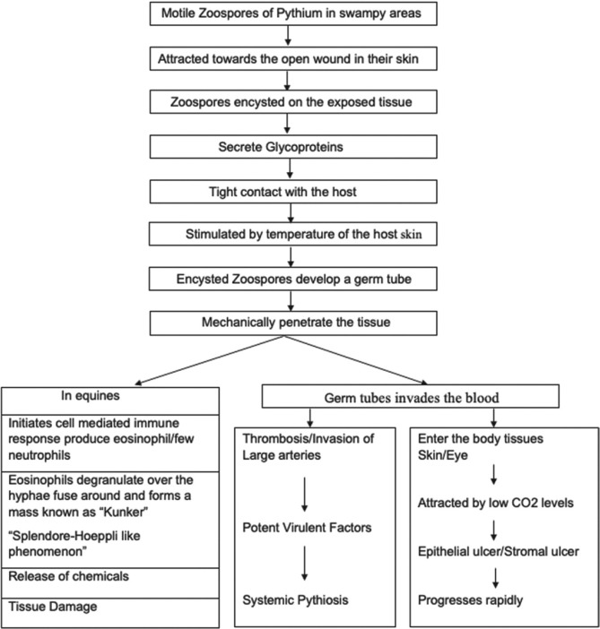
Pythium insidiosum is an oomycete and is also called "parafungus" as it closely mimics fungal keratitis. The last decade saw an unprecedented surge in Pythium keratitis cases, especially from Asia and India, probably due to growing research on the microorganism and improved diagnostic and treatment modalities. The clinical features such as subepithelial infiltrate, cotton wool-like fluffy stromal infiltrate, satellite lesions, corneal perforation, endoexudates, and anterior chamber hypopyon closely resemble fungus. The classical clinical features of Pythium that distinguish it from other microorganisms are reticular dots, tentacular projections, peripheral furrowing, and early limbal spread, which require a high index of clinical suspicion. Pythium also exhibits morphological and microbiological resemblance to fungus on routine smearing, revealing perpendicular or obtuse septate or aseptate branching hyphae. Culture on blood agar or any other nutritional agar is the gold standard for diagnosis. It grows as cream-colored white colonies with zoospores formation, further confirmed using the leaf incarnation method. Due to limited laboratory diagnostic modalities and delayed growth on culture, there was a recent shift toward various molecular diagnostic modalities such as polymerase chain reaction, confocal microscopy, ELISA, and immunodiffusion. As corneal scraping (10% KOH, Gram) reveals fungal hyphae, antifungals are started before the culture results are available. Recent in vitro molecular studies have suggested antibacterials as the first-line drugs in the form of 0.2% linezolid and 1% azithromycin. Early therapeutic keratoplasty is warranted in nonresolving cases. This review aims to describe the epidemiology, clinical features, laboratory and molecular diagnosis, and treatment of Pythium insidiosum keratitis.
Keywords: Keratitis; Pythium insidiosum; linezolid; parafungus; zoospore.
Conflict of interest statement
None
Figures


References
-
- Gaastra W, Lipman LJ, De Cock AW, Exel TK, Pegge RB, Scheurwater J, et al. Pythium insidiosum: An overview. Vet Microbiol. 2010;146:1–16. - PubMed
-
- Bagga B, Sharma S, Madhuri Guda SJ, Nagpal R, Joseph J, Manjulatha K, et al. Leap forward in the treatment of Pythium insidiosum keratitis. Br J Ophthalmol. 2018;102:1629–33. - PubMed
-
- Sharma S, Balne PK, Motukupally SR, Das S, Garg P, Sahu S, et al. Pythium insidiosum keratitis: Clinical profile and role of DNA sequencing and zoospore formation in diagnosis. Cornea. 2015;34:438–42. - PubMed
Publication types
MeSH terms
Substances
LinkOut - more resources
Full Text Sources
Research Materials
