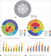
Corneal and corneal epithelial thickness distribution characteristics in healthy North Indian eyes using spectral domain optical coherence tomography
- PMID: 35326008
- PMCID: PMC9240488
- DOI: 10.4103/ijo.IJO_2259_21
Corneal and corneal epithelial thickness distribution characteristics in healthy North Indian eyes using spectral domain optical coherence tomography
Abstract
Purpose: To determine the pattern of corneal thickness and epithelial thickness distribution in healthy North Indian eyes by using spectral domain optical coherence tomography (SD-OCT).
Methods: The observational study measured total corneal and epithelial thickness in the central 2 mm zone and eight sectors each in paracentral 2-5 mm (ring 1) and midperipheral 5-7 mm (ring 2) zones on SD-OCT.
Results: The study included 67 eyes of 67 subjects with a male:female ratio of 32:35 and mean age of 25.04 ± 4.54 years. The mean central corneal and epithelial thicknesses were 505.97 ± 30.12 μm and 60.48 ± 8.37 μm, respectively. The epithelium of inferior and infero-nasal sectors in ring 1 and inferior sector in ring 2 was significantly thicker than the radially opposite sectors of the respective rings (P = 0.001; P = 0.01 and P = 0.02, respectively). Sector-wise analysis did not reveal any significant correlation between the total corneal thickness and epithelial thickness (all P > 0.05) except in the outer superior sector where there was a weak positive correlation (r = 0.28, P = 0.02). Central epithelial thickness in males (60.59 ± 9.28 μm) and females (60.37 ± 7.58 μm) was comparable (P = 0.91). Pachymetry was thinnest in the inferior, inferonasal, and inferotemporal sectors in 44.79% of eyes (n = 30), while thinnest epithelium was seen in the superior, superonasal, and superotemporal quadrants in 50.75% of eyes (n = 34).
Conclusion: The epithelial thickness distribution in this sample of topographically normal healthy North Indian eyes was nonuniform and independent of the underlying corneal thickness. Epithelium was thinner in the superior cornea, whereas total corneal thickness was minimum in the inferior part.
Keywords: Corneal epithelial thickness; corneal thickness; spectral-domain OCT.
Conflict of interest statement
None
Figures


Comment in
-
Commentary: Usefulness of corneal epithelial thickness measurement.Indian J Ophthalmol. 2022 Apr;70(4):1178-1179. doi: 10.4103/ijo.IJO_3062_21. Indian J Ophthalmol. 2022. PMID: 35326009 Free PMC article. No abstract available.
Similar articles
-
The Corneal Epithelial Thickness Profile in a Healthy Saudi Population.Cureus. 2024 Oct 9;16(10):e71135. doi: 10.7759/cureus.71135. eCollection 2024 Oct. Cureus. 2024. PMID: 39525125 Free PMC article.
-
Corneal epithelial thickness map in long-term soft contact lenses wearers.Optom Vis Sci. 2014 Dec;91(12):1455-61. doi: 10.1097/OPX.0000000000000410. Optom Vis Sci. 2014. PMID: 25303838
-
Evaluation of Corneal Epithelial Thickness Imaged by High Definition Optical Coherence Tomography in Healthy Eyes.Cornea. 2019 Jan;38(1):62-66. doi: 10.1097/ICO.0000000000001745. Cornea. 2019. PMID: 30211744
-
A cross-sectional study to determine the normal corneal epithelial thickness in Indian population using 9-mm wide optical coherence tomography scans.Indian J Ophthalmol. 2021 Sep;69(9):2425-2429. doi: 10.4103/ijo.IJO_244_21. Indian J Ophthalmol. 2021. PMID: 34427236 Free PMC article.
-
Corneal Epithelial Mapping Characteristics in Normal Eyes Using Anterior Segment Spectral Domain Optical Coherence Tomography.Transl Vis Sci Technol. 2022 Mar 2;11(3):6. doi: 10.1167/tvst.11.3.6. Transl Vis Sci Technol. 2022. PMID: 35258560 Free PMC article.
Cited by
-
Corneal Epithelial Thickness Mapping: A Major Review.J Ophthalmol. 2024 Jan 2;2024:6674747. doi: 10.1155/2024/6674747. eCollection 2024. J Ophthalmol. 2024. PMID: 38205099 Free PMC article. Review.
-
Blepharoptosis and corneal epithelial thickness alterations, is there any relation?BMC Ophthalmol. 2024 Jul 12;24(1):282. doi: 10.1186/s12886-024-03556-w. BMC Ophthalmol. 2024. PMID: 38997644 Free PMC article.
-
The Corneal Epithelial Thickness Profile in a Healthy Saudi Population.Cureus. 2024 Oct 9;16(10):e71135. doi: 10.7759/cureus.71135. eCollection 2024 Oct. Cureus. 2024. PMID: 39525125 Free PMC article.
-
Commentary: Usefulness of corneal epithelial thickness measurement.Indian J Ophthalmol. 2022 Apr;70(4):1178-1179. doi: 10.4103/ijo.IJO_3062_21. Indian J Ophthalmol. 2022. PMID: 35326009 Free PMC article. No abstract available.
-
Effect of Age and Gender on Corneal Epithelial Thickness in Refractive Surgery Candidates.Clin Ophthalmol. 2025 Aug 16;19:2765-2771. doi: 10.2147/OPTH.S528185. eCollection 2025. Clin Ophthalmol. 2025. PMID: 40852332 Free PMC article.
References
Publication types
MeSH terms
LinkOut - more resources
Full Text Sources

