

The Non-Specific Drp1 Inhibitor Mdivi-1 Has Modest Biochemical Antioxidant Activity
- PMID: 35326100
- PMCID: PMC8944504
- DOI: 10.3390/antiox11030450
The Non-Specific Drp1 Inhibitor Mdivi-1 Has Modest Biochemical Antioxidant Activity
Abstract
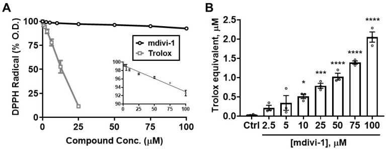
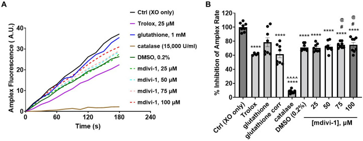
Mitochondrial division inhibitor-1 (mdivi-1), a non-specific inhibitor of Drp1-dependent mitochondrial fission, is neuroprotective in numerous preclinical disease models. These include rodent models of Alzheimer's disease and ischemic or traumatic brain injury. Among its Drp1-independent actions, the compound was found to suppress mitochondrial Complex I-dependent respiration but with less resultant mitochondrial reactive oxygen species (ROS) emission compared with the classical Complex I inhibitor rotenone. We employed two different methods of quantifying Trolox-equivalent antioxidant capacity (TEAC) to test the prediction that mdivi-1 can directly scavenge free radicals. Mdivi-1 exhibited moderate antioxidant activity in the 2,2'-azinobis (3-ethylbenzothiazoline 6-sulfonate) (ABTS) assay. Half-maximal ABTS radical depletion was observed at ~25 μM mdivi-1, equivalent to that achieved by ~12.5 μM Trolox. Mdivi-1 also showed antioxidant activity in the α, α-diphenyl-β-picrylhydrazyl (DPPH) assay. However, mdivi-1 exhibited a reduced capacity to deplete the DPPH radical, which has a more sterically hindered radical site compared with ABTS, with 25 μM mdivi-1 displaying only 0.8 μM Trolox equivalency. Both assays indicate that mdivi-1 possesses biochemical antioxidant activity but with modest potency relative to the vitamin E analog Trolox. Future studies are needed to evaluate whether the ability of mdivi-1 to directly scavenge free radicals contributes to its mechanisms of neuroprotection.
Keywords: Complex I; ROS; free radical; mitochondria; mitochondrial fission; neurons; oxidative stress; oxygen; scavenger; superoxide.
Conflict of interest statement
The authors declare no conflict of interest. The funders had no role in the design of the study; in the collection, analyses, or interpretation of data; in the writing of the manuscript, or in the decision to publish the results.
Figures


Similar articles
-
Inhibition of Mitochondrial Dynamics by Mitochondrial Division Inhibitor-1 Suppresses Cell Migration and Metastatic Markers in Colorectal Cancer HCT116 Cells.J Exp Pharmacol. 2025 Mar 18;17:143-157. doi: 10.2147/JEP.S510578. eCollection 2025. J Exp Pharmacol. 2025. PMID: 40124420 Free PMC article.
-
Mitochondrial Division Inhibitor 1 (mdivi-1) Protects Neurons against Excitotoxicity through the Modulation of Mitochondrial Function and Intracellular Ca2+ Signaling.Front Mol Neurosci. 2018 Jan 17;11:3. doi: 10.3389/fnmol.2018.00003. eCollection 2018. Front Mol Neurosci. 2018. PMID: 29386996 Free PMC article.
-
Mdivi-1 ameliorates early brain injury after subarachnoid hemorrhage via the suppression of inflammation-related blood-brain barrier disruption and endoplasmic reticulum stress-based apoptosis.Free Radic Biol Med. 2017 Nov;112:336-349. doi: 10.1016/j.freeradbiomed.2017.08.003. Epub 2017 Aug 5. Free Radic Biol Med. 2017. PMID: 28790012
-
To mdivi-1 or not to mdivi-1: Is that the question?Dev Neurobiol. 2017 Nov;77(11):1260-1268. doi: 10.1002/dneu.22519. Epub 2017 Aug 30. Dev Neurobiol. 2017. PMID: 28842943 Free PMC article. Review.
-
Advances in the analytical methods for determining the antioxidant properties of honey: a review.Afr J Tradit Complement Altern Med. 2011 Oct 2;9(1):36-42. doi: 10.4314/ajtcam.v9i1.5. eCollection 2012. Afr J Tradit Complement Altern Med. 2011. PMID: 23983317 Free PMC article. Review.
Cited by
-
Targeting mitochondrial shape: at the heart of cardioprotection.Basic Res Cardiol. 2023 Nov 13;118(1):49. doi: 10.1007/s00395-023-01019-9. Basic Res Cardiol. 2023. PMID: 37955687 Free PMC article.
-
A novel small molecule inhibitor of human Drp1.Sci Rep. 2022 Dec 13;12(1):21531. doi: 10.1038/s41598-022-25464-z. Sci Rep. 2022. PMID: 36513726 Free PMC article.
-
Mitochondrial Dynamics in Neurodegenerative Diseases: Unraveling the Role of Fusion and Fission Processes.Int J Mol Sci. 2023 Aug 22;24(17):13033. doi: 10.3390/ijms241713033. Int J Mol Sci. 2023. PMID: 37685840 Free PMC article. Review.
-
Detailed analysis of Mdivi-1 effects on complex I and respiratory supercomplex assembly.Sci Rep. 2024 Aug 24;14(1):19673. doi: 10.1038/s41598-024-69748-y. Sci Rep. 2024. PMID: 39187541 Free PMC article.
-
Modulating Mitochondrial Dynamics Mitigates Cognitive Impairment in Rats with Myocardial Infarction.Curr Neuropharmacol. 2024;22(10):1749-1760. doi: 10.2174/1570159X22666240131114913. Curr Neuropharmacol. 2024. PMID: 38362882 Free PMC article.
References
-
- Wu Q., Xia S.X., Li Q.Q., Gao Y., Shen X., Ma L., Zhang M.Y., Wang T., Li Y.S., Wang Z.F., et al. Mitochondrial division inhibitor 1 (Mdivi-1) offers neuroprotection through diminishing cell death and improving functional outcome in a mouse model of traumatic brain injury. Brain Res. 2016;1630:134–143. doi: 10.1016/j.brainres.2015.11.016. - DOI - PubMed
-
- Fan L.F., He P.Y., Peng Y.C., Du Q.H., Ma Y.J., Jin J.X., Xu H.Z., Li J.R., Wang Z.J., Cao S.L., et al. Mdivi-1 ameliorates early brain injury after subarachnoid hemorrhage via the suppression of inflammation-related blood-brain barrier disruption and endoplasmic reticulum stress-based apoptosis. Free Radic Biol. Med. 2017;112:336–349. doi: 10.1016/j.freeradbiomed.2017.08.003. - DOI - PubMed
-
- Wu Q., Gao C., Wang H., Zhang X., Li Q., Gu Z., Shi X., Cui Y., Wang T., Chen X., et al. Mdivi-1 alleviates blood-brain barrier disruption and cell death in experimental traumatic brain injury by mitigating autophagy dysfunction and mitophagy activation. Int. J. Biochem. Cell Biol. 2018;94:44–55. doi: 10.1016/j.biocel.2017.11.007. - DOI - PubMed
Grants and funding
LinkOut - more resources
Full Text Sources
Miscellaneous
