Review
. 2022 Feb 27;11(3):471.
doi: 10.3390/antiox11030471.

Shedding a New Light on Skin Aging, Iron- and Redox-Homeostasis and Emerging Natural Antioxidants

Affiliations
Review

Shedding a New Light on Skin Aging, Iron- and Redox-Homeostasis and Emerging Natural Antioxidants

Charareh Pourzand et al. Antioxidants (Basel). .

Abstract

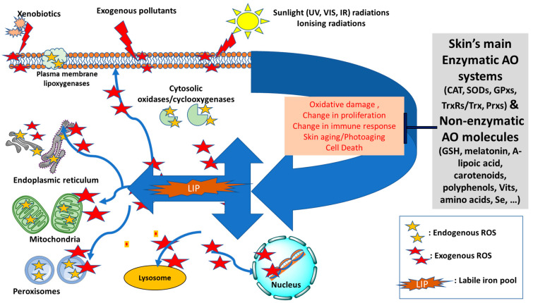
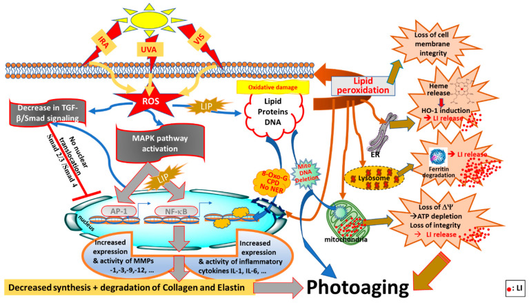
Reactive oxygen species (ROS) are necessary for normal cell signaling and the antimicrobial defense of the skin. However excess production of ROS can disrupt the cellular redox balance and overwhelm the cellular antioxidant (AO) capacity, leading to oxidative stress. In the skin, oxidative stress plays a key role in driving both extrinsic and intrinsic aging. Sunlight exposure has also been a major contributor to extrinsic photoaging of the skin as its oxidising components disrupt both redox- and iron-homeostasis, promoting oxidative damage to skin cells and tissue constituents. Upon oxidative insults, the interplay between excess accumulation of ROS and redox-active labile iron (LI) and its detrimental consequences to the skin are often overlooked. In this review we have revisited the oxidative mechanisms underlying skin damage and aging by focussing on the concerted action of ROS and redox-active LI in the initiation and progression of intrinsic and extrinsic skin aging processes. Based on these, we propose to redefine the selection criteria for skin antiaging and photoprotective ingredients to include natural antioxidants (AOs) exhibiting robust redox-balancing and/or iron-chelating properties. This would promote the concept of natural-based or bio-inspired bifunctional anti-aging and photoprotective ingredients for skincare and sunscreen formulations with both AO and iron-chelating properties.

Keywords: aging; anti-aging; antioxidant; photoaging; photoprotection; redox; skin; sunlight.


Conflict of interest statement

All authors except C.P. are full time employees of ASEA, LLC, Pleasant Grove, UT, USA. None has any financial interest in the company and the views expressed in this article are the personal views of the authors and may not be understood or quoted as being made on behalf of or reflecting the position of the company or organisation with which the authors are affiliated. ASEA, LLC, had no role in the design of the study; in the collection, analyses, or interpretation of data; in the writing of the manuscript, or in the decision to publish the results.

Figures

Figure 1
Figure 1
Diagram of the redox balance in the skin—Endogenous sources of reactive oxygen species (ROS, shown as yellow stars) can be generated intracellularly in mitochondria, peroxisomes, plasma membrane (lipoxygenases) or cytosolic enzymatic systems (oxidases and cyclooxygenases). The skin’s antioxidant (AO) defense will maintain the redox homeostasis by neutralising the excess ROS that are likely to form by exogenous factors, notably sunlight radiations, xenobiotics or pollution. The increase in the level of intracellular labile iron pool (LIP) disrupts the iron homeostasis and intensifies the oxidative damage to cell components due to formation of highly reactive oxygenated species via Fenton Chemistry (shown as red stars). As a result, the AO defense system can be overwhelmed and redox homeostasis disrupted leading to significant oxidative cell damage, change in cell proliferation and immune response, acceleration of aging processes and even cell death (Adapted and modified from references [4,5,6]). UV: ultraviolet; VIS: visible; IR: infrared; CAT: catalase; SODs: superoxide dismutases; GPxs: glutathione peroxidases; TrxRs/Trx: Thioredoxin reductases/Thioredoxin; Prxs: Peroxiredoxins; GSH: glutathione; Vits: vitamins.
Figure 2
Figure 2
Photoaging pathways through disruption of redox- and iron-homeostasis by oxidising components of sunlight (UVA, VIS, IRA). ROS (reactive oxygen species) generated by oxidising components of sunlight, notably UVA, can activate the MAPK (mitogen-activated protein kinase) and NF-κB (nuclear factor kappa B) signaling pathways, as well as AP-1 (activator protein 1) and NF-κB transcription factors leading to increased expression of proinflammatory cytokines (e.g., Interleukins IL-1β and IL-6) and MMPs (matrix metalloproteinases) that contribute to photoaging by changing concomitantly the composition of ECM (extracellular matrix) components by breaking down the collagen and elastin networks and regulating the TGF-β/Smad (tumour growth factor-B/Smad) signaling pathway to reduce collagen production. UVA-induced increase in LIP (labile iron pool) is also a major contributor to NF-κB activation and the related inflammatory response. Concomitantly with this, ROS generated by oxidising components of sunlight can regulate the TGF-β/Smad (tumour growth factor-B/Smad) signaling pathway to reduce collagen production, and ultimately accelerate skin photoaging. The presence of LIP in different compartments of cells, notably cytosol, lysosomes and mitochondria, makes the skin cell constitutents (i.e., lipid, protein and DNA) highly vulnerable to iron-catalysed ROS damage leading to lipid peroxidation, oxidative DNA 8-oxo-G (8-hydroxy guoanosine adducts) and CPDs (cyclopyrimidine dimers), and for mitochondrial DNA as deletions and finally for proteins as oxidation and degradation. The UVA-induced increase in cytosolic LIP also occurs as a result of proteolytic degradation of ferritin and release of heme from ER (endoplasmic reticulum) and activation of HO-1 (heme-oxygenase 1), all of which disrupt the cellular redox (and iron) homeostasis, leading to photoaging (taken and modified from references [5,78,112,113,114]).

References

    1. Proksch E., Brandner J.M., Jensen J.-M. The skin: An indispensable barrier. Exp. Dermatol. 2008;17:1063–1072. doi: 10.1111/j.1600-0625.2008.00786.x. - DOI - PubMed
    1. Madison K.C. Barrier function of the skin: “La raison d’être” of the epidermis. J. Investig. Dermatol. 2003;121:231–241. doi: 10.1046/j.1523-1747.2003.12359.x. - DOI - PubMed
    1. Krutmann J., Bouloc A., Sore G., Bernard B.A., Passeron T. The skin aging exposome. J. Dermatol. Sci. 2017;85:152–161. doi: 10.1016/j.jdermsci.2016.09.015. - DOI - PubMed
    1. Pastore S., Korkina L. Redox imbalance in T cell-mediated skin diseases. Mediat. Inflamm. 2010;2010:861949. doi: 10.1155/2010/861949. - DOI - PMC - PubMed
    1. Aroun A., Zhong J.L., Tyrrell R.M., Pourzand C. Iron, oxidative stress and the example of solar ultraviolet A radiation. Photochem. Photobiol. Sci. 2012;11:118–134. doi: 10.1039/C1PP05204G. - DOI - PubMed

LinkOut - more resources